. 2017 Apr 11;199(9):e00846-16.
doi: 10.1128/JB.00846-16. Print 2017 May 1.

Structural Analysis of Mycobacterium tuberculosis Homologues of the Eukaryotic Proteasome Assembly Chaperone 2 (PAC2)

Affiliations

Structural Analysis of Mycobacterium tuberculosis Homologues of the Eukaryotic Proteasome Assembly Chaperone 2 (PAC2)

Lin Bai et al. J Bacteriol. .

Abstract

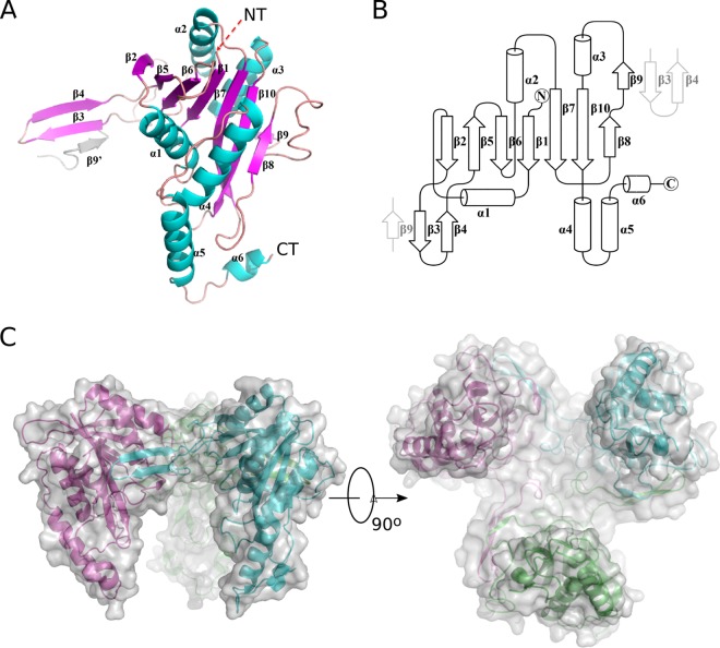
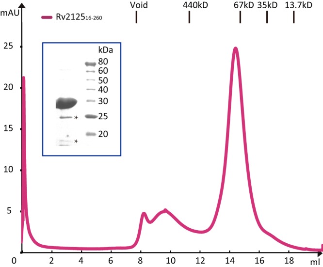
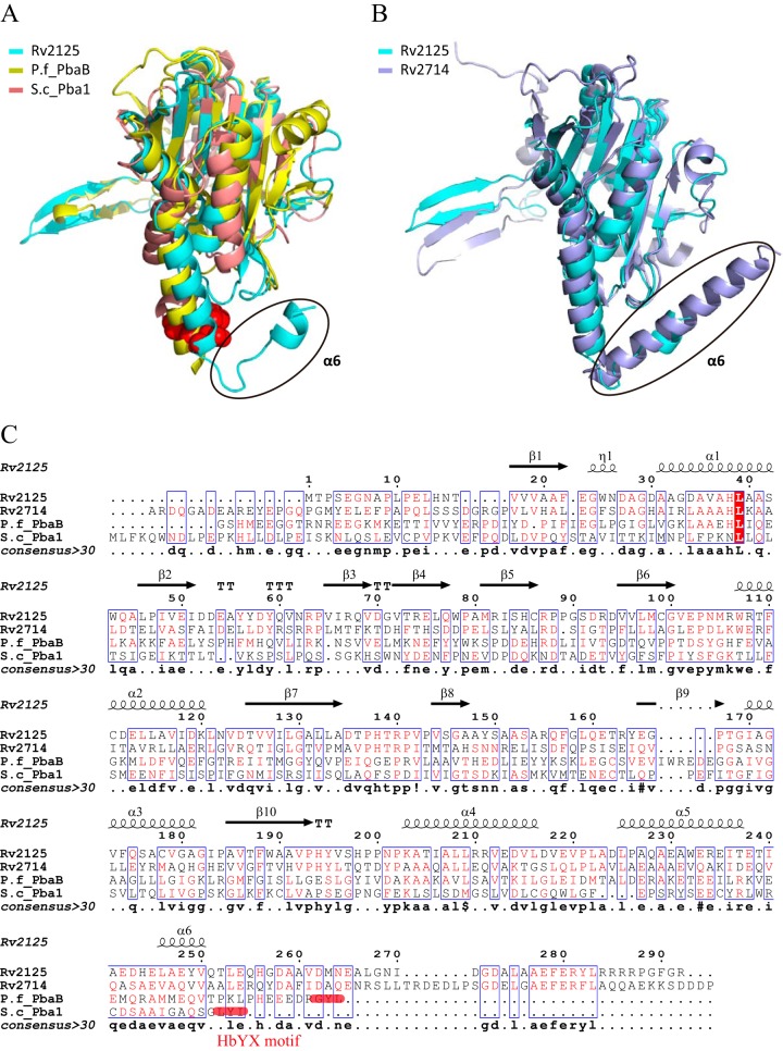
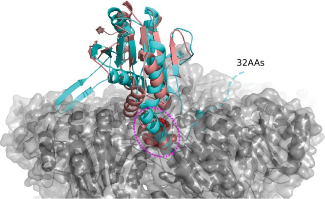
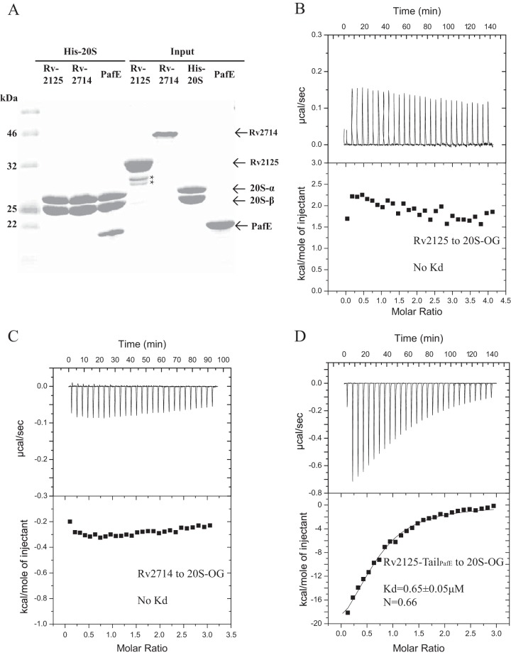
A previous bioinformatics analysis identified the Mycobacterium tuberculosis proteins Rv2125 and Rv2714 as orthologs of the eukaryotic proteasome assembly chaperone 2 (PAC2). We set out to investigate whether Rv2125 or Rv2714 can function in proteasome assembly. We solved the crystal structure of Rv2125 at a resolution of 3.0 Å, which showed an overall fold similar to that of the PAC2 family proteins that include the archaeal PbaB and the yeast Pba1. However, Rv2125 and Rv2714 formed trimers, whereas PbaB forms tetramers and Pba1 dimerizes with Pba2. We also found that purified Rv2125 and Rv2714 could not bind to M. tuberculosis 20S core particles. Finally, proteomic analysis showed that the levels of known proteasome components and substrate proteins were not affected by disruption of Rv2125 in M. tuberculosis Our work suggests that Rv2125 does not participate in bacterial proteasome assembly or function.IMPORTANCE Although many bacteria do not encode proteasomes, M. tuberculosis not only uses proteasomes but also has evolved a posttranslational modification system called pupylation to deliver proteins to the proteasome. Proteasomes are essential for M. tuberculosis to cause lethal infections in animals; thus, determining how proteasomes are assembled may help identify new ways to combat tuberculosis. We solved the structure of a predicted proteasome assembly factor, Rv2125, and isolated a genetic Rv2125 mutant of M. tuberculosis Our structural, biochemical, and genetic studies indicate that Rv2125 and Rv2714 do not function as proteasome assembly chaperones and are unlikely to have roles in proteasome biology in mycobacteria.

Keywords: Mycobacterium tuberculosis; proteasome; protein chaperone; structural biology.


Figures

FIG 1
FIG 1
Crystal structure of Rv2125. (A) The Rv2125 monomer. The secondary structural elements (β-strands, α-helices, and loops) are shown in magenta, cyan, and salmon, respectively. The N and C termini are labeled as NT and CT, respectively. The β3-β4 hairpin of the monomer interacts with the β9 strand of a neighboring monomer (β9′) within a trimer. (B) Secondary-structure topology of the Rv2125 monomer. The β3-β4 hairpin of the monomer interacts with the β9 strand of a neighboring monomer within a trimer. (C) The Rv2125 trimer in side (left) and bottom (right) views. Chains A, B, and C are colored cyan, magenta, and green, respectively. Surface representation of the trimer is in transparent light gray.
FIG 2
FIG 2
Size exclusion chromatography of Rv212516–260. Rv212516–260 was eluted as the major peak at about 14.2 ml, consistent with the expected elution volume of a globular protein of about 80 kDa, indicating that Rv2125 forms trimers. Size standards for calibration of the S200 size exclusion column are marked above the chromatographic panel. The inset is an SDS-PAGE gel of the elution peak. The major band at 27 kDa is the Rv2125 protein. The two lower bands marked by asterisks may be contaminants or breakdown products.
FIG 3
FIG 3
Structure comparison and sequence alignment of Rv2125, Rv2714 (PDB accession number 2WAM), archaeal PbaB (PDB accession number 3VR0), and yeast Pba1 (PDB accession number 4G4S). (A) Superimposition of the crystal structures of Rv2125, PbaB, and Pba1. The C-terminal HbYX motif of Pba1 is shown as red spheres. The black oval marks a hook-like C-terminal segment of Rv2125 that is absent in the other two proteins. (B) Superimposition of the crystal structures of Rv2125 and Rv2714. The hook-like C-terminal segments of both M. tuberculosis proteins are marked by the black oval. (C) Sequence alignment of four different PAC2 homologues. Alignment was done using MultAlin multiple sequence alignments (46) and ESPript 2.2 (47). The secondary structural elements of Rv2125 are drawn above the primary sequences. Amino acid sequences used in the alignment are those of M. tuberculosis Rv2125 (GI 15609262), M. tuberculosis Rv2714 (GI 15609851), P. furiosus PbaB (P.f_PbaB; GI 474452817), and Saccharomyces cerevisiae Pba1 (S.c_Pba1; GI 1023942167).
FIG 4
FIG 4
Superposition of the Rv212516–260 crystal structure with that of Pba1 in complex with a 20S CP (PDB accession number 4G4S). Rv212516–260 and Pba1 are shown in cyan and salmon, respectively. The C-terminal HbYX motif of Pba1 inserted in the binding pocket of the proteasome α-ring is shown as spheres and highlighted by a dashed magenta oval. The 20S CP is shown in a gray, semitransparent surface view. The dashed cyan line indicates the C-terminal 32 amino acids (32AAs) that were truncated in our Rv2125 construct. The C-terminal hook-like α-helix plus the additional 32-residue sequence are too long to be accommodated in the proteasome binding pocket.
FIG 5
FIG 5
Rv2125 and Rv2714 do not interact with the M. tuberculosis 20S CP. (A) SDS-PAGE of purified input proteins (lanes 5 to 8) and samples eluted off of the Ni2+ affinity column by 500 mM imidazole (lanes 2 to 4). Full-length Rv2125 or Rv2714 with the 6×His tag removed was mixed with purified and 6×His-tagged M. tuberculosis 20SOG before being loaded into the affinity column. Lane 1 shows the molecular mass markers. Lanes 2 and 3 contain only the 20S α subunit (PrcA) and β subunit (PrcB) because Rv2125 (lane 2) and Rv2714 (lane 3) did not bind the His-tagged 20S CP on the Ni2+ beads and were therefore washed off the column. Lane 4 is a positive control with PafE. We used an inactive 20SOG variant, which carries a T1A substitution at the catalytic N terminus of the β subunit, to prevent potential degradation of the input proteins by the CPs. The two light bands in lane 5 marked by asterisks are either breakdown products or contaminants. (B to D) ITC of Rv2125 (B), Rv2714 (C), and Rv2125-TailPafE (D) titrated against the M. tuberculosis 20SOG did not detect any heat signal.

References

    1. Nguyen L, Pieters J. 2009. Mycobacterial subversion of chemotherapeutic reagents and host defense tactics: challenges in tuberculosis drug development. Annu Rev Pharmacol Toxicol 49:427–453. doi:10.1146/annurev-pharmtox-061008-103123. - DOI - PubMed
    1. Darwin KH, Ehrt S, Gutierrez-Ramos JC, Weich N, Nathan CF. 2003. The proteasome of Mycobacterium tuberculosis is required for resistance to nitric oxide. Science 302:1963–1966. doi:10.1126/science.1091176. - DOI - PubMed
    1. Gandotra S, Lebron MB, Ehrt S. 2010. The Mycobacterium tuberculosis proteasome active site threonine is essential for persistence yet dispensable for replication and resistance to nitric oxide. PLoS Pathog 6:e1001040. doi:10.1371/journal.ppat.1001040. - DOI - PMC - PubMed
    1. Gandotra S, Schnappinger D, Monteleone M, Hillen W, Ehrt S. 2007. In vivo gene silencing identifies the Mycobacterium tuberculosis proteasome as essential for the bacteria to persist in mice. Nat Med 13:1515–1520. doi:10.1038/nm1683. - DOI - PMC - PubMed
    1. Samanovic MI, Darwin KH. 2016. Game of 'somes: protein destruction for Mycobacterium tuberculosis pathogenesis. Trends Microbiol 24:26–34. doi:10.1016/j.tim.2015.10.001. - DOI - PMC - PubMed

Publication types

MeSH terms

Substances

LinkOut - more resources