

Animal Models of Emerging Tick-Borne Phleboviruses: Determining Target Cells in a Lethal Model of SFTSV Infection
- PMID: 28194148
- PMCID: PMC5276813
- DOI: 10.3389/fmicb.2017.00104
Animal Models of Emerging Tick-Borne Phleboviruses: Determining Target Cells in a Lethal Model of SFTSV Infection
Abstract
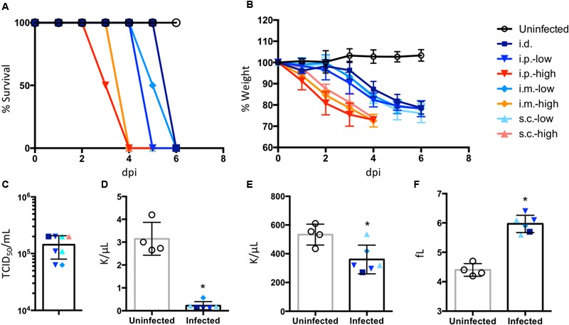
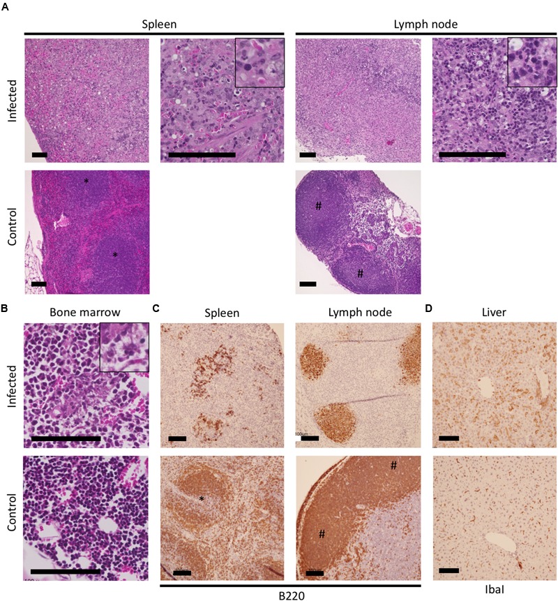
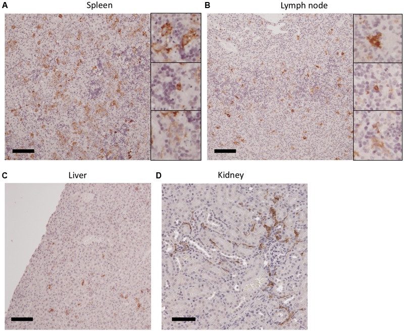
The pathogenesis of clinical manifestations caused by newly emerging tick-borne phleboviruses [i.e., Severe fever with thrombocytopenia syndrome virus (SFTSV) and Heartland virus (HRTV)], such as severe thrombocytopenia and lymphocytopenia, are not yet fully understood. In the present study, to establish an animal model mimicking the profile of fatal human cases, we examined the susceptibilities of adult mice from 12 strains, aged mice from two strains, and cynomolgus macaques to SFTSV and/or HRTV infections. However, none of these immunocompetent animals developed lethal diseases after infection with SFTSV or HRTV. Thus, we tested a lethal animal model of SFTSV infection using interferon-α/β receptor knock-out (IFNAR-/-) mice to identify the target cell(s) of virus infection, as well as lesions that are potentially associated with hematological changes. IbaI-positive macrophages and Pax5-positive immature B cells overlapped with SFTSV-positive cells in the spleen and lymph nodes of IFNAR-/- mice, and IbaI-SFTSV-double positive cells were also observed in the liver and kidney, thereby suggesting crucial roles for macrophages in the pathogenesis of SFTSV infection in mice. In the mandibular lymph nodes and spleens of infected mice, we observed extensive necrosis comprising B220-positive B cells, which may be associated with severe lymphocytopenia. The results of this study suggest a resemblance between the IFNAR-/- mouse model and lethal infections in humans, as well as roles for multiple cells during pathogenesis in mice.
Keywords: aged mouse; disease modeling; heartland virus; immunocompromised mouse; mouse; nonhuman primate; severe fever with thrombocytopenia syndrome virus.
Figures
Similar articles
-
Susceptibility of Type I Interferon Receptor Knock-Out Mice to Heartland Bandavirus (HRTV) Infection and Efficacy of Favipiravir and Ribavirin in the Treatment of the Mice Infected with HRTV.Viruses. 2022 Jul 28;14(8):1668. doi: 10.3390/v14081668. Viruses. 2022. PMID: 36016290 Free PMC article.
-
The pathogenesis of severe fever with thrombocytopenia syndrome virus infection in alpha/beta interferon knockout mice: insights into the pathologic mechanisms of a new viral hemorrhagic fever.J Virol. 2014 Feb;88(3):1781-6. doi: 10.1128/JVI.02277-13. Epub 2013 Nov 20. J Virol. 2014. PMID: 24257618 Free PMC article.
-
Single dose of a rVSV-based vaccine elicits complete protection against severe fever with thrombocytopenia syndrome virus.NPJ Vaccines. 2019 Jan 25;4:5. doi: 10.1038/s41541-018-0096-y. eCollection 2019. NPJ Vaccines. 2019. PMID: 30701094 Free PMC article.
-
Immune Modulation and Immune-Mediated Pathogenesis of Emerging Tickborne Banyangviruses.Vaccines (Basel). 2019 Sep 20;7(4):125. doi: 10.3390/vaccines7040125. Vaccines (Basel). 2019. PMID: 31547199 Free PMC article. Review.
-
Heartland Virus Epidemiology, Vector Association, and Disease Potential.Viruses. 2018 Sep 14;10(9):498. doi: 10.3390/v10090498. Viruses. 2018. PMID: 30223439 Free PMC article. Review.
Cited by
-
Vaccine Development for Severe Fever with Thrombocytopenia Syndrome.Viruses. 2021 Apr 6;13(4):627. doi: 10.3390/v13040627. Viruses. 2021. PMID: 33917632 Free PMC article. Review.
-
Severe fever with thrombocytopenia syndrome virus: emerging novel phlebovirus and their control strategy.Exp Mol Med. 2021 May;53(5):713-722. doi: 10.1038/s12276-021-00610-1. Epub 2021 May 6. Exp Mol Med. 2021. PMID: 33953322 Free PMC article. Review.
-
Fatal Tickborne Phlebovirus Infection in Captive Cheetahs, Japan.Emerg Infect Dis. 2018 Sep;24(9):1726-1729. doi: 10.3201/eid2409.171667. Emerg Infect Dis. 2018. PMID: 30124411 Free PMC article.
-
Infection Route Impacts the Pathogenesis of Severe Fever with Thrombocytopenia Syndrome Virus in Ferrets.Viruses. 2022 May 29;14(6):1184. doi: 10.3390/v14061184. Viruses. 2022. PMID: 35746656 Free PMC article.
-
Development and Characterization of a Reverse Genetics System for a Human-Derived Severe Fever With Thrombocytopenia Syndrome Virus Isolate From South Korea.Front Microbiol. 2021 Nov 11;12:772802. doi: 10.3389/fmicb.2021.772802. eCollection 2021. Front Microbiol. 2021. PMID: 34867909 Free PMC article.
References
-
- Flurkey K., Currer J. M., Harrison D. E. (2006). “The mouse in aging research,” in The Mouse in Biomedical Research ed. Fox J. G. (Burlington, MA: Elsevier; ) 637–672.
LinkOut - more resources
Full Text Sources
Other Literature Sources
Research Materials