

Case Reports
doi: 10.1155/2017/8919546.
Epub 2017 Jan 17.
Cystic Echinococcosis: A Case of Extrahepatic Intra-Abdominal Involvement
Affiliations
- PMID: 28194292
- PMCID: PMC5282424
- DOI: 10.1155/2017/8919546
Item in Clipboard
Case Reports
Cystic Echinococcosis: A Case of Extrahepatic Intra-Abdominal Involvement
Case Rep Radiol.
2017.
Abstract
Hydatid disease, or echinococcal disease, is a parasitic infestation caused by the larval stage of the Echinococcus tapeworm and it primarily affects the liver and lung but involvement of other organs is also possible secondary to peritoneal seeding or hematogeneous dissemination. We describe a rare case of extensive abdominal disease, with lesions affecting the liver, peritoneum, and lesser omentum, requiring aggressive surgical intervention. Complementary diagnostic exams were crucial to reach the diagnosis and evaluate the extension of the disease.
Conflict of interest statement
The authors declare that they have no competing interests.
Figures

Coronal contrast enhanced CT image shows multiple abdominal hypodense lesions within liver (orange arrowhead) and lesser omentum (blue arrowhead) and peritoneum and above the bladder (green arrowhead).

Axial contrast enhanced CT image nicely shows the hypodense lesions with water density and multiple smaller cysts at the periphery of the larger cysts (daughter cysts, arrowheads).
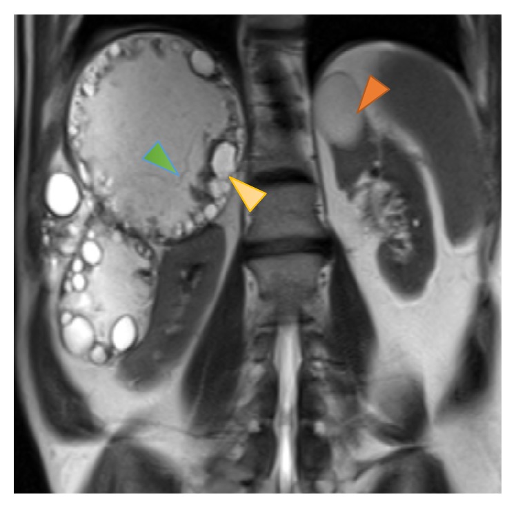
Coronal T2-weighted MR image shows multiple abdominal hyperintense lesions with smaller peripheral cysts compatible with daughter cysts (yellow arrow) and also thin linear septation, compatible with the detachment of the laminated membrane of the pericyst (green arrowhead). Note also a simple renal cyst (orange arrowhead), probably an incidental finding.

(a) DWI b = 50 and (b) ADC mapping at the levels of the liver hydatid cysts and renal cyst show higher signal intensity of the renal cyst at DWI, suggesting a simple cyst. There is also an area of water motion restriction identified as a focus of high signal intensity at DWI and low signal on the ADC map (arrows).
References
-
- Dandan I. S., Soweid A. M., Abiad F. Hydatid Cysts Differential Diagnoses. Medscape 2016, http://emedicine.medscape.com/article/178648-differential.
Publication types
LinkOut - more resources
Full Text Sources
Other Literature Sources
