

Review
doi: 10.4103/2152-7806.196919.
eCollection 2016.
Management of intracerebral hemorrhage in pediatric neurosurgery
Affiliations
- PMID: 28194298
- PMCID: PMC5299152
- DOI: 10.4103/2152-7806.196919
Item in Clipboard
Review
Management of intracerebral hemorrhage in pediatric neurosurgery
Surg Neurol Int.
.
No abstract available
Keywords: Arteriovenous malformation; cavernoma; cavernous malformation; intracerebral hemorrhage; intracranial hemorrhage; neurovascular.
Conflict of interest statement
There are no conflicts of interest.
Figures
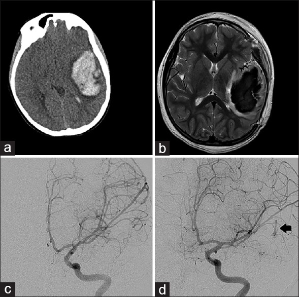
Left frontal hemorrhagic lesion from Case 1. (a) Computed tomography of the head at presentation showed a 9 × 4 × 4 cm hematoma with midline shift. (b) Magnetic resonance imaging brain, T2 sequence showed surrounding vessels but no definite arteriovenous malformation nidus around the large hematoma. (c) Left internal carotid artery digital subtraction angiogram at presentation, without evidence of vascular malformation. (d) Left internal carotid artery digital subtraction angiogram at 4 months post-hemorrhage reveals a left temporal Grade 1 arteriovenous malformation (arrow)

Right cerebellar hemorrhagic lesion from Case 2. (a) Computed tomography of the head at presentation showed a large hemorrhage of the right cerebellar hemisphere with displacement of the fourth ventricle. (b) Magnetic resonance imaging brain, T2 sequence showed perilesional edema and some associated vessels. (c) Right vertebral artery injection DynaCT, coronal view, showed an arteriovenous malformation with feeders from the right anterior and posterior cerebellar arteries. (d) Postoperative angiogram, AP projection, showed no residual malformation

Left frontal hemorrhage from Case 3. (a) Computed tomography of the head showed a large left frontal intracerebral hemorrhage with subfalcine shift and perilesional edema. (b) Computed tomography with contrast showed partial enhancement of the lesion without any vascular feeders. (c) Magnetic resonance imaging brain, gradient echo sequence, showed a central lesion with surrounding hemorrhage, suggestive of cavernous malformation. (d) Postoperative magnetic resonance imaging brain with contrast showed complete resection of the lesion and hematoma, with resolution of mass effect
References
-
- Abla AA, Rutledge WC, Seymour ZA, Guo D, Kim H, Gupta N, et al. A treatment paradigm for high-grade brain arteriovenous malformations: Volume-staged radiosurgical downgrading followed by microsurgical resection. J Neurosurg. 2015;122:419–32. - PubMed
-
- Al-Holou WN, O’Lynnger TM, Pandey AS, Gemmete JJ, Thompson BG, Muraszko KM, et al. Natural history and imaging prevalence of cavernous malformations in children and young adults. J Neurosurg Pediatr. 2012;9:198–205. - PubMed
-
- Bilginer B, Narin F, Hanalioglu S, Oguz KK, Soylemezoglu F, Akalan N. Cavernous malformations of the central nervous system (CNS) in children: Clinico-radiological features and management outcomes of 36 cases. Childs Nerv Syst. 2014;30:1355–66. - PubMed
-
- Blauwblomme T, Bourgeois M, Meyer P, Puget S, Di Rocco F, Boddaert N, et al. Long-term outcome of 106 consecutive pediatric ruptured brain arteriovenous malformations after combined treatment. Stroke. 2014;45:1664–71. - PubMed
-
- Clatterbuck RE, Moriarity JL, Elmaci I, Lee RR, Breiter SN, Rigamonti D. Dynamic nature of cavernous malformations: A prospective magnetic resonance imaging study with volumetric analysis. J Neurosurg. 2000;93:981–6. - PubMed
Publication types
LinkOut - more resources
Full Text Sources
Other Literature Sources