. 2017 Feb 8:5:e2947.
doi: 10.7717/peerj.2947. eCollection 2017.

Tracking the virus-like particles of Macrobrachium rosenbergii nodavirus in insect cells

Affiliations

Tracking the virus-like particles of Macrobrachium rosenbergii nodavirus in insect cells

Ummi Fairuz Hanapi et al. PeerJ. .

Abstract

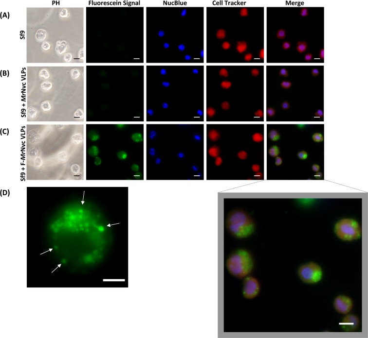
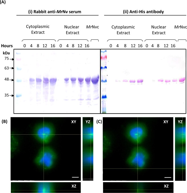
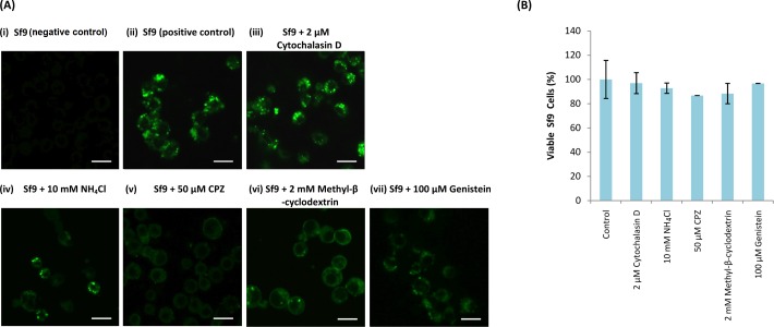
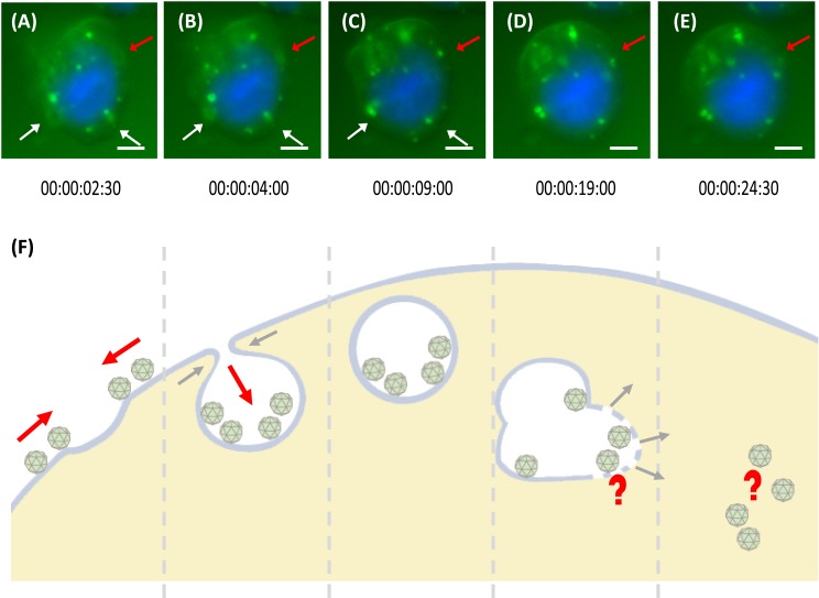
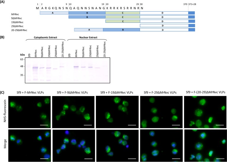
Macrobrachium rosenbergii nodavirus (MrNv) poses a major threat to the prawn industry. Currently, no effective vaccine and treatment are available to prevent the spread of MrNv. Its infection mechanism and localisation in a host cell are also not well characterised. The MrNv capsid protein (MrNvc) produced in Escherichia coli self-assembled into virus-like particles (VLPs) resembling the native virus. Thus, fluorescein labelled MrNvc VLPs were employed as a model to study the virus entry and localisation in Spodoptera frugiperda, Sf9 cells. Through fluorescence microscopy and sub-cellular fractionation, the MrNvc was shown to enter Sf9 cells, and eventually arrived at the nucleus. The presence of MrNvc within the cytoplasm and nucleus of Sf9 cells was further confirmed by the Z-stack imaging. The presence of ammonium chloride (NH4Cl), genistein, methyl-β-cyclodextrin or chlorpromazine (CPZ) inhibited the entry of MrNvc into Sf9 cells, but cytochalasin D did not inhibit this process. This suggests that the internalisation of MrNvc VLPs is facilitated by caveolae- and clathrin-mediated endocytosis. The whole internalisation process of MrNvc VLPs into a Sf9 cell was recorded with live cell imaging. We have also identified a potential nuclear localisation signal (NLS) of MrNvc through deletion mutagenesis and verified by classical-NLS mapping. Overall, this study provides an insight into the journey of MrNvc VLPs in insect cells.

Keywords: Endosome; Nodavirus; Nuclear translocation; Sub-cellular localisation; Virus-like particle.


Conflict of interest statement

The authors declare there are no competing interests.

Figures

Figure 1
Figure 1. Triple fluorescence labelling and detection of MrNvc VLPs in Sf9 cells.
(A) Sf9 cells in the absence of MrNvc VLPs, (B) Sf9 cells incubated with non-labelled MrNvc VLPs and (C) Sf9 cells incubated with F-MrNvc VLPs. The cell nucleus and cytoplasm were labelled with NucBlue Live Ready Probe Reagent (blue) and Cell Tracker Orange (red), respectively. PH indicates images captured under white light. Merge images represent the superimposed green, blue and red signals. (D) Small granular appearance in a Sf9 cell incubated with F-MrNvc VLPs is indicated by the arrows. Bars, 10 µm.
Figure 2
Figure 2. MrNvc distribution in Sf9 cytoplasmic and nuclear components.
(A) Western blot analysis of the cytoplasmic and nuclear components of Sf9 cells incubated with MrNvc VLPs (25 µg/ml) at different time (0, 4, 8, 12 and 16 h). Both cytoplasmic and nuclear extracts were probed with rabbit anti-MrNv serum (A(i)) and anti-His antibody (A(ii)). (B–C) The orthogonal view from the Z-stack images of the green fluorescence in the nucleus (B) and cytoplasm (C) of Sf9 cells incubated with MrNvc VLPs. Blue fluorescence indicates the cell nucleus. Yellow lines interception shows the location of the green fluorescent spots (F-MrNvc VLPs). Bars, 5 µm.
Figure 3
Figure 3. Effect of endosomal inhibitors on the entry of MrNvc VLPs into Sf9 cells.
Sf9 cells were pre-incubated with different endosomal inhibitors: (A(iii)) cytochalasin D (2 µM), (A(iv)) NH4Cl (10 mM), (A(v)) CPZ (50 µM), (A(vi)) methyl-β-cyclodextrin (2 mM) and (A(vii)) genistein (100 µM). MrNvc VLPs labelled with NHS-fluorescein (F-MrNvc VLPs; 25 mg/ml) were added to each pre-treated sample and incubated for 16 h in the presence of endosomal inhibitors. Bars, 20 µm. (A(ii)) Sf9 cells added with F-MrNvc VLPs but without any inhibitor serve as positive control, whereas (A(i)) Sf9 cells without any inhibitor nor F-MrNvc VLPs serve as negative control. (B) MTT assay showing the viability of Sf9 cells in the presence of inhibitors.
Figure 4
Figure 4. Trafficking mechanism of MrNvc VLPs into Sf9 cells.
(A–E) Live cell imaging of the formation of endosomes and endosomal escape of F-MrNvc VLPs. Sf9 cells were incubated with F-MrNvc VLPs at 4 °C for 1 h and shifted to RT for 30 min before they were viewed under a live cell imaging system. Each image was captured in 30 s time lapse for 1 h (Video S1). White arrows indicate endosome formation and red arrows show endosome formation to endosomal escape of VLPs. The live cell imaging image shows (A) the attached F-MrNvc VLPs were gathered around a hollow membrane pit, and (B) accumulated inside the pit. (C) Endosome enclosing the VLPs was formed. (D) The size and shape of the endosome become disproportionate and (E) F-MrNvc was released into the cytosol. These images were captured at the specified duration of time lapse (Video S1). (F) A schematic diagram summarising the whole process. Bars, (A–E) 5 µm.
Figure 5
Figure 5. Sub-cellular localisation of the N-terminal deletion mutants of MrNvc in Sf9 cells
(A) Four mutants and the full length MrNvc were used to infect Sf9 cells for 16 h. (B) The cytoplasm and nuclear components were extracted and analysed with Western blotting by using rabbit anti-MrNv serum to examine the localisation sites. Sf9 cells served as negative controls. (C) The N-terminal mutants and the full length capsid proteins were labelled with NHS-fluorescein and viewed under a fluorescence microscope. Blue colour indicates the cell nucleus. The merged images are the superimposed images of blue and green signals. Bars, 20 µm.

Similar articles

Cited by

References

    1. Bennett SM, Zhao L, Bosard C, Imperiale MJ. Role of a nuclear localization signal on the minor capsid proteins VP2 and VP3 in BKPyV nuclear entry. Virology. 2015;474:110–116. doi: 10.1016/j.virol.2014.10.013. - DOI - PMC - PubMed
    1. Bonami J-R, Widada JS. Viral diseases of the giant fresh water prawn Macrobrachium rosenbergii: a review. Journal of Invertebrate Pathology. 2011;106:131–142. doi: 10.1016/j.jip.2010.09.007. - DOI - PubMed
    1. Doyle M, Badertscher L, Jaskiewicz L, Guttinger S, Jurado S, Hugenschmidt T, Kutay U, Filipowicz W. The double-stranded RNA-binding domain of human Dicer functions as a nuclear localization signal. RNA. 2013;19:1238–1252. doi: 10.1261/rna.039255.113. - DOI - PMC - PubMed
    1. Goh ZH, Mohd NA, Tan SG, Bhassu S, Tan WS. RNA-binding region of Macrobrachium rosenbergii nodavirus capsid protein. Journal of General Virology. 2014;95:1919–1928. doi: 10.1099/vir.0.064014-0. - DOI - PubMed
    1. Goh ZH, Tan SG, Bhassu S, Tan WS. Virus-like particles of Macrobrachium rosenbergii nodavirus produced in bacteria. Journal of Virological Methods. 2011;175:74–79. doi: 10.1016/j.jviromet.2011.04.021. - DOI - PubMed

LinkOut - more resources