Review
. 2017 Jan 30:4:2.
doi: 10.3389/fcvm.2017.00002. eCollection 2017.

Eating the Dead to Keep Atherosclerosis at Bay

Affiliations
Review

Eating the Dead to Keep Atherosclerosis at Bay

Megan L Brophy et al. Front Cardiovasc Med. .

Abstract

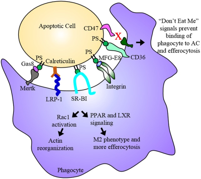
Atherosclerosis is the primary cause of coronary heart disease (CHD), ischemic stroke, and peripheral arterial disease. Despite effective lipid-lowering therapies and prevention programs, atherosclerosis is still the leading cause of mortality in the United States. Moreover, the prevalence of CHD in developing countries worldwide is rapidly increasing at a rate expected to overtake those of cancer and diabetes. Prominent risk factors include the hardening of arteries and high levels of cholesterol, which lead to the initiation and progression of atherosclerosis. However, cell death and efferocytosis are critical components of both atherosclerotic plaque progression and regression, yet, few currently available therapies focus on these processes. Thus, understanding the causes of cell death within the atherosclerotic plaque, the consequences of cell death, and the mechanisms of apoptotic cell clearance may enable the development of new therapies to treat cardiovascular disease. Here, we review how endoplasmic reticulum stress and cholesterol metabolism lead to cell death and inflammation, how dying cells affect plaque progression, and how autophagy and the clearance of dead cells ameliorates the inflammatory environment of the plaque. In addition, we review current research aimed at alleviating these processes and specifically targeting therapeutics to the site of the plaque.

Keywords: apoptosis; atherosclerosis; autophagy; efferocytosis; macrophages.


Figures

Figure 1
Figure 1
Atherosclerotic plaque progression. (A) Endothelial cells activated by disturbed flow and oxidized low-density lipoprotein (oxLDL) uptake express adhesion molecules and chemoattractant proteins that recruit monocytes and promote their adhesion to the endothelium and transmigration into the subendothelium. These monocytes differentiate into macrophages. (B) Macrophages uptake oxLDL sequestered in the subendothelium eventually becoming lipid-laden foam cells. Vascular smooth muscle cells (VSMCs) can migrate to the subendothelium where they lose expression of SMC markers and gain expression of macrophage markers. This allows them to ingest lipids and eventually become foam cells contributing to plaque progression. (C) These foam cells eventually undergo apoptosis and necroptosis, and, if not effectively cleared by M2 macrophages via efferocytosis, undergo secondary necrosis contributing to the formation of the necrotic core. As the necrotic core grows and the fibrous cap thins, the plaque is vulnerable to rupture, which may result in acute cardiovascular events such as thrombosis. (D) VSMCs near the cap of the plaque secrete extracellular matrix components that contribute to the formation of a fibrous cap that protects the plaque from rupturing. M2 macrophages express anti-inflammatory markers that act to reduce the inflammation of the plaque. They also perform efferocytosis, thereby reducing the apoptotic and necrotic cells within the plaque and promoting plaque stability.
Figure 2
Figure 2
Efferocytosis. Efferocytosis is the phagocytosis of dying cells by macrophages and is an essential process for the maintenance or regression of the atherosclerotic plaque. Phosphatidylserine (PS) among other molecules are “eat me” signals expressed on the dying cell, which are recognized by receptors on the phagocyte. Binding of PS by these receptors results in Rac1 activation and actin reorganization as well as PPAR and LXR signaling. PPAR and LXR signaling result in the upregulation of proteins involved in binding the dying cell as well as proteins involved in cholesterol efflux and anti-inflammatory cytokines ultimately promoting an anti-inflammatory M2 macrophage phenotype. The dying cell is engulfed and digested, its inflammatory contents cleared from the subendothelium. In advanced plaques, apoptosis and efferocytosis become dysregulated and imbalanced. Apoptotic cells that express “don’t eat me” signals evade uptake by phagocytes and eventually undergo secondary necrosis contributing to the development of the necrotic core.

References

    1. Writing Group M. Mozaffarian D, Benjamin EJ, Go AS, Arnett DK, Blaha MJ, et al. Heart disease and stroke statistics-2016 update: a report from the American Heart Association. Circulation (2016) 133(4):e38–360.10.1161/CIR.0000000000000350 - DOI - PubMed
    1. Weber C, Noels H. Atherosclerosis: current pathogenesis and therapeutic options. Nat Med (2011) 17(11):1410–22.10.1038/nm.2538 - DOI - PubMed
    1. Libby P. Inflammation in atherosclerosis. Arterioscler Thromb Vasc Biol (2012) 32(9):2045–51.10.1161/ATVBAHA.108.179705 - DOI - PMC - PubMed
    1. Charo IF, Taub R. Anti-inflammatory therapeutics for the treatment of atherosclerosis. Nat Rev Drug Discov (2011) 10(5):365–76.10.1038/nrd3444 - DOI - PMC - PubMed
    1. Virmani R, Burke AP, Farb A, Kolodgie FD. Pathology of the vulnerable plaque. J Am Coll Cardiol (2006) 47(8 Suppl):C13–8.10.1016/j.jacc.2005.10.065 - DOI - PubMed

LinkOut - more resources