. 2017 Feb 9;2(3):e89473.
doi: 10.1172/jci.insight.89473.

Real-time genomic profiling of histiocytoses identifies early-kinase domain BRAF alterations while improving treatment outcomes

Affiliations

Real-time genomic profiling of histiocytoses identifies early-kinase domain BRAF alterations while improving treatment outcomes

Lynn H Lee et al. JCI Insight. .

Abstract

Many patients with histiocytic disorders such as Langerhans cell histiocytosis (LCH) or Erdheim-Chester disease (ECD) have treatment-refractory disease or suffer recurrences. Recent findings of gene mutations in histiocytoses have generated options for targeted therapies. We sought to determine the utility of prospective sequencing of select genes to further characterize mutations and identify targeted therapies for patients with histiocytoses. Biopsies of 72 patients with a variety of histiocytoses underwent comprehensive genomic profiling with targeted DNA and RNA sequencing. Fifteen patients (21%) carried the known BRAF V600E mutation, and 11 patients (15%) carried various mutations in MAP2K1, which we confirm induce constitutive activation of extracellular signal-regulated kinase (ERK) and were sensitive to inhibitors of mitogen-activated protein kinase kinase (MEK, the product of MAP2K1). We also identified recurring ALK rearrangements, and 4 LCH patients with an uncommon in-frame deletion in BRAF (N486_P490del or N486_T491>K), resulting in constitutive activation of ERK with resistance to V600E-specific inhibitors. We subsequently describe clinical cases where patients with aggressive multisystem LCH experience dramatic and sustained responses to monotherapy with either dabrafenib or trametinib. These findings support our conclusion that comprehensive genomic profiling should be regularly applied to these disorders at diagnosis, and can positively impact clinical care.


Conflict of interest statement

S. Ali, M. Bailey, P. Stephens, V.A. Miller, and J.S. Ross are employees of Foundation Medicine Inc.

Figures
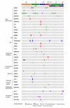
Figure 1
Figure 1. Comutation plot demonstrating the variety of likely pathogenic mutations and gene fusions detected through targeted sequencing.
Single columns represent individual patients with the diagnosis identified in the first row. Samples are grouped by disease type and age group as indicated in horizontal bars. Asterisks at the bottom represent 2 previously reported patients as described in the text. Basic plot created using cBioPortal (39, 40).
Figure 2
Figure 2. Mutant BRAF is detected in non-Langerhans bone marrow histiocytes and responds to targeted therapy.
Patient 1: (A) Most of the cellularity in the bone marrow aspirate was comprised of mature-appearing histiocytes, many of which contain hemosiderin. Few hematopoietic cells were present. (B) Frequent hemophagocytic cells were present in the aspirate. (C) Histiocytes filled much of the bone marrow in the biopsy. (D) The bone marrow was diffusely positive for CD163 and negative for CD1a and S100 (not shown). (E) The histiocytes were positive for BRAF V600E using a mutation-specific antibody (pink), whereas other hematopoietic cells in the marrow were BRAF negative (arrows). (F) Positive control BRAF immunohistochemistry on a Langerhans cell histiocytosis skin biopsy containing BRAF V600E; note specific cytoplasmic staining (pink) of the neoplastic histiocytes in the lower one-half of the field. (G) Blood counts and soluble IL-2 receptor levels before and after initiation of dabrafenib by day of admission (x axis). Red asterisks represent the patient’s last required transfusion (blood or platelets) or dose of granulocyte colony-stimulating factor, as in the case of absolute neutrophil count. (H) Coronal view of fluorodeoxyglucose–avid basilar skull lesion with orbital involvement (arrow) is markedly improved on followup PET/CT obtained 2 months later (I) after 6 weeks on dabrafenib. Original magnification, ×400 (A and F), ×1,000 (B), ×200 (CE).
Figure 3
Figure 3. Responses to targeted therapy in Langerhans cell histiocytosis by functional imaging.
(A) Patient 2: PET-CT at diagnosis (left) demonstrated markedly fluorodeoxyglucose-avid (FDG-avid) lesions in the left neck and groin (latter not pictured). (B) Follow-up PET-CT after 4 months of therapy demonstrates near-total resolution at sites of disease in the neck. (C) Patient 3: PET-CT obtained upon transfer to our institution demonstrated marked FDG avidity in the skeletal medullary spaces (white arrows) secondary to disease activity, which demonstrated near-total resolution on follow-up imaging (D) 3 months after the start of therapy.
Figure 4
Figure 4. Identification of cells expressing mutant BRAF protein in hepatic involvement by Langerhans cell histiocytosis.
(A) Patient 4: Immunohistochemistry for BRAF V600E (brown) and CD163 (red) in representative liver biopsy section demonstrates presence of BRAF V600E mutant protein, but not in cells of macrophage/monocyte lineage (original magnification, ×400). (B) Immunostain for CD1a is negative.
Figure 5
Figure 5. MEK (MAP2K1) mutants demonstrate altered activation kinetics and exhibit signs of transformation in vitro.
(A) NIH/3T3 cells were transduced with retroviruses carrying empty vector or HA-tagged MEK constructs. Shown is a representative immunoblot using antibodies against the HA-tag or GAPDH (control) with the various mutations labeled on the top of each lane. WT, wild type. (B) Transduced cells were treated with murine epidermal growth factor (mEGF) and lysates were analyzed by immunoblotting. Representative blots depict phospho-ERK (pERK) and total ERK (tERK) levels in the various 3T3 clones labeled on the top. Antibodies are labeled to the right with GAPDH used as internal control. (C) 3T3 cells expressing WT and mutant MEK constructs were stimulated with EGF for the indicated time periods; mutant enzymes exhibit prolonged pERK activation in response to sustained EGF stimulation compared with WT MEK. (D) Fibroblasts were plated in monolayer in conventional liquid culture and foci were counted after 21 days. Representative micrographs of cultures expressing the various MEK mutants are shown, labeled on the top, shown at ×40 (upper row) and ×400 (lower row) original magnification. (E) Bar graph depicting the number of foci observed in each condition. Middle horizontal bars represent mean number of foci; error bars represent standard deviation of 3 separate experiments. No foci were seen with WT MEK or vector-only transduced cells. Statistical analysis performed using 1-way ANOVA with Tukey’s test post-hoc. *P < 0.001, **P < 0.0001.
Figure 6
Figure 6. Activating mutations in MEK are sensitive to allosteric inhibition.
(A) Dose-response curves quantifying phospho-ERK (pERK) activation in transduced NIH/3T3 cells in response to drug treatment. Following drug treatment, cells were fixed and probed simultaneously with fluorescent pERK and ERK antibodies, and imaged with an Odyssey scanner (LiCor Biosciences). Relative pERK fluorescence (y axis) was calculated and reflects the percentage change relative to DMSO-treated cells; all values are normalized to total ERK (tERK) expression. Data represent mean of 3 separate experiments; error bars represent standard deviation. (B) Western blot demonstrates stable transduction of BaF/3 cells with pBABE-puro-based empty vector, or WT/mutant HA-tagged MEK constructs. (C) Immunoblots for pERK after 30 minutes of incubation with the indicated drug and concentration show a similar dose-dependent response.

References

    1. Delprat C, Aricò M. Blood spotlight on Langerhans cell histiocytosis. Blood. 2014;124(6):867–872. doi: 10.1182/blood-2014-02-556407. - DOI - PubMed
    1. Diamond EL, et al. Consensus guidelines for the diagnosis and clinical management of Erdheim-Chester disease. Blood. 2014;124(4):483–492. doi: 10.1182/blood-2014-03-561381. - DOI - PMC - PubMed
    1. Grois N, et al. Central nervous system disease in Langerhans cell histiocytosis. J Pediatr. 2010;156(6):873–81, 881.e1. - PubMed
    1. Gadner H, et al. Therapy prolongation improves outcome in multisystem Langerhans cell histiocytosis. Blood. 2013;121(25):5006–5014. doi: 10.1182/blood-2012-09-455774. - DOI - PubMed
    1. Donadieu J, et al. Endocrine involvement in pediatric-onset Langerhans’ cell histiocytosis: a population-based study. J Pediatr. 2004;144(3):344–350. doi: 10.1016/j.jpeds.2003.12.030. - DOI - PubMed

Publication types

MeSH terms