. 2017 Nov;177(5):1337-1348.
doi: 10.1111/bjd.15379. Epub 2017 Nov 1.

Age-related reduction of dermal fibroblast size upregulates multiple matrix metalloproteinases as observed in aged human skin in vivo

Affiliations

Age-related reduction of dermal fibroblast size upregulates multiple matrix metalloproteinases as observed in aged human skin in vivo

Z Qin et al. Br J Dermatol. 2017 Nov.

Abstract

Background: Fragmentation of collagen fibrils, the major structure protein in skin, is a hallmark of dermal ageing. Matrix metalloproteinases (MMPs) are largely responsible for the fragmentation of collagen fibrils.

Objectives: To quantify gene expression of all 23 known mammalian MMPs in sun-protected young and aged human skin in vivo and to investigate the potential mechanism underlying age-related alteration of multiple MMPs.

Methods: MMP mRNA expression levels and MMP activity in sun-protected young and aged human skin in vivo were determined by real-time reverse transcription polymerase chain reaction (RT-PCR) and in situ zymography, respectively. The relative contributions to elevated MMPs in epidermis and dermis were quantified by laser capture microdissection coupled real-time RT-PCR. Dermal fibroblast morphology and collagen fibril fragmentation in human skin in vivo were assessed by second-harmonic generation microscopy and atomic force microscopy, respectively. In vitro cell morphology was assessed by CellTracker® fluorescent dye (Molecular Probes, Eugene, OR, U.S.A.) and phalloidin staining. Protein levels were determined by ProteinSimple capillary electrophoresis immunoassay (ProteinSimple, Santa Clare, CA, U.S.A.).

Results: Multiple MMPs are elevated in aged human skin dermis. Increased MMP activity and collagen fibril fragmentation were observed in aged skin dermis. As dermal fibroblasts are the major MMP-producing cells in the dermis, reduction of dermal fibroblast size, which is observed in aged human skin, contributes to the elevation of age-related multiple MMPs. Reduction of fibroblast size upregulates c-Jun/c-Fos and activates AP-1.

Conclusions: Combined actions of the wide variety of MMPs that are constitutively elevated in aged dermis may be involved in the progressive degradation of dermal collagen fibrils. Age-related elevations of multiple MMPs are likely to be a result of the reduction of fibroblast size via activation of AP-1.


Figures

Figure 1
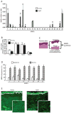
Figure 1. Elevated expression of multiple MMPs in aged human dermis
Adult human skin punch biopsies were obtained from sun-protected buttock skin. (A) Elevated multiple MMPs in aged (83±4 years) compared to young (26±3 years) human skin. N=12 each group. Mean ± SEM. *p<0.05. (B) No change in TIMP mRNA expression in young and aged human skin. N=12 each group. Mean ± SEM. (C) Schematic representation of the dissection of human skin epidermis and dermis by Laser Capture Microdissection (LCM, see Methods for details). (D) Elevated MMPs in the dermis of aged human skin. Epidermis and dermis were captured by LCM and total RNA was prepared form epidermis and dermis. N=8, Mean ± SEM. All MMPs mRNA levels were quantified by real-time RT-PCR and were normalized by the housekeeping gene (36B4, internal control). (E) Elevated collagenase activity in the dermis of aged human skin determined by in situ zymography (see Methods for details). Loss of green fluorescence in aged dermis indicates degradation of fluorescein-collagen substrate. White lines indicate boundary between the epidermis (top) and dermis (bottom). N=6.
Figure 2
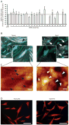
Figure 2
(A) MMPs are similarly expressed in the fibroblasts isolated from aged (83±4 years) and young (26±3 years) skin. Dermal fibroblasts were isolated from sun-protected young and aged buttock skin. MMPs mRNA expression levels were quantified by real-time RT-PCR and were normalized by the housekeeping gene (36B4, internal control). N=6. Mean ± SEM. (B) Reduced dermal fibroblast size is a prominent feature of aged dermal fibroblasts in human skin in vivo. Representative images of dermal fibroblasts in aged (78 years, right panel) and young (26 years, left panel) human skin. Skin was sectioned, and dermal fibroblasts were identified by immunostaining with collagen chaperone heat shock protein 47 (blue). Gray/white color is collagen fibrils and green color is elastin. Note spread fibroblasts in young skin versus contracted fibroblasts in aged human skin. Images were obtained by multiphoton laser scanning fluorescence microscopy. N=6 for each group. The black and white arrow heads indicate dermal fibroblasts in young and aged human skin, respectively Bars=25μm. (C) Collagen fibrils in aged skin dermis were fragmented and disorganized. Nanoscale collagen fibrils were imaged by atomic force microscopy. The black and white arrow heads indicate intact and fragmented/disorganized collagen fibrils, respectively. Images are representative of six independent experiments. Bars=100 nm (D) The morphology of the dermal fibroblasts from young (left panel) and aged (right panel) were similar in standard monolayer culture in vitro. Cells were stained with CellTracker® fluorescent dye and were imaged by fluorescence microscopy. Red fluorescence delineates cell cytoplasm; blue fluorescence delineates nuclei. Bars=100 μm. Images are representative of the dermal fibroblasts from five young and aged individuals.
Figure 3
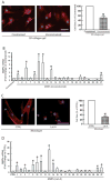
Figure 3. Age-related reduction of dermal fibroblast size up-regulates multiple MMPs observed in aged human skin
(A) To model young and aged dermal fibroblasts, dermal fibroblasts from young skin (26±3 years) were cultured under conditions of constrained (left panel) and unconstrained (right panel) 3D collagen lattices (see Methods for details). Cells were stained with CellTracker® fluorescent dye and were imaged by fluorescence microscopy. Red fluorescence delineates cell cytoplasm; blue fluorescence delineates nuclei. Relative cell surface areas were quantified using ImageJ software and data were expressed as % of control (non-contracted gel). Mean ± SEM, N =3, *p<0.05. Bars = 100 μm (B) The majority age-related MMPs were elevated by reduction of fibroblasts size. Total RNA was extracted from dermal fibroblasts cultured from 3D collagen lattices. MMPs mRNA expression levels were quantified by real-time RT-PCR and were normalized by the housekeeping gene (36B4, internal control). N=4. Mean ± SEM. *p<0.05. (C) Dermal fibroblasts from young skin (26±3 years) were treated with Lat-A (30 nM) for 24 hours. Dermal fibroblasts were stained with phalloidin and were imaged by fluorescence microscopy. Red fluorescence delineates cell cytoplasm; blue fluorescence delineates nuclei. Relative cell surface areas were quantified using ImageJ software and data were expressed as % of control (DMSO). Mean ± SEM, N=6, *p<0.05. Bars = 100 μm. (D) The majority age-related MMPs were elevated by reduction of fibroblast size via disruption of the actin cytoskeleton. Total RNA was extracted from dermal fibroblasts. MMPs mRNA expression levels were quantified by real-time RT-PCR and were normalized by the housekeeping gene (36B4, internal control). N=5. Mean ± SEM. *p<0.05. (E) Restoration of dermal fibroblast size after withdrawn Lat-A. Lat-A was withdrawn (right panel) 24 hours after Lat-A (30 nM) treatment (middle panel) by replacing with fresh culture medium, and the cells were further incubated for 48 hours. Dermal fibroblasts were stained with phalloidin and relative cell surface areas were quantified using ImageJ software. Data were expressed as % of control (DMSO, left panel). Mean ± SEM, N=6, *p<0.05. Bars = 100 μm. (F) Restoration of dermal fibroblast size reverses elevated MMPs. MMPs mRNA expression levels were quantified by real-time RT-PCR and were normalized by the housekeeping gene (36B4, internal control). MMPs mRNA expression levels after Lat-A withdrawn were expressed as % of control cells (DMSO). N=5. Mean ± SEM.
Figure 4
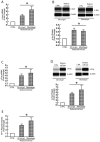
Figure 4. Reduced fibroblast size up-regulates c-Jun/c-Fos and activates AP-1, the major regulator of multiple MMPs
Dermal fibroblasts from young skin (26±3 years) were cultured in 3D collagen lattices or monolayer under conditions of reduced cell size, as described in Method. (A) Reduction of fibroblast size upregulates c-Jun mRNA expression. (B) Reduction of fibroblast size upregulates c-Jun protein expression. (C) Reduction of fibroblast size upregulates c-Fos mRNA expression. (D) Reduction of fibroblast size upregulates c-Fos protein expression. mRNA levels were quantified by real-time RT-PCR and were normalized by the housekeeping gene (36B4, internal control). Mean ± SEM, N=3, *p<0.05. Protein levels were determined by ProteinSimple capillary electrophoresis immunoassay (see Materials and methods for details) and normalized by β-actin (loading control). Insert shows representative digital images. Mean ± SEM, N=3, *p<0.05. (E) Dermal fibroblasts were transfected with AP-1 reporter construct (pAP-1-TA-Luc) or empty vector (CTRL). Cell lysates were prepared 48 hours after transfection. Reporter activity was determined by luciferase assay. Data are Mean ± SEM, N=3, *p<0.05.
Figure 5
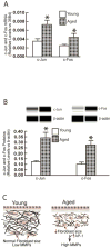
Figure 5. Elevated expression of c-Jun and c-Fos expression in aged human skin dermis
Adult human skin punch biopsies were obtained from young (26±3 years) and aged (83±4 years) sun-protected buttock skin. Dermis was prepared by cutting off epidermis at a depth of 1 mm by cryostat. (A) Total RNA was prepared from dermis and mRNA levels were quantified by real-time RT-PCR and were normalized by the housekeeping gene (36B4, internal control). Mean ± SEM, N=6, *p<0.05. (B) Dermal protein levels were determined by ProteinSimple capillary electrophoresis immunoassay (see Methods for details) and normalized by β-actin (loading control). Insert shows representative digital images. Mean ± SEM, N=6, *p<0.05. (C) Reduced fibroblast size elevates age-related MMPs (see details in Discussion).

Comment in

Similar articles

Cited by

  • The Role of Probiotics in Skin Photoaging and Related Mechanisms: A Review.
    Teng Y, Huang Y, Danfeng X, Tao X, Fan Y. Teng Y, et al. Clin Cosmet Investig Dermatol. 2022 Nov 16;15:2455-2464. doi: 10.2147/CCID.S388954. eCollection 2022. Clin Cosmet Investig Dermatol. 2022. PMID: 36420112 Free PMC article. Review.
  • The Skin Microbiome: Current Landscape and Future Opportunities.
    Smythe P, Wilkinson HN. Smythe P, et al. Int J Mol Sci. 2023 Feb 16;24(4):3950. doi: 10.3390/ijms24043950. Int J Mol Sci. 2023. PMID: 36835363 Free PMC article. Review.
  • Molecular Mechanisms of Changes in Homeostasis of the Dermal Extracellular Matrix: Both Involutional and Mediated by Ultraviolet Radiation.
    Zorina A, Zorin V, Kudlay D, Kopnin P. Zorina A, et al. Int J Mol Sci. 2022 Jun 15;23(12):6655. doi: 10.3390/ijms23126655. Int J Mol Sci. 2022. PMID: 35743097 Free PMC article. Review.
  • YAP/TAZ regulates TGF-β/Smad3 signaling by induction of Smad7 via AP-1 in human skin dermal fibroblasts.
    Qin Z, Xia W, Fisher GJ, Voorhees JJ, Quan T. Qin Z, et al. Cell Commun Signal. 2018 Apr 25;16(1):18. doi: 10.1186/s12964-018-0232-3. Cell Commun Signal. 2018. PMID: 29695252 Free PMC article.
  • Biomarkers of aging.
    Aging Biomarker Consortium; Bao H, Cao J, Chen M, Chen M, Chen W, Chen X, Chen Y, Chen Y, Chen Y, Chen Z, Chhetri JK, Ding Y, Feng J, Guo J, Guo M, He C, Jia Y, Jiang H, Jing Y, Li D, Li J, Li J, Liang Q, Liang R, Liu F, Liu X, Liu Z, Luo OJ, Lv J, Ma J, Mao K, Nie J, Qiao X, Sun X, Tang X, Wang J, Wang Q, Wang S, Wang X, Wang Y, Wang Y, Wu R, Xia K, Xiao FH, Xu L, Xu Y, Yan H, Yang L, Yang R, Yang Y, Ying Y, Zhang L, Zhang W, Zhang W, Zhang X, Zhang Z, Zhou M, Zhou R, Zhu Q, Zhu Z, Cao F, Cao Z, Chan P, Chen C, Chen G, Chen HZ, Chen J, Ci W, Ding BS, Ding Q, Gao F, Han JJ, Huang K, Ju Z, Kong QP, Li J, Li J, Li X, Liu B, Liu F, Liu L, Liu Q, Liu Q, Liu X, Liu Y, Luo X, Ma S, Ma X, Mao Z, Nie J, Peng Y, Qu J, Ren J, Ren R, Song M, Songyang Z, Sun YE, Sun Y, Tian M, Wang S, Wang S, Wang X, Wang X, Wang YJ, Wang Y, Wong CCL, Xiang AP, Xiao Y, Xie Z, Xu D, Ye J, Yue R, Zhang C, Zhang H, Zhang L, Zhang W, Zhang Y, Zhang YW, Zhang Z, Zhao T, Zhao Y, Zhu D, Zou W, Pei G, Liu GH. Aging Biomarker Consortium, et al. Sci China Life Sci. 2023 May;66(5):893-1066. doi: 10.1007/s11427-023-2305-0. Epub 2023 Apr 11. Sci China Life Sci. 2023. PMID: 37076725 Free PMC article. Review.

References

    1. Di Lullo GA, Sweeney SM, Korkko J, et al. Mapping the ligand-binding sites and disease-associated mutations on the most abundant protein in the human, type I collagen. J Biol Chem. 2002;277:4223–31. - PubMed
    1. Uitto J. Connective tissue biochemistry of the aging dermis. Age-related alterations in collagen and elastin. Dermatol Clin. 1986;4:433–46. - PubMed
    1. Fisher GJ, Varani J, Voorhees JJ. Looking older: fibroblast collapse and therapeutic implications. Arch Dermatol. 2008;144:666–72. - PMC - PubMed
    1. Quan T, Fisher GJ. Role of Age-Associated Alterations of the Dermal Extracellular Matrix Microenvironment in Human Skin Aging: A Mini-Review. Gerontology. 2015;61:427–34. - PMC - PubMed
    1. Ma W, Wlaschek M, Tantcheva-Poor I, et al. Chronological ageing and photoageing of the fibroblasts and the dermal connective tissue. Clin Exp Dermatol. 2001;26:592–9. - PubMed