. 2017 Feb 14;18(1):28.
doi: 10.1186/s13059-017-1151-0.

Comparative genomics reveals high biological diversity and specific adaptations in the industrially and medically important fungal genus Aspergillus

Ronald P de Vries  1   2 Robert Riley  3 Ad Wiebenga  4   5 Guillermo Aguilar-Osorio  6 Sotiris Amillis  7 Cristiane Akemi Uchima  8   9 Gregor Anderluh  10 Mojtaba Asadollahi  11 Marion Askin  12   13 Kerrie Barry  3 Evy Battaglia  4   5 Özgür Bayram  14   15 Tiziano Benocci  4   5 Susanna A Braus-Stromeyer  14 Camila Caldana  8   16 David Cánovas  17   18 Gustavo C Cerqueira  19 Fusheng Chen  20 Wanping Chen  20 Cindy Choi  3 Alicia Clum  3 Renato Augusto Corrêa Dos Santos  8 André Ricardo de Lima Damásio  8   21 George Diallinas  7 Tamás Emri  22 Erzsébet Fekete  11 Michel Flipphi  11 Susanne Freyberg  14 Antonia Gallo  23 Christos Gournas  24   25 Rob Habgood  26 Matthieu Hainaut  27 María Laura Harispe  28   29 Bernard Henrissat  27   30   31 Kristiina S Hildén  32 Ryan Hope  26 Abeer Hossain  33   34 Eugenia Karabika  35   36 Levente Karaffa  11 Zsolt Karányi  37 Nada Kraševec  10 Alan Kuo  3 Harald Kusch  14   38   39 Kurt LaButti  3 Ellen L Lagendijk  12 Alla Lapidus  3   40 Anthony Levasseur  41   42 Erika Lindquist  3 Anna Lipzen  3 Antonio F Logrieco  43 Andrew MacCabe  44 Miia R Mäkelä  32 Iran Malavazi  45 Petter Melin  46   47 Vera Meyer  48 Natalia Mielnichuk  17   49 Márton Miskei  22   50 Ákos P Molnár  11 Giuseppina Mulé  43 Chew Yee Ngan  3 Margarita Orejas  44 Erzsébet Orosz  4   22 Jean Paul Ouedraogo  12   51 Karin M Overkamp  33 Hee-Soo Park  52 Giancarlo Perrone  43 Francois Piumi  41   53 Peter J Punt  12   33 Arthur F J Ram  12 Ana Ramón  54 Stefan Rauscher  55 Eric Record  41 Diego Mauricio Riaño-Pachón  8 Vincent Robert  4 Julian Röhrig  55 Roberto Ruller  8 Asaf Salamov  3 Nadhira S Salih  26   56 Rob A Samson  4 Erzsébet Sándor  57 Manuel Sanguinetti  54 Tabea Schütze  12   58 Kristina Sepčić  59 Ekaterina Shelest  60 Gavin Sherlock  61 Vicky Sophianopoulou  24 Fabio M Squina  8 Hui Sun  3 Antonia Susca  43 Richard B Todd  62 Adrian Tsang  63 Shiela E Unkles  35 Nathalie van de Wiele  4 Diana van Rossen-Uffink  12   64 Juliana Velasco de Castro Oliveira  8 Tammi C Vesth  65 Jaap Visser  4 Jae-Hyuk Yu  66 Miaomiao Zhou  4   5 Mikael R Andersen  65 David B Archer  26 Scott E Baker  67 Isabelle Benoit  4   5   68 Axel A Brakhage  69 Gerhard H Braus  14 Reinhard Fischer  55 Jens C Frisvad  65 Gustavo H Goldman  70 Jos Houbraken  4 Berl Oakley  71 István Pócsi  22 Claudio Scazzocchio  72   73 Bernhard Seiboth  74 Patricia A vanKuyk  4   12 Jennifer Wortman  75   76 Paul S Dyer  26 Igor V Grigoriev  3
Affiliations

Comparative genomics reveals high biological diversity and specific adaptations in the industrially and medically important fungal genus Aspergillus

Ronald P de Vries et al. Genome Biol. .

Abstract

Background: The fungal genus Aspergillus is of critical importance to humankind. Species include those with industrial applications, important pathogens of humans, animals and crops, a source of potent carcinogenic contaminants of food, and an important genetic model. The genome sequences of eight aspergilli have already been explored to investigate aspects of fungal biology, raising questions about evolution and specialization within this genus.

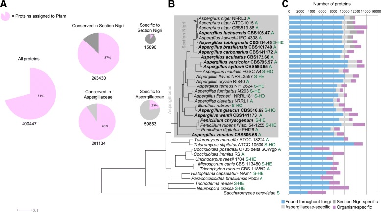
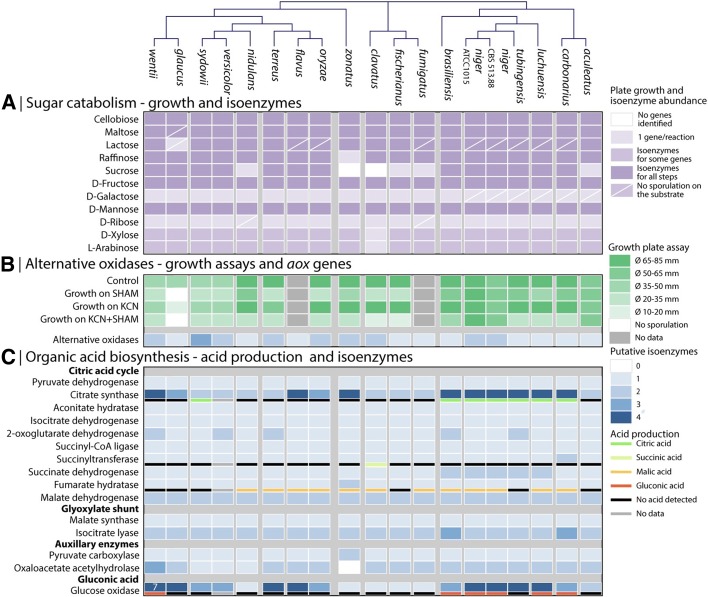
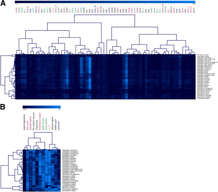
Results: We have generated genome sequences for ten novel, highly diverse Aspergillus species and compared these in detail to sister and more distant genera. Comparative studies of key aspects of fungal biology, including primary and secondary metabolism, stress response, biomass degradation, and signal transduction, revealed both conservation and diversity among the species. Observed genomic differences were validated with experimental studies. This revealed several highlights, such as the potential for sex in asexual species, organic acid production genes being a key feature of black aspergilli, alternative approaches for degrading plant biomass, and indications for the genetic basis of stress response. A genome-wide phylogenetic analysis demonstrated in detail the relationship of the newly genome sequenced species with other aspergilli.

Conclusions: Many aspects of biological differences between fungal species cannot be explained by current knowledge obtained from genome sequences. The comparative genomics and experimental study, presented here, allows for the first time a genus-wide view of the biological diversity of the aspergilli and in many, but not all, cases linked genome differences to phenotype. Insights gained could be exploited for biotechnological and medical applications of fungi.

Keywords: Aspergillus; Comparative genomics; Fungal biology; Genome sequencing.


Figures

Fig. 1
Fig. 1
Genome overview of Aspergillus and comparative species. a Core genes, based on MCL clustering of protein sequences, and cluster membership in section Nigri and the Aspergillaceae. Pink area of pie charts indicates proteins assignable to one or more Pfam domain; white or gray areas indicate proteins with no Pfam domain. The majority of proteins conserved in Section Nigri, Aspergillaceae, or the full set of comparative fungi could be assigned to a Pfam. Contrastingly, most clade-specific proteins (occurring only in Section Nigri or Aspergillaceae) were not assignable to any Pfam. b Maximum likelihood phylogeny inferred from 149 conserved protein sequences. Organisms newly sequenced for this study are indicated in bold. All bootstrap values are 100 except where indicated. Section Nidulantes is inferred to be a sister group to section Nigri, in contrast to previous studies. Letters in green behind the strain numbers indicate the reproductive state: A asexual, S-HO sexual homothallic, S-HE sexual heterothallic. c Protein conservation, inferred from MCL clustering of proteins, indicates that the majority of proteins in Aspergillaceae have homologs in other fungi, bars showing number of proteins being aligned to individual species to the left-hand side in (b). Some 21% of proteins are specific to the Aspergillaceae, while 14% of proteins in section Nigri are specific to that clade. Organism-specific proteins make up 9% for the Aspergillaceae as a whole and 8% for section Nigri
Fig. 2
Fig. 2
Regulatory pathway of asexual sporulation in A. nidulans. a Central regulators, upstream activators, negative regulators, velvet regulators, and light-responsive regulators are illustrated by green, light purple, blue, dark purple, and red icons, respectively. b Distribution of central regulators for asexual sporulation in 85 fungi. These fungi are representatives from the phyla Ascomycota, Basidiomycota, Chytridiomycota, and Zygomycota. Their genome protein sequences were searched for homologs of AbaA, BrlA, and WetA by BlastP using sequences of A. nidulans AbaA, BrlA, and WetA as queries. Details of these fungi are presented in Central Regulator Strain Information. As shown, BrlA seems to be limited to the Eurotiales group, suggesting a specific role for conidiation in Eurotiales fungi. By contrast, WetA is widely distributed in Pezizomycotina fungi, which suggests a general function for the synthesis of cell wall layers to make conidia mature and impermeable. Surprisingly, AbaA is widespread being found in the phyla Ascomycota, Basidiomycota, and Zygomycota, suggesting that AbaA is involved not only in conidial development, but also has other general functions in fungal development
Fig. 3
Fig. 3
Experimental and in silico analysis of central metabolism. Clustering of the species was performed based on gpdA and orthologs from all genomes. a Catabolism of 11 sugars correlated to the number of isoenzymes investigated for each of the 11 catabolic pathways. A cross line marks all conditions where not growth was observed. b Copy numbers of (putative) alternative oxidases (aox) compared to growth assays on plates with agents inhibiting either the standard oxidative phosphorylation (KCN) or Aox (SHAM). c Correlation of putative isoenzymes for activities involved in organic acid formation and produced organic acids
Fig. 4
Fig. 4
HCL (a) the number of genes per CAZy family related to plant biomass degradation in the tested genomes and (b) the relative growth of the tested species on polymeric substrates. Color coding in (a) refers to the polysaccharide these families act on. Black families either contain multiple activities or are active on multiple polysaccharides. For the other colors: pink = guar gum (galactomannan), purple = starch, red = xylan, green = pectin, orange = inulin, dark blue = cellulose, pale blue = xyloglucan
Fig. 5
Fig. 5
Comparative proteomics of aspergilli during growth on wheat bran (a) and sugar beet pulp (b). Results are summarized by the polysaccharide that the enzymes act upon. Only CAZy families specific for one polysaccharide are included in the comparison. Cellulose-active enzymes: GH6, GH7, GH12, GH45, GH74, AA9; xylan-active enzymes: CE1, CE15, GH10, GH11, GH62, GH67, GH115; pectin-active enzymes: CE8, CE12, GH28, GH35, GH51, GH53, GH54, GH78, GH88, GH93, GH105, PL1, PL3, PL4, PL9, PL11; starch-active enzymes: GH13, GH15; other (CAZy families with multiple activities or minor activities on the substrates): CE16, GH1, GH2, GH3, GH5, GH26, GH27, GH31, GH32, GH36, GH43, GH95. Extracellular proteins following trypsin digestion were analyzed by LC-MS/MS using a LTQ-Orbitrap Velos mass analyzer (Thermo-Fisher). Quantification was based on MS precursor ion signal. Extracted ion chromatograms were used to determine the peptide area value associated to each identified precursor ion. A protein area value was calculated as the average of the three most intense, distinct peptides assigned to a protein. The amounts of proteins associated with each enzyme activity were expressed as percentage of the amount of total extracellular proteins present in each culture condition
Fig. 6
Fig. 6
Conservation of penicillin (a) and pseurotin (b) biosynthesis gene clusters in Aspergillus species
Fig. 7
Fig. 7
Molecular phylogenetic tree of (a) CYP51F1 (CYP51B) family and inhibition of growth on MBFA agar supplemented by 0.02 mM ketoconazole (K) and (b) CYP53 family and inhibition of growth on 2 mM benzoic acid (BA). Inhibition of growth (Inhib.) as per cent of the control without supplementation (Contr.) is shown in relation to phylogenetic trees, with growth culture plates shown to the right-hand side of the accompanying tree branch. Key to Aspergillus species: Aspfo = A. luchuensis (formerly A. foetidus); Asptu = A. tubingensis; Aspka = A. luchuensis (A. kawachii); Aspbr = A. brasiliensis; Aspni = A. nidulans; Aspca = A. carbonarius; Aspac = A. aculeatus; Aspfl = A. flavus; Aspor = A. oryzae; Aspwe = A. wentii; Aspcl; = A. clavatus; Aspfu = A. fumigatus; Neofi = A. fischeri; Aster = A. terreus; Aszo = A. zonatus; Asgl = A. glaucus; Aspsy = A. sydowii; Aspve = A. versicolor
Fig. 8
Fig. 8
a Phylogenetic tree of Aspergillus species and summarized phenotypes from this study (modified from [308]). All Aspergillus species were grown on malt extract agar (MEA) (blue) at 30 °C. Yellow squares stand for induced conidiation in white light. Abiotic and biotic stress determinants were colored in green for resistance (r), red for sensitive (s), no color for insensitive (-) and not determined (nd) different abiotic and biotic stressors: 2,4-diacetylphloroglucinol (DAPG), Hydrogen peroxide (OX), nitric oxide (NO), calcofluor white (CFW), caspofungin (CA), voriconazole (VO), amphotericin B (AmB), and Aspergillus-Pseudomonas fluorescens co-cultivation (Biotic). Species used in this study are framed in black. b Qualitative descriptive statistics of the exoproteome of four selected Aspergillus strains. Exoproteomes were enriched from filtered culture supernatants (Aspergillus MM, 30 °C) and analyzed by shotgun proteomics (LC-MS). Proteins were identified using draft genomic databases of the respective strains. For prediction of subcellular localization of the proteins the programs “SignaIP” and “WoLF PSORT” were used. For further analysis, sequences with an extracellular score >12 (WoLF PSORT) were defined as putatively secreted proteins (Additional file 18). Proteins with higher spectral count values after 0.2 mM hydrogen peroxide treatment (“upregulated after oxidative stress”) were selected statistically using MARVIS Filter (s/l > 0.5). c Exoproteomic heterogeneity in the genus Aspergillus. Comparative MARVIS cluster analysis of PFAM domains predicted in identified exoproteins (WoLF PSORT columns in bar chart B) of four selected Aspergillus species (WoLF PSORT: extracellular score >12) (Additional file 19). Colors represent the normalized frequency of occurrence of PFAM-domains in the respective exoproteome. Indicated by the color scale on the right, the color scale ranges from blue (no domain) via green (few domains) to red (more domains). Each column of the cluster image resembles one PFAM-domain. The proteome data is available at http://wwwuser.gwdg.de/~hkusch/GBIO_DeVries/
Fig. 9
Fig. 9
Linking species-level differences in selected Aspergillus stress-defense proteins to major stress tolerance phenotypes. The remarkable Cd(II) tolerance of A. fumigatus and A. sydowii (a), the outstanding menadione sodium bisulfite (MSB) tolerance of A. brasiliensis (b) and the osmophility of A. glaucus and A. wentii (c) were attributed to variations in the cadmium transporting P-type ATPases (a), Cu/Zn-superoxide dismutases (b), and NAD-dependent glycerol-3-phosphate dehydrogenases (c), respectively. In this Figure, the stress sensitivities of A. nidulans are also presented for comparison (ac), although this species lacks any Pca1 ortholog (Additional file 15). In the dendograms, putative orthologs of baker’s yeast Pca1 cadmium transporting P-type ATPase (a), Aspergillus nidulans SodA Cu/Zn-superoxide dismutase (b), and A. nidulans GfdA/B NAD-dependent glycerol-3-phosphate dehydrogenases (c) are shown. In (c), S. cerevisiae Gpd1/2 paralogs are also presented. Putative A. sydowii (formula image
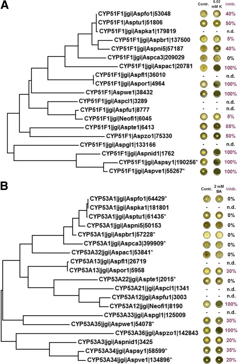
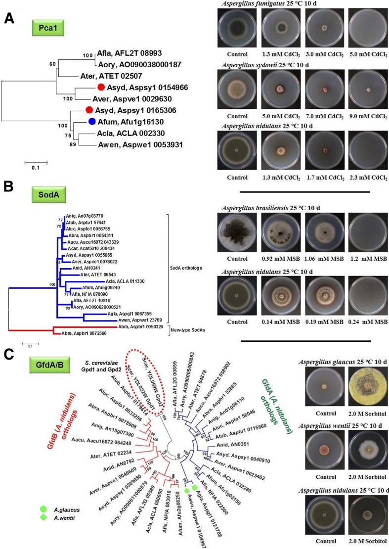
) and A. fumigatus (formula image) Pca1 orthologs (a) and putative A. glaucus (formula image) and A. wentii (formula image) GfdA orthologs (c) are indicated by symbols presented in the parentheses. Hypothetical new-type SodA enzymes found in A. brasiliensis are indicated by red lines in (b). Pca1, SodA, and GfdA/B orthologs are indicated by four-letter species name identifiers (listed in Additional file 15) and locus IDs as found in AspGD (http://www.aspergillusgenome.org/) for the aspergilli. The relevant JGI locus IDs are listed in Additional file 16. The Gpd1/2 paralogs of budding yeast are from the Saccharomyces Genome Database (http://www.yeastgenome.org/). Photographs on the tress tolerance/sensitivity phenotypes were taken from the Fungal Stress Database (http://www.fung-stress.org/). Further details of the phylogenetic and evolutionary calculations and more information on the stress response proteins can be found in Additional file 15
Fig. 10
Fig. 10
Overview of sugar transporters. The evolutionary history of 940 PF00083 sequences identified in the genomes of A. clavatus, A. flavus, A. fumigatus, A. nidulans, A. niger, A. oryzae, A. terreus, and A. fischeri was inferred using the neighbor-joining method. The optimal tree is shown. Nodes with 50–69% (∆) or 70–100% (○) bootstrap support (1000 replicates) are indicated. Evolutionary distances were computed using the Poisson correction method and are in the units of the number of amino acid substitutions per site. All ambiguous positions were removed for each sequence pair. Evolutionary analyses were conducted in MEGA6. A small number of sequences could not be assigned to the main clades identified; referring to the smallest genome analyzed (A. clavatus), these correspond to loci ACLA023780, ACLA026890, ACLA031920, and ACLA041390. Potential substrates for members of the clades are suggested based on experimentally determined transporter functions
Fig. 11
Fig. 11
Inhibition of growth by NO. The table depicts the number of homologs of the flavohaemoglobin genes (fhbA and fhbB), the P450 nitric oxide reductase, and the S-nitrosoglutathione (GSNO) reductase in each species. Strains are listed in the same order as in the phylogenetic tree shown in Fig. 1. Strains were grown in the presence or in the absence of the NO-releasing compound nitroprusside. Growth was monitored at different time intervals. The graph shows the percentage of growth in the presence of 128 mM nitroprusside compared to the control samples grown in the absence of nitroprusside. The time and temperature of growth was optimized for each strain and it was as follows (species name, strain, temperature, and incubation time for growth inhibition calculation): A. glaucus: 22 °C for 72 h; A. tubingensis: 30 °C for 96 h; A. zonatus: 30 °C for 96 h; A. brasiliensis: 30 °C for 72 h; A. versicolor: 30 °C for 96 h; A. sydowii: 30 °C for 96 h; A. niger ATCC1015: 30 °C for 48 h; A. luchuensis: 30 °C for 72 h; A. niger NRRL3: 30 °C for 72 h; A. wentii: 30 °C for 96 h; A. niger CBS 513.88: 30 °C for 96 h; A. fischeri: 30 °C for 96 h; A. terreus: 30 °C for 96 h; A. flavus: 30 °C for 96 h; A. fumigatus: 30 °C for 96 h; A. clavatus: 30 °C for 96 h; A. nidulans: 30 °C for 96 h; A. carbonarius: 30 °C for 72 h; A. aculeatus: 30 °C for 96 h
Fig. 12
Fig. 12
a Proportion of species-specific genes of the different signal transduction protein-classes in Eurotiomycetes. b Number of protein phosphatases in fungi. Eurotiomycetes appear in red

References

    1. Samson RA, Visagie CM, Houbraken J, Hong SB, Hubka V, Klaassen CH, et al. Phylogeny, identification and nomenclature of the genus Aspergillus. Stud Mycol. 2014;78:141–73. doi: 10.1016/j.simyco.2014.07.004. - DOI - PMC - PubMed
    1. Scazzocchio C. Aspergillus, a multifacted genus In: Schaechter M, editor. Encyclopaedia of Microbiology. Amsterdam: Elsevier; 2009. p. 401-420.
    1. Geiser DM, Samson RA, Varga J, Rokas A, Witiak SM. A review of molecular phylogenetics in Aspergillus, and prospects for a robust genus-wide phylogeny. In: Varga J, Samson RA, editors. Aspergillus in the genomic era. Wageningen: Wageningen Academic Publishers; 2008. pp. 17–32.
    1. Galagan JE, Calvo SE, Cuomo C, Ma LJ, Wortman JR, Batzoglou S, et al. Sequencing of Aspergillus nidulans and comparative analysis with A. fumigatus and A. oryzae. Nature. 2005;438:1105–15. doi: 10.1038/nature04341. - DOI - PubMed
    1. Machida M, Asai K, Sano M, Tanaka T, Kumagai T, Terai G, et al. Genome sequencing and analysis of Aspergillus oryzae. Nature. 2005;438:1157–61. doi: 10.1038/nature04300. - DOI - PubMed

Publication types

MeSH terms