

Imaging of unilateral adrenal hemorrhages in patients after blunt abdominal trauma: Report of two cases
- PMID: 28196654
- PMCID: PMC5343102
- DOI: 10.1016/j.cjtee.2016.05.002
Imaging of unilateral adrenal hemorrhages in patients after blunt abdominal trauma: Report of two cases
Abstract
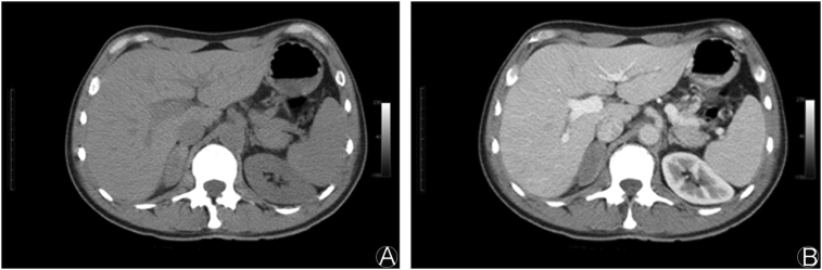
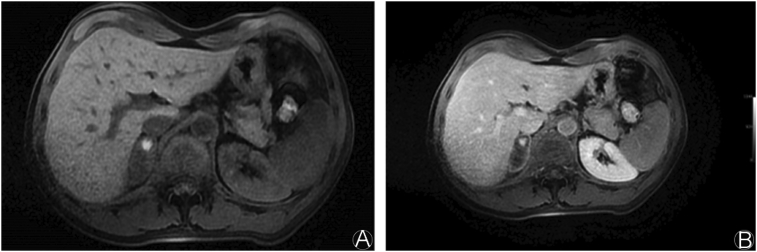
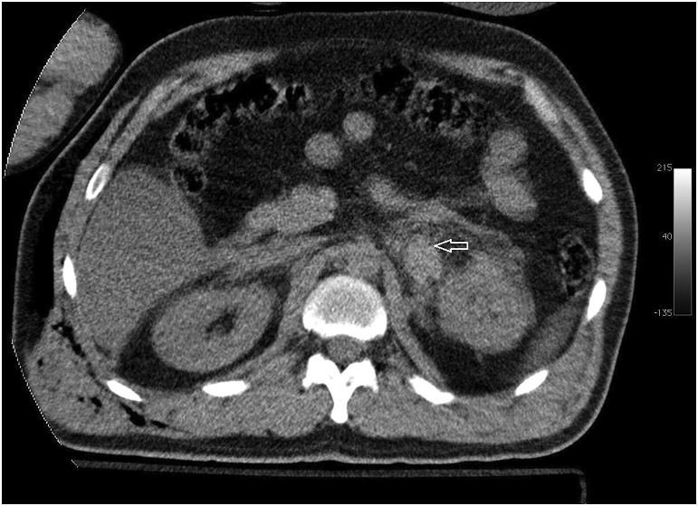
Adrenal hemorrhage following blunt abdominal trauma is extremely rare. Most of the lesions are unilateral and right sided. Although often asymptomatic, life-threatening adrenal insufficiency may develop in the bilateral adrenal gland hemorrhage. Isolated adrenal injuries are very rare. They are often associated with other organ injuries. The mortality rates of patients range from 7% to 32%. In this report, we present the computed tomography and magnetic resonance imaging findings of unilateral adrenal hemorrhages in two patients with a history of fall from a height.
Keywords: Adrenal glands; Blunt abdominal trauma; Computed tomography; Magnetic resonance imaging.
Copyright © 2017 Daping Hospital and the Research Institute of Surgery of the Third Military Medical University. Production and hosting by Elsevier B.V. All rights reserved.
Figures

Similar articles
-
Isolated unilateral adrenal gland hemorrhage following motor vehicle collision: a case report and review of the literature.J Med Case Rep. 2017 Dec 26;11(1):358. doi: 10.1186/s13256-017-1506-x. J Med Case Rep. 2017. PMID: 29277157 Free PMC article. Review.
-
Traumatic adrenal injury in children.Isr Med Assoc J. 2000 Feb;2(2):132-4. Isr Med Assoc J. 2000. PMID: 10804936
-
Bilateral adrenal haemorrhage and acute adrenal insufficiency in a blunt abdominal trauma: a case-report and literature review.Eur J Emerg Med. 2004 Jun;11(3):164-7. doi: 10.1097/01.mej.0000127646.23587.cb. Eur J Emerg Med. 2004. PMID: 15167178 Review.
-
Posttraumatic adrenal hemorrhage in children: CT findings in 34 patients.AJR Am J Roentgenol. 1992 Jun;158(6):1299-302. doi: 10.2214/ajr.158.6.1590128. AJR Am J Roentgenol. 1992. PMID: 1590128
-
Acute adrenal injury after blunt abdominal trauma: CT findings.AJR Am J Roentgenol. 1992 Mar;158(3):503-7. doi: 10.2214/ajr.158.3.1738984. AJR Am J Roentgenol. 1992. PMID: 1738984
Cited by
-
Adrenal hemorrhage and hemorrhagic masses; diagnostic workup and imaging findings.Br J Radiol. 2021 Nov 1;94(1127):20210753. doi: 10.1259/bjr.20210753. Epub 2021 Aug 31. Br J Radiol. 2021. PMID: 34464549 Free PMC article. Review.
-
Multi-detector computed tomography in the diagnosis and characterization of adrenal gland traumatic injuries.Gland Surg. 2019 Apr;8(2):164-173. doi: 10.21037/gs.2019.01.07. Gland Surg. 2019. PMID: 31183326 Free PMC article. Review.
-
Precocious ischemia preceding bilateral adrenal hemorrhage: A case report.Radiol Case Rep. 2020 Apr 21;15(6):803-807. doi: 10.1016/j.radcr.2020.03.013. eCollection 2020 Jun. Radiol Case Rep. 2020. PMID: 32346458 Free PMC article.
-
Bilateral adrenal haematoma complicated by adrenal insufficiency in a patient treated with bevacizumab.BMJ Case Rep. 2021 Feb 22;14(2):e239689. doi: 10.1136/bcr-2020-239689. BMJ Case Rep. 2021. PMID: 33619141 Free PMC article.
-
Dabigatran Etexilate Related Unilateral Adrenal Hemorrhage.Cureus. 2024 Feb 29;16(2):e55254. doi: 10.7759/cureus.55254. eCollection 2024 Feb. Cureus. 2024. PMID: 38558587 Free PMC article.
References
-
- Sinelnikov A.O., Abujudeh H.H., Chan D. CT manifestations of adrenal trauma: experience with 73 cases. Emerg Radiol. 2007;13:313–318. - PubMed
-
- To'o K.J., Duddalwar V.A. Imaging of traumatic adrenal injury. Emerg Radiol. 2012;19:499–503. - PubMed
-
- Roupakias S., Papoutsakis M., Tsikopoulos G. Adrenal injuries following blunt abdominal trauma in children: report of two cases. Ulus Travma Acil Cerrahi Derg. 2012;18:171–174. - PubMed
-
- Jordan E., Poder L., Courtier J. Imaging of nontraumatic adrenal hemorrhage. AJR. 2012;199:w91–w98. - PubMed
-
- Lee M.J., Kim A.G., Jang J.E. A case of traumatic bilateral adrenal hemorrhage mimicking bilateral adrenal adenomas. YUJM. 2012;29:35–37.
Publication types
MeSH terms
LinkOut - more resources
Full Text Sources
Other Literature Sources
Medical