. 2017 Feb 14;6(2):e004878.
doi: 10.1161/JAHA.116.004878.

Bone Marrow-Derived MicroRNA-223 Works as an Endocrine Genetic Signal in Vascular Endothelial Cells and Participates in Vascular Injury From Kawasaki Disease

Affiliations

Bone Marrow-Derived MicroRNA-223 Works as an Endocrine Genetic Signal in Vascular Endothelial Cells and Participates in Vascular Injury From Kawasaki Disease

Maoping Chu et al. J Am Heart Assoc. .

Abstract

Background: Kawasaki disease (KD) is now the most common cause of acquired cardiac disease in children due to permanent coronary artery damage with unknown etiology. The study sought to determine the role of blood microRNA miR-223 in KD and KD-induced injuries in vascular endothelial cells (ECs) as well as the mechanisms involved.

Methods and results: MicroRNA profiles in serum from patients with KD and from healthy controls were assessed by microarray analysis. We noted that multiple serum microRNAs were aberrantly expressed in KD, among them miR-223, which was the most upregulated abundant serum microRNA. We found that bone marrow-derived blood cells (leukocytes and platelets) were able to secrete miR-223 into serum. Vascular ECs had no endogenous miR-223; however, the blood cell-secreted serum miR-223 could enter into the vascular ECs in the vascular walls. The exogenous miR-223 had strong biological effects on EC functions via its target genes such as IGF1R. Interestingly, KD-induced EC injuries were related to increased miR-223 because they were inhibited by miR-223 knockdown. Finally, these observations were verified using miR-223 knockout mice and the chimeric mice generated by transplantation of bone marrow from miR-223 knockout mice into wild-type mice.

Conclusions: In KD patients, the levels of blood cell-derived miR-223 in ECs are significantly increased. The increased miR-223 in ECs could work as a novel endocrine genetic signal and participate in vascular injury of KD. MiR-223 may provide a novel mechanism and a new therapeutic target for vascular complication of KD.

Keywords: Kawasaki disease; blood cells; endothelial cells; miR‐223; microRNA; vascular inflammation.


Figures

Figure 1
Figure 1
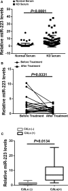
Serum microRNA miR‐223 levels are increased in patients with Kawasaki disease (KD). A, Serum levels of miR‐223 in 78 KD patients and 103 healthy control participants. B, Changes in serum miR‐223 levels in 22 KD patients before and after treatment with high‐dose intravenous immunoglobulin. C, Serum levels of miR‐223 in 12 KD patients with coronary artery lesions (CALs) and in 12 sex‐ and age‐matched KD patients without CALs.
Figure 2
Figure 2
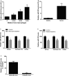
Vascular endothelial cell (EC) microRNA miR‐223 is from bone marrow–derived, blood cell–released serum miR‐223. A, No expression was found in passaged vascular ECs cultured in serum‐free medium. In this experiment, 2 known EC‐expressed microRNAs, miR‐34a and miR‐222, were used as positive controls. Another positive control was the human monocyte line THP‐1 cells, which have high levels of endogenous miR‐223. Water (H2O) was used a negative control (its mean threshold cycle level was similar to miR‐223). The mean level of miR‐34a in ECs (normalized by its U6) was expressed as 100%. The relative levels of miR‐222, miR‐223 in ECs, and miR‐223 in THP‐1 cells (normalized by their U6) are shown. Note: n=6; *P<0.05 compared with miR‐34a levels. B, Platelet‐derived growth factor (PDGF; 20 ng/mL) stimulation does not induce any miR‐223 expression in passaged ECs (normalized by its U6) compared with vehicle‐treated control, for which the level of miR‐223 was expressed as 100% (P>0.05). C, A significant amount of miR‐223 was found in freshly isolated ECs (n=6). *P<0.05 compared with the miR‐34a levels. D, A significant amount of miR‐223 was found in healthy vascular walls. Two well‐known miRNAs in vascular walls, miR‐222, and miR‐34a, were used as positive controls. The mean level of miR‐223 in vascular walls was expressed as 100%. Note: n=6; *P<0.05 compared with the level of miR‐223. E, miR‐223 levels in serum (normalized by spiked‐in cel‐miR‐39), blood cells (platelets, leukocytes; normalized by U6), and freshly isolated aortic ECs (normalized by U6) were almost undetectable in chimeric mice. The mean level of miR‐223 in serum, blood cells (platelets, leukocytes), and freshly isolated aortic ECs from wild‐type mice were expressed as 100%. Note: n=6; *P<0.05 compared with wild‐type control mice.
Figure 3
Figure 3
Blood cell–secreted microRNA miR‐223 in extracellular space, vascular endothelial cells (ECs), and vascular walls. A, Culture medium of THP‐1 macrophages was added into culture medium of ECs. The miR‐223 levels in ECs were increased with miR‐223–containing THP‐1 macrophage medium. B, MiR‐223 levels were higher in the group in which fresh serum isolated from mice was added into a culture medium (at 10%) of ECs for 24 hours followed by washing and then kept in serum‐free medium for another 24 hours. C, The levels of miR‐223 in serum, vascular ECs, and aortas were decreased in mice with neutrophil depletion. D, The levels of miR‐223 in serum (normalized by spiked‐in cel‐miR‐39), vascular ECs, and aortas (normalized by U6) were decreased in mice with platelet depletion. E, Only a very low level of miR‐223 was identified in aortas from the chimeric mice compared with wild‐type control mice (normalized by U6). Note: n=6 to 8; *P<0.05 compared with control groups.
Figure 4
Figure 4
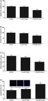
Cellular functions of microRNA miR‐223 in endothelial cells (ECs). The effect of miR‐223 on platelet‐derived growth factor–induced EC proliferation determined by (A) cell counting, (B) MTT assay, and (C) 5‐ethynyl‐2′‐deoxyuridine (EdU) assay. D, The effect of miR‐223 on vascular smooth muscle cell apoptosis induced by H2O2 (100 μmol/L) and representative images determined by terminal deoxynucleotidyl transferase dUTP nick end labeling analysis. Note: n=6; *P<0.05 compared with control groups. Oligo indicates Oligonucleotides.
Figure 5
Figure 5
The effect of microRNA miR‐223 on endothelial cell (EC) injury induced by Kawasaki disease (KD). A, The levels of miR‐223 in KD serum–cultured human vascular ECs are significantly increased in cultured human arterial ECs and could be inhibited by miR‐223 inhibitor (30 nmol/L). B, EC apoptosis was induced by KD serum and inhibited by miR‐223 inhibitor (30 nmol/L). C, Representative terminal deoxynucleotidyl transferase dUTP nick end labeling (TUNEL) images of different groups. Note: n=6; *P<0.05 compared with 20% KD serum without an miR‐223 inhibitor group.
Figure 6
Figure 6
IGF1R is a direct target gene of microRNA miR‐223 in endothelial cells (ECs), and Kawasaki disease (KD) serum could affect its target gene expression in ECs. A, miR‐223, but not scramble control (control oligo) or vehicle, inhibited luciferase activity in ECs. In the mutated control groups, the inhibitory effect of miR‐223 mimics disappeared. B, Overexpression of miR‐223 decreased the expression of IGF1R at the protein level in ECs. C, Representative Western blots showing the IGF1R protein levels from different groups. D, Overexpression of miR‐223 decreased the expression of IGF1R at the mRNA level in ECs. E, In KD serum–treated ECs, the IGF1R protein level was decreased; that effect could be partially blocked by miR‐223 inhibitor (50 nmol/L). F, In KD serum–treated ECs, the IGF1R mRNA level was decreased; that effect could be partially blocked by miR‐223 inhibitor (50 nmol/L). G, KD serum decreased the expression of FBXW7 in ECs. H, KD serum decreased the expression SEMA6A in ECs. I, IGF1R protein levels in freshly isolated vascular ECs from miR‐223 knockout mice and wild‐type control mice. J, Representative Western blots of IGF1R protein in freshly isolated vascular ECs from miR‐223 knockout mice and wild‐type control mice. K, IGF1R mRNA levels in freshly isolated vascular ECs from miR‐223 knockout mice and wild‐type control mice. Note: n=6; *P<0.05 compared with KD groups in (D and F) and with control groups in others.
Figure 7
Figure 7
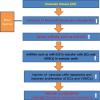
Bone marrow–derived, blood cell–released microRNAs (miRNAs) such as miR‐223 work as endocrine genetic signals in vascular cells and have a role in vascular injury from Kawasaki disease (KD). The inflammatory blood cells such as leukocytes and platelets, which originate from hematopoietic cells in bone marrow, could secrete the hematopoietic lineage cell–specific miRNAs such as miR‐223 into circulating serum. The blood cell–secreted serum miRNAs such as miR‐223 could then enter vascular cells such as endothelial cells (ECs) and vascular smooth muscle cells (VSMCs) in vascular walls and work as a novel endocrine genetic signal (like a hormone does) in ECs and VSMCs to regulate their biological functions such as proliferation and apoptosis. With the pathological condition of KD, blood cells are activated to release more miRNAs such as miR‐223 into serum and result in increased levels of miRNAs such as miR‐223 in ECs, VSMCs, and vascular walls. The increased miRNAs such as miR‐223 in vascular cells in KD could increase vascular cell injuries such as enhanced apoptosis and impaired proliferation, which could finally induce the vascular damage in KD such as vascular thrombosis and artery aneurysms.

Similar articles

Cited by

References

    1. Newburger JW, Takahashi M, Burns JC. Kawasaki disease. J Am Coll Cardiol. 2016;67:1738–1749. - PubMed
    1. Chen KY, Curtis N, Dahdah N, Kowalski R, Cheung M, Burgner DP, Kowalski R. Kawasaki disease and cardiovascular risk: a comprehensive review of subclinical vascular changes in the longer term. Acta Paediatr. 2016;105:752–761. - PubMed
    1. Liang M. MicroRNA: a new entrance to the broad paradigm of systems molecular medicine. Physiol Genomics. 2009;38:113–115. - PMC - PubMed
    1. Zhang C. MicroRNAs in vascular biology and vascular disease. J Cardiovasc Transl Res. 2010;3:235–240. - PMC - PubMed
    1. Qin S, Zhang C. MicroRNAs in vascular disease. J Cardiovasc Pharmacol. 2011;57:8–12. - PMC - PubMed

Publication types

MeSH terms

LinkOut - more resources