. 2017 Mar 31;292(13):5325-5334.
doi: 10.1074/jbc.M116.730523. Epub 2017 Feb 14.

S-Palmitoylation of Junctional Adhesion Molecule C Regulates Its Tight Junction Localization and Cell Migration

Affiliations

S-Palmitoylation of Junctional Adhesion Molecule C Regulates Its Tight Junction Localization and Cell Migration

Pornpun Aramsangtienchai et al. J Biol Chem. .

Abstract

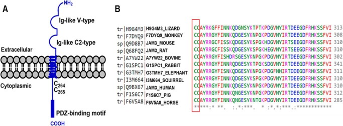
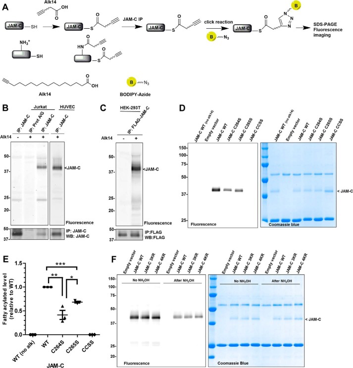
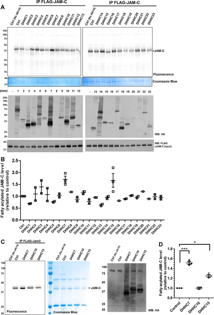
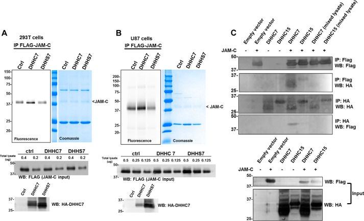
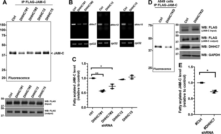
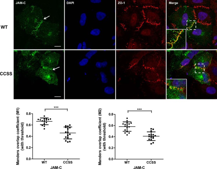
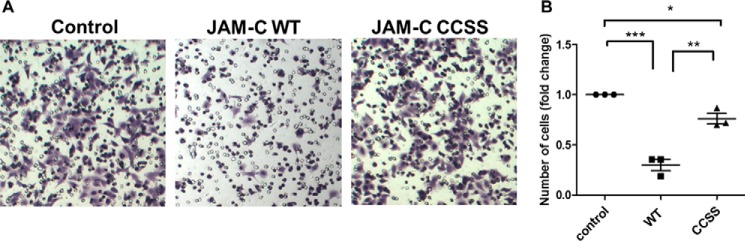
Junctional adhesion molecule C (JAM-C) is an immunoglobulin superfamily protein expressed in epithelial cells, endothelial cells, and leukocytes. JAM-C has been implicated in leukocyte transendothelial migration, angiogenesis, cell adhesion, cell polarity, spermatogenesis, and metastasis. Here, we show that JAM-C undergoes S-palmitoylation on two juxtamembrane cysteine residues, Cys-264 and Cys-265. We have identified DHHC7 as a JAM-C palmitoylating enzyme by screening all known palmitoyltransferases (DHHCs). Ectopic expression of DHHC7, but not a DHHC7 catalytic mutant, enhances JAM-C S-palmitoylation. Moreover, DHHC7 knockdown decreases the S-palmitoylation level of JAM-C. Palmitoylation of JAM-C promotes its localization to tight junctions and inhibits transwell migration of A549 lung cancer cells. These results suggest that S-palmitoylation of JAM-C can be potentially targeted to control cancer metastasis.

Keywords: cell adhesion; cell migration; chemical biology; post-translational modification (PTM); protein palmitoylation; protein-lipid interaction; tight junction.


Conflict of interest statement

The authors declare that they have no conflicts of interest with the contents of this article

Figures

FIGURE 1.
FIGURE 1.
JAM-C is an immunoglobulin superfamily protein containing two conserved cysteine residues (Cys-264 and Cys-265, human sequence). A, schematic structure of JAM-C transmembrane protein with two Ig-like domains in the extracellular region and one PDZ-binding motif in the cytoplasmic region. B, multiple sequence alignment by Clustal Omega showing the two cytoplasmic cysteine residues that are conserved across multiple species.
FIGURE 2.
FIGURE 2.
S-Palmitoylation of JAM-C depends on Cys-264 and Cys-265. A, method for the detection of S-palmitoylation in JAM-C with Alk14, the palmitic acid analogue. Cells were cultured with Alk14 for metabolic labeling. JAM-C was immunoprecipitated from total lysate, and BODIPY-azide was then conjugated to the alkyne group using click chemistry. The fluorescence signal was imaged after SDS-PAGE. B, endogenous JAM-C in both Jurkat and human umbilical vein endothelial cells (HUVEC) was labeled by Alk14. C, overexpressed FLAG-tagged JAM-C in HEK-293T cells also contained fatty acylation. D, the C264S and C265S mutations of JAM-C decreased the Alk14 labeling signal. Compared with the wild type JAM-C, palmitoylation in the single cysteine mutants (C264S and C265S) was reduced, whereas it was abolished in the double cysteine mutant (CCSS). E, quantified fatty acylation level of the JAM-C mutants relative to WT (n = 3; error bars represent S.D.). The palmitoylation signal was quantified and normalized by the protein level on the Coomassie Blue gel using Quantity One software. *, p < 0.05; **, p < 0.01; ***, p < 0.001. F, the cytosolic lysine to arginine mutants 3KR and 4KR of JAM-C did not significantly reduce the Alk14 labeling signal. Representative results from two independent experiments are shown. IP, immunoprecipitation; WB, Western blotting; alk, Alk14.
FIGURE 3.
FIGURE 3.
DHHC7 overexpression enhances the palmitoylation of JAM-C. A, the palmitoylation levels of FLAG-tagged JAM-C co-overexpressed with different HA-tagged DHHC1–23 in HEK-293T cells. B, quantification of the results shown in A (mean ± S.D., n = 2). The palmitoylation signal was quantified and normalized with the protein levels on the Coomassie Blue gel using Quantity One software. The signal in control cells without DHHC overexpression was set to 1.00 and served as the reference point for all other samples. C, DHHC7 overexpression most significantly increased JAM-C palmitoylation. A representative result from two independent experiments is shown. D, the quantified fatty acylation level of JAM-C co-overexpressed with DHHC7, DHHC10, or DHHC15 relative to control (n = 2; error bars represent S.D.). *, p < 0.05; ***, p < 0.001. IP, immunoprecipitation; WB, Western blotting; Ctrl, control.
FIGURE 4.
FIGURE 4.
DHHC7 interacts with JAM-C, and its catalytic activity is required for JAM-C S-palmitoylation. A, overexpression of DHHC7, but not DHHS7, increased JAM-C S-palmitoylation in HEK-293T cells. HA-tagged DHHC7 or HA-tagged DHHS7 was co-overexpressed with FLAG-tagged JAM-C. FLAG-tagged JAM-C was pulled down for palmitoylation detection. B, the same experiment as in A was performed in U87 cells. The full FLAG JAM-C input Western blot can be found in supplemental Fig. S5. C, DHHC7 interacts with JAM-C. HA-tagged DHHC7 was co-overexpressed with FLAG-tagged JAM-C in HEK-293T cells. HA-tagged DHHC7 was pulled down, and FLAG-tagged JAM-C was detected by Western blotting and vice versa. In the two lanes labeled with mixed lysate, JAM-C and DHHC7 or DHHC15 were expressed in different cells, and then the cell lysate was mixed before IP. A representative result from two independent experiments is shown. IP, immunoprecipitation; WB, Western blotting; Ctrl, control.
FIGURE 5.
FIGURE 5.
JAM-C palmitoylation level decreases in DHHC7 knockdown cells. A, HEK-293T cells were infected with lentiviruses containing scrambled shRNA (control) or DHHC7, DHHC12, or DHHC15 shRNAs. Puromycin-resistant cells were selected for stable DHHC knockdown cells and used for FLAG-tagged JAM-C overexpression. The FLAG-tagged JAM-C in the DHHC knockdown cells was then immunoprecipitated and detected for palmitoylation levels by fluorescence labeling. B, semiquantitative RT-PCR showing the mRNA levels of Dhhc7, Dhhc12, and Dhhc15 in the stable knockdown HEK-293T cells. C, quantified fatty acylation levels of FLAG-tagged JAM-C expressed in DHHC7, DHHC12, and DHHC15 knockdown HEK-293T cells relative to control (mean ± S.D., n = 2). The palmitoylation level from each group was quantified and normalized with the corresponding protein level on the Coomassie Blue gel using Quantity One software. The signal from FLAG-tagged JAM-C in the control knockdown cells was set to 1.00 and served as the reference point for the other samples. D, DHHC7 knockdown in A549 cells also decreased the palmitoylation of JAM-C. The experiments were carried out similar to that described in A. E, quantified fatty acylation levels of JAM-C in A549 cells (n = 2; error bars represent S.D.). *, p < 0.05; **, p < 0.01. IP, immunoprecipitation; WB, Western blotting; Ctrl, control.
FIGURE 6.
FIGURE 6.
S-Palmitoylation promotes JAM-C localization to the tight junction. Top, FLAG-tagged JAM-C WT and the CCSS mutant were ectopically expressed in A549 cells. The cells were immunofluorescently stained with anti-FLAG and anti-ZO1 antibodies after fixation. JAM-C WT was more co-localized with ZO-1 at the tight junction (white arrows), whereas the CCSS mutant had much less co-localization with ZO-1. The cells were visualized at room temperature with a Zeiss LSM 710 confocal microscope with a 63×/1.4 oil immersion objective. Images were viewed and analyzed using ZEN 2012 imaging software. These representative images were from three independent experiments with at least 15 FLAG-JAM-C-containing cells analyzed in each experiment. Scale bars, 10 μm. Bottom, the co-localization between JAM-C and ZO-1 was analyzed by the Coloc2 plug-in in Fiji software and is presented as the mean of Manders' coefficient (n = 15; error bars represent S.D.). ***, p ≤ 0.0001, Student's t test.
FIGURE 7.
FIGURE 7.
JAM-C S-palmitoylation affects cell migration. A549 cells were transfected with FLAG-tagged JAM-C WT or the CCSS mutant for 24 h and then cultured in RPMI 1640 serum-free medium for 14 h. The cell migration assay was then performed in a 24-well Transwell plate with 8-μm polycarbonate sterile membranes. A total of 3.5 × 10⁴ cells in 200 μl of RPMI 1640 serum-free medium were plated into each upper chamber and placed in wells containing 600 μl of RPMI 1640 medium supplemented with 10% FBS. After 24 h, cells on the upper surface were detached with a cotton swab. The chambers were fixed, and cells in the lower filter were stained with 0.1% crystal violet for 15 min and counted. The quantified results were calculated by counting three random fields of migrated cells. The control cells were transfected with an empty pCMV4a vector. A, representative images of migrated cells from three independent experiments are shown. B, the number of migrated cells per field was quantified and normalized by the value of the control (n = 3; error bars represent S.D.). *, p < 0.05; **, p < 0.01; ***, p < 0.001.

References

    1. Ebnet K., Suzuki A., Ohno S., and Vestweber D. (2004) Junctional adhesion molecules (JAMs): more molecules with dual functions? J. Cell Sci. 117, 19–29 - PubMed
    1. Keiper T., Santoso S., Nawroth P. P., Orlova V., and Chavakis T. (2005) The role of junctional adhesion molecules in cell-cell interactions. Histol. Histopathol. 20, 197–203 - PubMed
    1. Bazzoni G. (2003) The JAM family of junctional adhesion molecules. Curr. Opin. Cell Biol. 15, 525–530 - PubMed
    1. Weber C., Fraemohs L., and Dejana E. (2007) The role of junctional adhesion molecules in vascular inflammation. Nat. Rev. Immunol. 7, 467–477 - PubMed
    1. Gliki G., Ebnet K., Aurrand-Lions M., Imhof B. A., and Adams R. H. (2004) Spermatid differentiation requires the assembly of a cell polarity complex downstream of junctional adhesion molecule-C. Nature 431, 320–324 - PubMed

MeSH terms