. 2017 Jul 15;23(14):3906-3917.
doi: 10.1158/1078-0432.CCR-16-2530. Epub 2017 Feb 14.

MUC16 Regulates TSPYL5 for Lung Cancer Cell Growth and Chemoresistance by Suppressing p53

Affiliations

MUC16 Regulates TSPYL5 for Lung Cancer Cell Growth and Chemoresistance by Suppressing p53

Imayavaramban Lakshmanan et al. Clin Cancer Res. .

Abstract

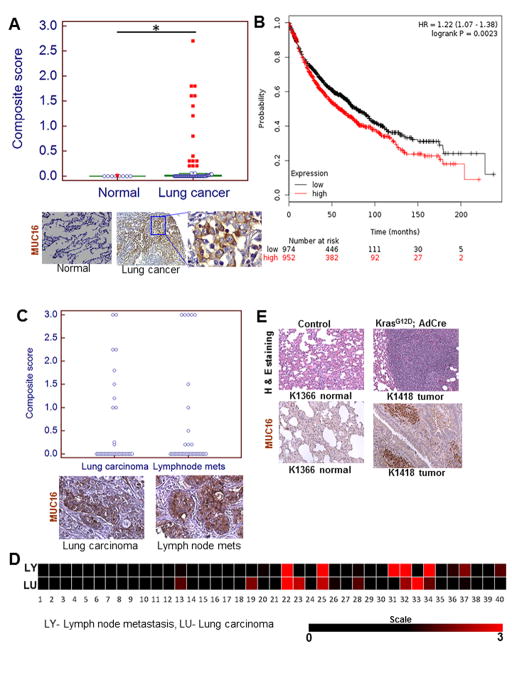
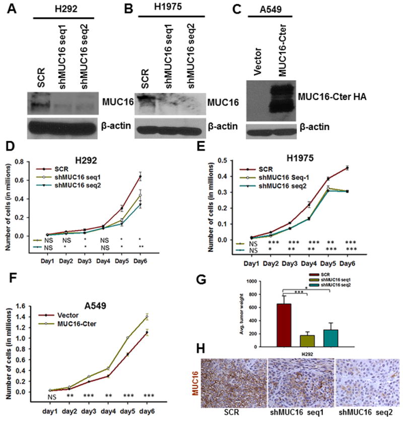
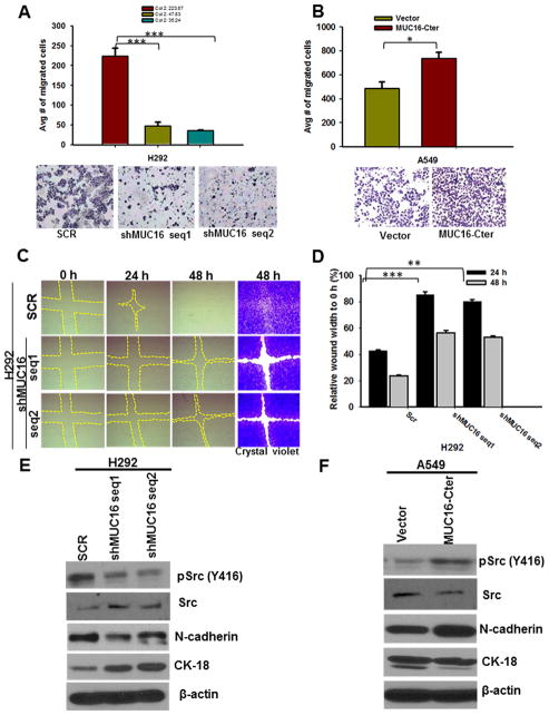
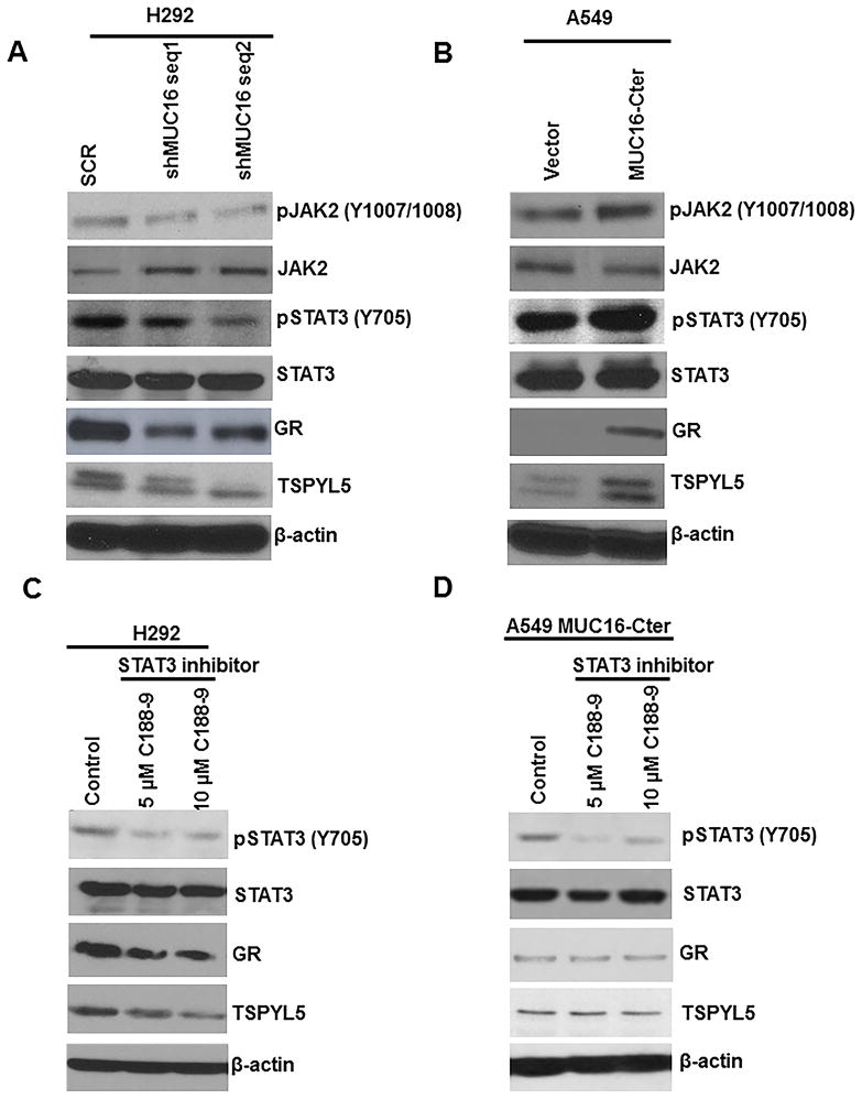
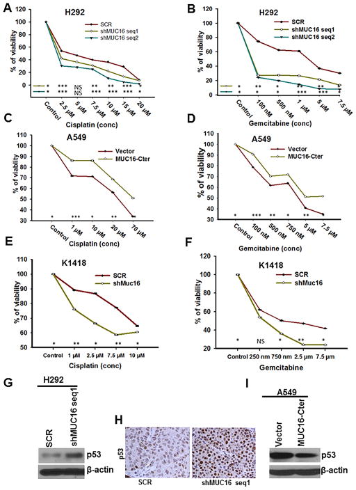
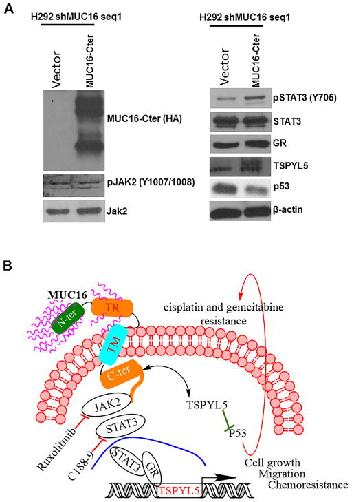
Purpose: MUC16, a tumor biomarker and cell surface-associated mucin, is overexpressed in various cancers; however, its role in lung cancer pathogenesis is unknown. Here, we have explored the mechanistic role of MUC16 in lung cancer.Experimental Design: To identify the functional role of MUC16, stable knockdown was carried in lung cancer cells with two different shRNAs. Clinical significance of MUC16 was evaluated in lung cancer patient tissues using IHC. We have generated genetically engineered mouse model (KrasG12D; AdCre) to evaluate the preclinical significance of MUC16.Results: MUC16 was overexpressed (P = 0.03) in lung cancer as compared with normal tissues. MUC16 knockdown (KD) in lung cancer cell lines decreased the in vitro growth rate (P < 0.05), migration (P < 0.001), and in vivo tumor growth (P = 0.007), whereas overexpression of MUC16-carboxyl terminal (MUC16-Cter) resulted in increased growth rate (P < 0.001). Transcriptome analysis of MUC16 KD showed a downregulation (P = 0.005) of TSPYL5 gene, which encodes for a testis-specific Y-like protein. Rescue studies via overexpression of MUC16-Cter in MUC16 KD cells showed activation of signaling proteins, such as JAK2 (Y1007/1008), STAT3 (Y705), and glucocorticoid receptor (GR), which constitutes an important axis for the regulation of TSPYL5 for oncogenic process. Further, inhibition of STAT3 (Y705) led to decreased GR and TSPYL5, suggesting that MUC16 regulates TSPYL5 through the JAK2/STAT3/GR axis. Also, MUC16 overexpression induced cisplatin and gemcitabine resistance by downregulation of p53.Conclusions: Our findings indicate a significant role of MUC16 in tumorigenesis and metastasis of lung cancer cells possibly via regulation of TSPYL5 through the JAK2/STAT3/GR axis. Clin Cancer Res; 23(14); 3906-17. ©2017 AACR.


Conflict of interest statement

Disclosure of Potential Conflicts of Interest

The authors declare that they have no conflicts of interest.

Figures

Figure 1
Figure 1
MUC16 expression in lung carcinoma and association of MUC16 with lung cancer patient’s survival. A, MUC16 was observed in a smaller proportion of normal bronchial tissues (1/10) but in a higher proportion of lung carcinoma (31/101, 30.69%; P=0.03). B, MUC16 expression is associated with worse outcomes in lung cancer patients. C, MUC16 expression was retained in both primary and metastatic lymph node tissues. D, Heat map of composite score represents that MUC16 expression in both primary lung carcinoma and matched lymph node tissues. E, Immunohistochemical results show that Muc16 is strongly overexpressed in mouse lung adenocarcinoma tissues (KrasG12D; AdCre) as compared to littermate control lung tissues (KrasG12D). *P<0.05. A, C & E, Figure magnification 20X and A, lower right higher magnification 40X.
Figure 2
Figure 2
Stable of knockdown of MUC16 and ectopic overexpression of MUC16-Cter in lung cancer cells and its role in lung cancer cell growth and tumorigenicity. A & B, MUC16 is endogenously present in both H292 and H1975 lung cancer cells and its expression was silenced using pSUPER-Retro shRNA method with two different targets (shMUC16 seq1 and shMUC16 seq2). D & E, The growth of MUC16 knockdown cells (shMUC16 seq1 and shMUC16 seq2) was significantly (P<0.05) reduced. C, We ectopically overexpressed MUC16-Cter (F114HA) in MUC16 negative lung cancer cell A549. F, MUC16-Cter overexpressed lung cancer cells (A549-F114HA) had a higher growth rate (P<0.05) than vector-transfected (A549-CMV9) cells. G, We performed tumorigenic assay by sub-cutaneously injecting MUC16 knockdown and scramble in athymic mice. MUC16 knockdown cells (H292-shMUC16 seq1 (P=0.007) and seq2 (P=0.04) had significantly less tumorigenic capacity than scramble (H292-SCR) cells. H, MUC16 expression was low in tumors induced by MUC16 knockdown (H292-shMUC16 seq1 and seq2) cells as compared to scramble (H292-SCR) cells. β-actin was used as loading control. *P<0.05, **P<0.01 and ***P<0.001, and NS non-significant. H, Figure magnification 20X.
Figure 3
Figure 3
Effect of MUC16 on the migration of lung cancer cells. A, Transwell migration assay demonstrates that migration of MUC16 knockdown (H292-shMUC16 seq1 (P=0.001) and seq2 (P=0.0006)) cells was significantly decreased. B, Similarly, MUC16-Cter overexpressed cells have more migratory (P=0.02) capacity as compared to vector cells. C, Further, MUC16 knockdown (H292-shMUC16 seq1 and seq2) cells have less migratory capacity than scramble (H292-SCR) cells as demonstrated by a wound healing assay. The wound area was stained with crystal violet for better visualization. D, The area was quantitatively calculated and normalized with wound area at 0 hrs of respective controls. E, The phosphorylation of Src (Y416) was decreased in MUC16 knockdown (H292-shMUC16 seq1 and seq2). E, Expression of mesenchymal marker N-cadherin was decreased in MUC16 knockdown (H292-shMUC16 seq1 and seq2) cells, whereas epithelial marker CK-18 was increased. F, Increased phosphorylation of Src (Y416), increased expression of N-cadherin and downregulation of CK18 was observed in MUC16-Cter overexpressed (A549-F114HA). β-actin was used as loading control. *P<0.05, **P<0.01 and ***P<0.001. A, B & C Figure magnification 10X.
Figure 4
Figure 4
MUC16 mediated downstream oncogenic signaling and inhibition of JAK2/STAT3 pathway for TSPYL5 gene expression. A, Upon MUC16 knockdown, phosphorylation of JAK2 (Y1007/1008) and STAT3 (Y705) was decreased. B, Similarly, MUC16-Cter overexpressed cells also had increased phosphorylation of JAK2 (Y1007/1008) and STAT3 (Y705). A & B, Similarly, GR and TSPYL5 were decreased in MUC16 knockdown and or increased in MUC16-Cter overexpressed lung cancer cells respectively. C, We inhibited STAT3 (Y705) using specific Inhibitor XIII, C188-9 in lung cancer. Phosphorylation of STAT3 (Y705) was decreased following pharmacological inhibition of STAT3 (Y705) using two different concentrations (5 μM and 10 μM) of C188-9. C, Total STAT3 expression remained the same. Further, as a result of STAT3 (Y705) inhibition, GR and TSPYL5 were decreased as compared to untreated cells. D, Inhibition of phospho-STAT3 inhibition in MUC16-Cter overexpressed cells confirmed the above findings. β-actin was used as loading control.
Figure 5
Figure 5
Role of MUC16 in cisplatin and gemcitabine resistance. A & B, We treated MUC16 knockdown (H292-shMUC16 seq1 and seq2) and scramble (H292-SCR) cells with various concentrations of cisplatin and gemcitabine. MTT assay results show that MUC16 knockdown (P<0.05) cells were more responsive to cisplatin (A) and gemcitabine (B). C & D, Similarly, MUC16-Cter overexpressed cells (P<0.05) were more resistant to cisplatin (C) and gemcitabine (D). E & F, Muc16 knockdown (K1418-shMuc16) mouse GEMM tumor cells (P<0.05) were more sensitive to cisplatin (E) and gemcitabine (F) than scramble cells. Mechanism of MUC16 mediated chemoresistance. G, Upon MUC16 knockdown, expression of p53 was increased as compared to scramble. H, The expression of p53 was high in tumors derived by subcutaneous injection of MUC16 knockdown cells as compared to tumors derived by injection of scramble cells. I, Similarly, MUC16-Cter overexpressed cell had a lower p53 expression. β-actin was used as loading control. *P<0.05, **P<0.01, ***P<0.001, and NS non-significant. I, Figure magnification 20X.
Figure 6
Figure 6
Restoration of MUC16 mediated pathways in lung cancer. A, To determine the recue effect of MUC16, we transfected MUC16-Cter (F114HA) in MUC16 knockdown (H292-shMUC16) cells. Restoration of phospho STAT3 (Y705), GR and TSPYL5 was observed in MUC16-Cter overexpressed in MUC16 knockdown cells as compared to vector transfected MUC16 knockdown cells. As expected, p53 expression was low in MUC16-Cter overexpressed in MUC16 knockdown cells. Schematic representation for MUC16 signaling in lung cancer cell growth and mechanistic role of MUC16 in chemoresistance in lung cancer. B, MUC16 phosphorylates JAK2 (Y1007/1008) and STAT3 (Y705) leading to translocation of STAT3 into the nucleus, where it recruits the glucocorticoid receptor (GR). The GR regulates TSPYL5 gene for lung cancer cell growth and metastasis. Inhibition of STAT3 phosphorylation by C188-9 leads to decreased expression of its target gene TSPYL5. In summary, MUC16 promotes JAK2/STAT3/GR signaling axis for TSPYL5 gene expression. This in turn promotes lung cancer cell growth and metastasis. MUC16/TSPYL5 downregulates p53 (wild-type) leading to chemoresistance of lung cancer cells.

Similar articles

Cited by

References

    1. Hollingsworth MA, Swanson BJ. Mucins in cancer: protection and control of the cell surface. Nat Rev Cancer. 2004;4(1):45–60. - PubMed
    1. Kufe DW. Mucins in cancer: function, prognosis and therapy. Nat Rev Cancer. 2009;9(12):874–85. - PMC - PubMed
    1. Das S, Batra SK. Understanding the Unique Attributes of MUC16 (CA125): Potential Implications in Targeted Therapy. Cancer Res. 2015;75(22):4669–74. - PMC - PubMed
    1. Das S, Rachagani S, Torres-Gonzalez MP, Lakshmanan I, Majhi PD, Smith LM, et al. Carboxyl-terminal domain of MUC16 imparts tumorigenic and metastatic functions through nuclear translocation of JAK2 to pancreatic cancer cells. Oncotarget. 2015;6(8):5772–87. - PMC - PubMed
    1. Gubbels JA, Belisle J, Onda M, Rancourt C, Migneault M, Ho M, et al. Mesothelin-MUC16 binding is a high affinity, N-glycan dependent interaction that facilitates peritoneal metastasis of ovarian tumors. Mol Cancer. 2006;5(1):50. - PMC - PubMed

MeSH terms