Review
. 2017 Jan 31:8:49.
doi: 10.3389/fimmu.2017.00049. eCollection 2017.

Avian Interferons and Their Antiviral Effectors

Affiliations
Review

Avian Interferons and Their Antiviral Effectors

Diwakar Santhakumar et al. Front Immunol. .

Abstract

Interferon (IFN) responses, mediated by a myriad of IFN-stimulated genes (ISGs), are the most profound innate immune responses against viruses. Cumulatively, these IFN effectors establish a multilayered antiviral state to safeguard the host against invading viral pathogens. Considerable genetic and functional characterizations of mammalian IFNs and their effectors have been made, and our understanding on the avian IFNs has started to expand. Similar to mammalian counterparts, three types of IFNs have been genetically characterized in most avian species with available annotated genomes. Intriguingly, chickens are capable of mounting potent innate immune responses upon various stimuli in the absence of essential components of IFN pathways including retinoic acid-inducible gene I, IFN regulatory factor 3 (IRF3), and possibility IRF9. Understanding these unique properties of the chicken IFN system would propose valuable targets for the development of potential therapeutics for a broader range of viruses of both veterinary and zoonotic importance. This review outlines recent developments in the roles of avian IFNs and ISGs against viruses and highlights important areas of research toward our understanding of the antiviral functions of IFN effectors against viral infections in birds.

Keywords: antivirals; avian; innate immunity; interferon-stimulated genes; interferons; viruses.


Figures

Figure 1
Figure 1
Induction of interferons (IFNs) and establishment of an antiviral state in a model chicken cell. The double-stranded RNA (dsRNA), detected by either chicken retinoic acid-inducible gene I (RIG-I)-like helicase (RLH) [melanoma differentiation-associated gene 5 (MDA5) or laboratory of genetics and physiology 2 (LGP2) individually or in cooperation] or toll-like receptor (TLR)3 (endosomal, phagosomal, or transmembrane) initiates downstream signaling mediated through mitochondrial antiviral-signaling protein (MAVS) or TRIF, respectively. These adaptor molecules then activate the transcription factors IFN regulatory factor (IRF)7, nuclear factor kappa B (NF-κB), and activating protein 1 (AP-1) (ATF2/JUN) by orchestrating the assembly of multi-protein complexes. Once activated, IRF7, NF-κB, and AP-1 translocate to the nucleus where they stimulate the transcription of, among others, type I IFNs (e.g., IFN-β). The transcribed, translated, and secreted type I IFNs initiate the JAK–STAT pathway by both autocrine (depicted in the figure) and paracrine signaling through cognate type I IFN receptor recognition. Activated JAK–STAT leads to the phosphorylation of STAT1 and STAT2 molecules, which (together with factors that are currently unknown in chicken) results in the formation of the IFN-stimulated gene factor 3 (ISGF3) transcription factor complex. This multifunctional transcription factor then scans and recognizes unique IFN-stimulated response element (ISRE) sequences to initiate the transcription of hundreds of chicken IFN-stimulated genes (chISGs), which subsequently establish the antiviral state against the invading viruses. Few examples of IFN-stimulated genes (ISGs) along with a summarized description of their functions are enlisted in the right panel of the figure. Abbreviations used in the figure and are not described in the main text are as follows: IκB kinase (IKK) epsilon (IKKε), alpha (IKKα), beta (IKKβ), and gamma (IKKγ); NF-κB essential modulator (NEMO); TANK-binding kinase 1 (TBK1); inhibitors of NF-κB (IκB), NF-κB subunits p50 and p65; activating transcription factor 2 (ATF2); tyrosine kinase 2 (TYK2); Janus kinase 1 (JAK1); signal transducer and activator of transcription 1 (STAT1), and STAT2. “P” represents the phosphorylation state of the protein, and dotted lines indicate the involvement of multiple intermediary steps.
Figure 2
Figure 2
(A) Genomic architecture along with relative loci around the IRF7 gene in human, mouse, dog, chicken, and fish. The IRF7 genes in the compared species are flanked upstream with LRDD gene and downstream with KIAA1542 and RSSF7 genes. Direct comparison of previously identified chicken IRF3 with these species indicates that this gene is in fact IRF7. (B) Phylogenetic analysis of IRF3 and IRF7 genes in different species. Based on the clustering patterns and sequence homologies, the gene previously identified as “chicken IRF3” clustered closer to IRF7 of other mammals compared to mammalian IRF3. It is therefore proposed to rename “chicken IRF3” to “chicken IRF7.” Gene abbreviations used in the figure are dual specificity phosphatase 8 (DSUP8), patatin-like phospholipase domain containing 2 (PNPLA2), leucine-rich repeats and death domain containing (LRDD), interferon regulatory factor 7 (IRF7), CTD-binding SR-like protein rA9 (KIAA1542); Ras association (RalGDS/AF-6) domain family 7 (RSSF7); leucine-rich repeat containing 56 (LRRC56); plakophilin 3 (PKP3).
Figure 3
Figure 3
(A) Genomic architecture of interferon regulatory factor (IRF)10 loci and phylogenetic analysis of IRF9 and IRF10 in human, mouse, dog, chicken, and fish. Upstream and downstream genes architecture in the IRF10 of chicken, dog, and fish indicate that this locus is similar to the corresponding locus in human and mice, which lack IRF10. Based on this and phylogenetic analysis (B), it is evident that the currently annotated chicken IRF9 is in fact an ortholog of IRF10. Gene abbreviations used in the figure are microtubule-associated protein homolog (Xenopus laevis) (TPX2); myosin, light polypeptide kinase 2, skeletal muscle (MYLK2); forkhead-like 18 (Drosophila) (FLKHL18); dual specificity phosphatase-like 15 (DUSP15); X Kell blood group precursor-related family member 7 homolog (XKR7); chromosome 20 open reading frame 160 (C20orf160); protein O-fucosyltransferase 1 (POFUT1); kinesin family member 3B (KIF3B).
Figure 4
Figure 4
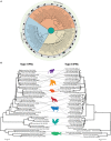
Phylogenetic analysis of interferon (IFN) genes of mammals (including rodents, primates, and domestic animals), avian, and fish species. (A) The open reading frames of type I, II, and III IFN genes were manually extracted from public databases and were aligned in BioEdit. The phylogenetic tree was constructed by MEGA6 software using the Kimura-2 model with 1,000 bootstrap replicates. The three types of IFNs clustered distantly and were labeled according to their clustering patterns. Approximate branching position was marked with a representative animal in the class. Only bootstrap values higher than 50 are shown. (B) Parallel comparison of type I and type II IFNs. Type III IFNs have been identified in limited numbers of species, and thus direct comparison was avoided. Clustering pattern of type I IFNs were linked to the type II IFN gene of the corresponding species for comparison purposes, and a representative animal image was shown to illustrate the clustering pattern.
Figure 5
Figure 5
(A) Structural and amino acid sequence homologies between type I interferons (IFNs) in different avian species. Alignment and sequence homology of avian IFN-alpha (IFN-α) [(A), top panel] and avian IFN-beta (IFN-β) [(A), bottom panel] amino acid sequences. Putative sites for IFN-α binding to IFNAR1 are marked with heart symbol, whereas the sites that are important for binding to IFNAR2 are marked with star sign. In comparison to mammals, sites required for interaction of IFNs with IFNARs are more variable among avian species (54). The previously reported signal peptides are underlined in both IFN-α (top) and IFN-β (bottom) sequences (54). (B) A modeled cartoon structure of human and chicken IFN-α. IFN-α protein structures were predicted using I-TASSER online tool and were annotated and aligned in MacPyMOL. Similar to human IFN-α (PBD ID: 1ITF), chicken IFN-α carries five helices and is structurally similar to human IFN-α. Direct structure comparison between human (mammalian) and chicken (avian) IFN-α proteins indicate that the chicken IFN-α protein carries five alpha-helices, which are considered crucial for the functionality of type I IFNs in mammals.
Figure 6
Figure 6
Structural and amino acid sequence homologies between type II and type III interferons (IFNs) in different avian species. (A) Protein sequence alignment of avian type II IFN (IFN-γ). (B) Predicted structure of chicken IFN-γ. (C) Protein sequence alignment of avian type III IFN (IFN-λ). (D) Predicted structure of chicken IFN-λ. Sequence alignments show that type II and type III IFNs are significantly conserved among avian species and may indicate interspecies cross-reactivity. Previously identified or predicted signal peptides are underlined in both IFN-γ (A) and IFN-λ (C) sequence alignments (54). These structures were predicted using I-TASSER online tool and were annotated using MacPyMOL. Structurally, these IFNs are well aligned with that of human IFNs (data not shown).

References

    1. Randall RE, Goodbourn S. Interferons and viruses: an interplay between induction, signalling, antiviral responses and virus countermeasures. J Gen Virol (2008) 89(Pt 1):1–47.10.1099/vir.0.83391-0 - DOI - PubMed
    1. Baron S, Dianzani F. The interferons: a biological system with therapeutic potential in viral infections. Antiviral Res (1994) 24(2–3):97–110.10.1016/0166-3542(94)90058-2 - DOI - PubMed
    1. Fensterl V, Sen GC. Interferons and viral infections. Biofactors (2009) 35(1):14–20.10.1002/biof.6 - DOI - PubMed
    1. Rathinam VA, Fitzgerald KA. Innate immune sensing of DNA viruses. Virology (2011) 411(2):153–62.10.1016/j.virol.2011.02.003 - DOI - PMC - PubMed
    1. Schoggins JW, Wilson SJ, Panis M, Murphy MY, Jones CT, Bieniasz P, et al. A diverse range of gene products are effectors of the type I interferon antiviral response. Nature (2011) 472(7344):481–5.10.1038/nature09907 - DOI - PMC - PubMed