Herbal Compound "Jiedu Huayu" Reduces Liver Injury in Rats via Regulation of IL-2, TLR4, and PCNA Expression Levels
- PMID: 28197212
- PMCID: PMC5288544
- DOI: 10.1155/2017/9819350
Herbal Compound "Jiedu Huayu" Reduces Liver Injury in Rats via Regulation of IL-2, TLR4, and PCNA Expression Levels
Abstract
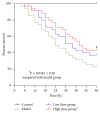
Aim of the Study. To investigate the preventative effects of Jiedu Huayu (JDHY) on D-galactosamine (D-GalN) and lipopolysaccharide-induced acute liver failure (ALF) and to evaluate the possible mechanisms of action. Materials and Methods. ALF was induced in Wistar rats by administrating D-GalN (900 mg/kg) and lipopolysaccharide (10 μg/kg). After treatment with JDHY granules, the levels of blood alanine aminotransferase, aspartate aminotransferase, total bilirubin, and prothrombin time were determined. Proliferating cell nuclear antigen was detected by immunohistochemistry staining. The expression of interleukin-2 (IL-2) and toll-like receptor 4 (TLR4) was examined by fluorescence quantitative reverse transcription polymerase chain reaction (qRT-PCR) and Western blot. Results. JDHY treatment dramatically improved liver function and increased survival rates in an ALF model in rats. We observed a decrease in IL-2 and TLR4 expression following treatment with JDHY in liver cells from ALF rats using qRT-PCR and Western blot analysis. Conclusion. We hypothesize that the therapeutic potential of JDHY for treating ALF is due to its modulatory effect on the suppression of inflammation and by promoting hepatocyte regeneration. Our results contribute towards validation of the traditional use of JDHY in the treatment of liver disease.
Conflict of interest statement
The authors declare that no conflict of interests exists.
Figures


Similar articles
-
The underlying mechanisms of Jie-Du-Hua-Yu granule for protecting rat liver failure.Drug Des Devel Ther. 2019 Feb 11;13:589-600. doi: 10.2147/DDDT.S180969. eCollection 2019. Drug Des Devel Ther. 2019. PMID: 30809090 Free PMC article.
-
Chinese Herb Jiedu Huayu Granules Inhibiting Immune and Inflammatory Response of Rats with Acute Liver Failure by Regulating the NF-κB Signaling Pathway.Biomed Res Int. 2022 May 11;2022:4479885. doi: 10.1155/2022/4479885. eCollection 2022. Biomed Res Int. 2022. Retraction in: Biomed Res Int. 2023 Dec 29;2023:9853907. doi: 10.1155/2023/9853907. PMID: 35601154 Free PMC article. Retracted.
-
Jie-Du-Hua-Yu Granules Promote Liver Regeneration in Rat Models of Acute Liver Failure: miRNA-mRNA Expression Analysis.Evid Based Complement Alternat Med. 2020 Dec 30;2020:8180959. doi: 10.1155/2020/8180959. eCollection 2020. Evid Based Complement Alternat Med. 2020. PMID: 33456491 Free PMC article.
-
Improved prescription of taohechengqi-tang alleviates D-galactosamine acute liver failure in rats.World J Gastroenterol. 2016 Feb 28;22(8):2558-65. doi: 10.3748/wjg.v22.i8.2558. World J Gastroenterol. 2016. PMID: 26937143 Free PMC article.
-
Integrated proteomics and metabolomics analysis of liver tissue reveal the therapeutic effect of Jiedu Huayu Granules on acute liver failure in rats.J Pharm Biomed Anal. 2025 Dec 15;266:117072. doi: 10.1016/j.jpba.2025.117072. Epub 2025 Jul 23. J Pharm Biomed Anal. 2025. PMID: 40737744
Cited by
-
ErHuang Formula Improves Renal Fibrosis in Diabetic Nephropathy Rats by Inhibiting CXCL6/JAK/STAT3 Signaling Pathway.Front Pharmacol. 2020 Jan 24;10:1596. doi: 10.3389/fphar.2019.01596. eCollection 2019. Front Pharmacol. 2020. Retraction in: Front Pharmacol. 2024 Dec 02;15:1530375. doi: 10.3389/fphar.2024.1530375. PMID: 32038260 Free PMC article. Retracted.
-
The underlying mechanisms of Jie-Du-Hua-Yu granule for protecting rat liver failure.Drug Des Devel Ther. 2019 Feb 11;13:589-600. doi: 10.2147/DDDT.S180969. eCollection 2019. Drug Des Devel Ther. 2019. PMID: 30809090 Free PMC article.
-
Chinese Medicine Jiedu Huayu Granules Reduce Liver Injury in Rats by Regulating T-Cell Immunity.Evid Based Complement Alternat Med. 2019 Nov 27;2019:1873541. doi: 10.1155/2019/1873541. eCollection 2019. Evid Based Complement Alternat Med. 2019. PMID: 31885638 Free PMC article.
-
[3-Methyladenine alleviates early renal injury in diabetic mice by inhibiting AKT signaling].Nan Fang Yi Ke Da Xue Xue Bao. 2024 Jul 20;44(7):1236-1242. doi: 10.12122/j.issn.1673-4254.2024.07.03. Nan Fang Yi Ke Da Xue Xue Bao. 2024. PMID: 39051069 Free PMC article. Chinese.
References
-
- Nie X.-H., Han T., Ha F.-U., et al. Comparison of the effects of the pretreatment and treatment with RhIL-11 on acute liver failure induced by D-galactosamine. European Review for Medical and Pharmacological Sciences. 2014;18(8):1142–1150. - PubMed
LinkOut - more resources
Full Text Sources
Other Literature Sources
Miscellaneous

