

The Triply Twisted Heart: Cyanosis in an Adult With Situs Inversus, Levocardia, Double Outlet Right Ventricle, and Malposition of the Great Arteries
- PMID: 28197259
- PMCID: PMC5295551
- DOI: 10.14740/cr440w
The Triply Twisted Heart: Cyanosis in an Adult With Situs Inversus, Levocardia, Double Outlet Right Ventricle, and Malposition of the Great Arteries
Abstract
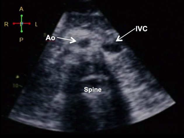
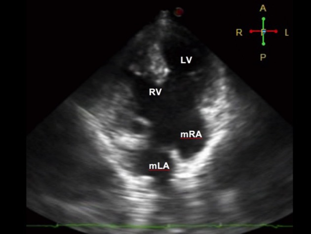
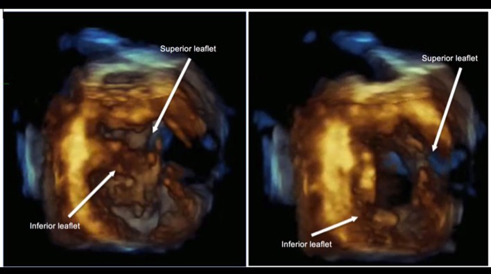
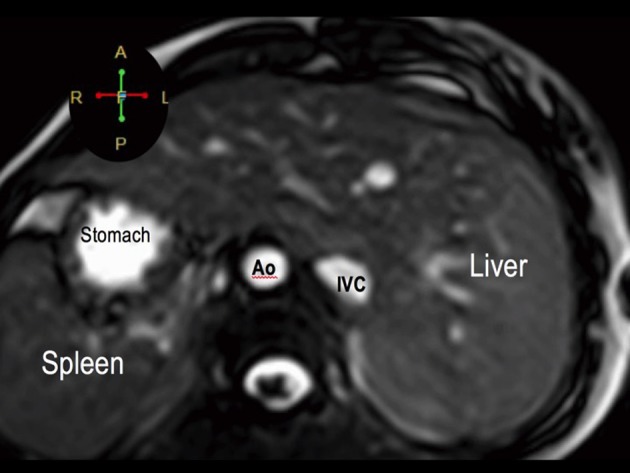
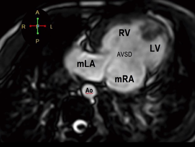
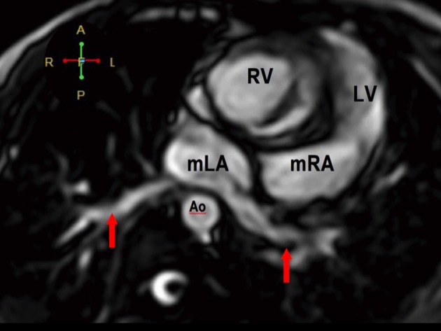
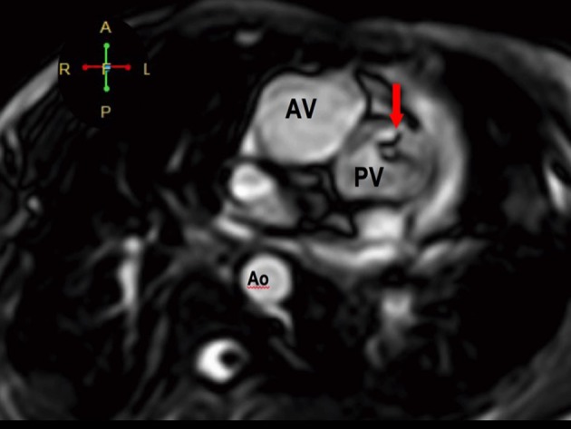
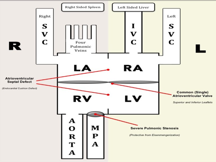
We present a case of a 19-year-old female presenting with cyanosis since birth. The major anomaly demonstrated was a "triply twisted heart" with a balanced physiology, allowing her to survive into adulthood. Non-invasive imaging was done using 2D and real-time 3D (or 4D) echocardiography with multi-slice imaging from 4D volume datasets. Findings were confirmed using cardiac magnetic resonance imaging (MRI). A segmental approach revealed atrial and visceral situs inversus, levocardia, atrioventricular discordance, and ventriculoarterial discordance. Both the aorta and pulmonary artery were malposed and arise from the right ventricle (double outlet right ventricle or DORV). There was also a complete atrioventricular septal defect (CAVSD) associated with a functional single atrium and a functional univentricle (single ventricle). Other findings include a severe pulmonic stenosis (PS), preserved right and left ventricular systolic function, and a normal pulmonary arterial pressure. She also had a persistent left superior vena cava (SVC) that drains into the morphologic right atrium, while the right-sided SVC drains into the morphologic left atrium. A multidisciplinary team deemed that management be palliative. She is on regular follow-up at our clinics for non-invasive monitoring. To our knowledge, this is the first reported case in an adult with this combination of anomalies.
Keywords: Atrioventricular septal defect; Malposition of the great arteries; Situs inversus.
Figures
Similar articles
-
Atrioventricular Discordance with Double-Outlet Right Ventricle in Mirror Imagery and Levocardia: A Very Rare Case Report.J Cardiovasc Echogr. 2020 Oct-Dec;30(4):227-230. doi: 10.4103/jcecho.jcecho_65_20. Epub 2021 Jan 20. J Cardiovasc Echogr. 2020. PMID: 33828947 Free PMC article.
-
[Double outlet right ventricle with discordant atrioventricular connection. Clinical study].Arch Inst Cardiol Mex. 1987 May-Jun;57(3):199-206. Arch Inst Cardiol Mex. 1987. PMID: 2959219 Spanish.
-
[Atrioventricular discordance without ventriculo-arterial discordance. Apropos of 3 cases].Arch Mal Coeur Vaiss. 1983 May;76(5):504-12. Arch Mal Coeur Vaiss. 1983. PMID: 6411022 French.
-
Prenatal Diagnosis of Isolated Atrioventricular Discordance and Ventriculoarterial Concordance and Double-Outlet Right Ventricle in Situs Inversus: Case Report and Review of the Literature.Pediatr Cardiol. 2020 Dec;41(8):1807-1810. doi: 10.1007/s00246-020-02467-z. Epub 2020 Sep 24. Pediatr Cardiol. 2020. PMID: 32970245 Review.
-
[Fontan-type procedure for an adult case of double-outlet right ventricle (S, D, L)].Nihon Kyobu Geka Gakkai Zasshi. 1995 Apr;43(4):553-61. Nihon Kyobu Geka Gakkai Zasshi. 1995. PMID: 7608612 Review. Japanese.
Cited by
-
Atrioventricular Discordance with Double-Outlet Right Ventricle in Mirror Imagery and Levocardia: A Very Rare Case Report.J Cardiovasc Echogr. 2020 Oct-Dec;30(4):227-230. doi: 10.4103/jcecho.jcecho_65_20. Epub 2021 Jan 20. J Cardiovasc Echogr. 2020. PMID: 33828947 Free PMC article.
References
-
- Neill. et al. The segmental approach to CHD. 2006;2:3–15.
-
- Goyal V, Devgarha S, Srivastava C. Atrial and visceral situs inversus with congenitally corrected transposition of the great arteries in a patient with dectrocardia, ventricular septal defect and pulmonary stenosis: A rare presentation. Turk Gogus Kalp Damar Cerrahisi Dergisi. 2012;20(3):622–624. doi: 10.5606/tgkdc.dergisi.2012.120. - DOI
Publication types
LinkOut - more resources
Full Text Sources
Other Literature Sources