. 2016 Dec 14;6(1):e1257452.
doi: 10.1080/2162402X.2016.1257452. eCollection 2017.

Tumor-infiltrating lymphocyte composition, organization and PD-1/ PD-L1 expression are linked in breast cancer

Affiliations

Tumor-infiltrating lymphocyte composition, organization and PD-1/ PD-L1 expression are linked in breast cancer

Laurence Buisseret et al. Oncoimmunology. .

Abstract

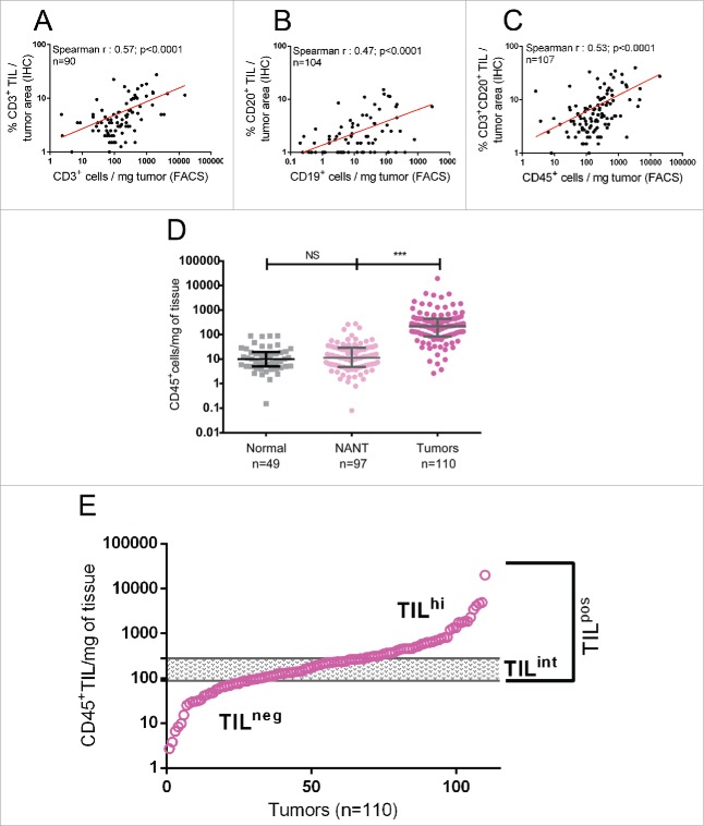
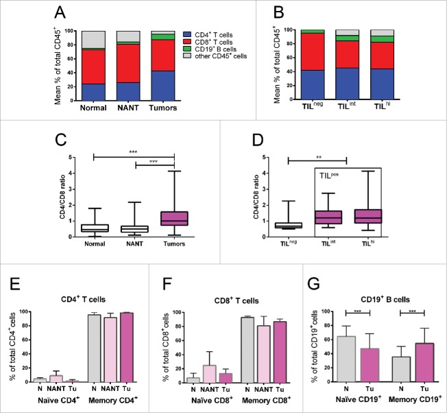
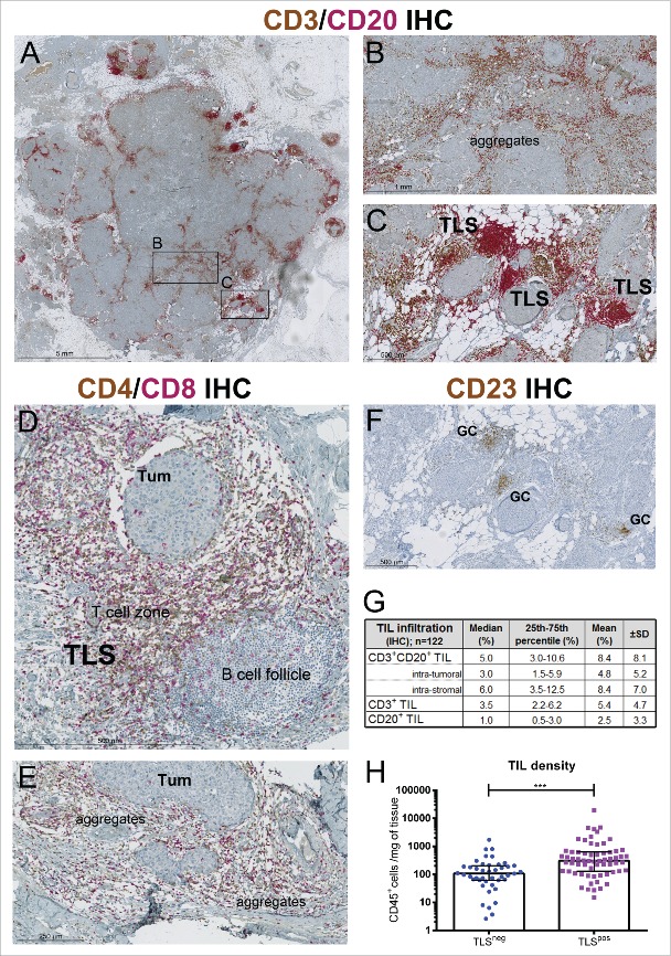
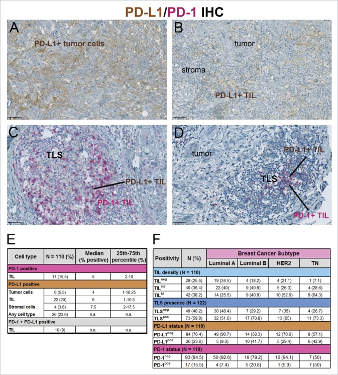
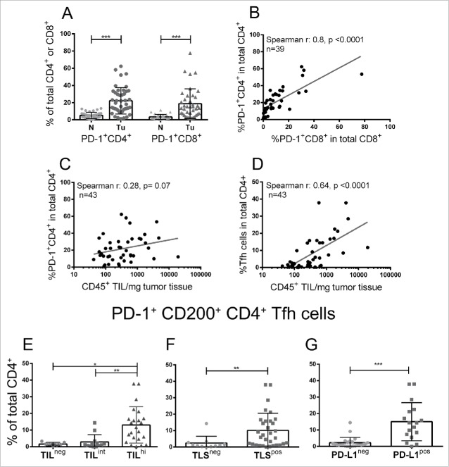
The clinical relevance of tumor-infiltrating lymphocytes (TIL) in breast cancer (BC) has been clearly established by their demonstrated correlation with long-term positive outcomes. Nevertheless, the relationship between protective immunity, observed in some patients, and critical features of the infiltrate remains unresolved. This study examined TIL density, composition and organization together with PD-1 and PD-L1 expression in freshly collected and paraffin-embedded tissues from 125 patients with invasive primary BC. Tumor and normal breast tissues were analyzed using both flow cytometry and immunohistochemistry. TIL density distribution is a continuum with 25% of tumors identified as TIL-negative at a TIL density equivalent to normal breast tissues. TIL-positive tumors (75%) were equally divided into TIL-intermediate and TIL-high. Tumors had higher mean frequencies of CD4+ T cells and CD19+ B cells and a lower mean frequency of CD8+ T cells compare with normal tissues, increasing the CD4+/CD8+ T-cell ratio. Tertiary lymphoid structures (TLS), principally located in the peri-tumoral stroma, were detected in 60% of tumors and correlated with higher TIL infiltration. PD-1 and PD-L1 expression were also associated with higher TIL densities and TLS. TIL density, TLS and PD-L1 expression were correlated with more aggressive tumor characteristics, including higher proliferation and hormone receptor negativity. Our findings reveal an important relationship between PD-1/PD-L1 expression, increased CD4+ T and B-cell infiltration, TIL density and TLS, suggesting that evaluating not only the extent but also the nature and location of the immune infiltrate should be considered when evaluating antitumor immunity and the potential for benefit from immunotherapies.

Keywords: Breast cancer; PD-1; PD-L1; tertiary lymphoid structures; tumor-infiltrating lymphocytes.


Figures

Figure 1.
Figure 1.
The density (cells/mg of tissue) of (A) CD3+ TIL (T cells) and (B) CD19+ TIL (B cells) quantified by flow cytometry (x-axis) was correlated with the percentage of tumor surface area occupied by CD3+ TIL or CD19+ TIL evaluated by trained pathologists on IHC-stained full-face tissue sections (y-axis), respectively. (C) The density of total CD45+ TIL quantified by flow cytometry was correlated with the percentage of tumor surface area occupied by CD3+ plus CD19+ TIL evaluated on IHC-stained tissue sections. (D) CD45+ cell density was compared between normal breast tissues, NANT and breast tumor tissues. Median and interquartile ranges are represented for each group. The level of significance is shown as a p value (*p < 0.05, **p < 0.01, ***p < 0.001). (E) The distribution of CD45+ cell density determined by flow cytometry for 110 breast tumors is shown graphically. The 99th percentile of CD45+ cell density in normal breast tissue is identified by the lower line, whereas the upper line delimits that for NANT. These thresholds are used to define TIL-negative (TILneg; below the lower threshold) and TIL-high tumors (TILhi; above the upper threshold), respectively, with TIL-intermediate tumors (TILint) falling between the two thresholds.
Figure 2.
Figure 2.
(A) Flow cytometry data represented as the mean percentage of CD4+ T cells, CD8+ T cells and B cells among total CD45+ cells in breast tumors compared with normal breast tissues or NANT. (B) The same lymphocyte subsets are shown for breast tumors divided into TILneg, TILint and TILhi density groups. (C) CD4+/CD8+ ratio in breast tumors compared with normal breast tissues or NANT. (D) CD4+/CD8+ ratio in breast tumors divided into TILneg, TILint and TILhi density groups. (E-G) CD4+ T cell, CD8+ T cell and CD19+ B-cell TIL in normal, NANT or tumor tissues separated into naïve vs. memory cell CD45RA/RO (CD45RA+/− for T cells and CD27+/− for B cells). The level of significance is shown as a p value (*p < 0.05, **p < 0.01, ***p < 0.001).
Figure 3.
Figure 3.
(A) A representative TILhi breast tumor IHC-stained for CD3+ T cells (brown) and CD20+ B cells (red). Enlargement of areas showing random aggregates of CD3+ and CD20+ TIL (B) or highly organized infiltrates in TLS (C) with the latter characterized a B-cell follicle adjacent to T-cell zone. (D, E) A different section from the same TILhi tumor IHC-stained for CD4+ (brown) and CD8+ T cells showing their location in TLS (D) and TIL aggregates (E). (F) CD23+ IHC staining of follicular dendritic cells (FDC) and germinal center B cells in the same TILhi tumor. (G) Table showing mean and median values for the percent TIL in different regions of the tumor evaluated on CD3 plus CD20 IHC-stained slides. (H) CD45+ TIL density determined by flow cytometry in TLSneg and TLSpos tumors, scored within the tumor area on CD3 plus CD20 IHC-stained slides.
Figure 4.
Figure 4.
(A–D) Representative tumors were IHC-stained for PD-1 (red) plus PD-L1 (brown) tumor cells (TC) and/or immune cells (IC), showing PD-L1+ tumor cells (A), PD-L1+ IC at the tumor-stroma interface (B) and PD-1+ plus PD-L1+ IC in TLS (C, D). (E) PD-1 and PD-L1 expression on different cell types is represented numerically for PD-L1+ tumors (>1% PD-L1+ among all cell types). (F) The four BC molecular subtypes (Luminal A (HR+, Ki67 <20%), Luminal B (HR+, Ki67 ≥20%), HER2+ (HR+ or HR−) and TN (HR− HER2−)) are numerically divided based on different TIL densities, TLS, PD-1 and PD-L1. “n.a” for not applicable.
Figure 5.
Figure 5.
(A) Graphic representation of the proportion of PD-1+ T cells determined by flow cytometry for tumors (Tu) compare with normal breast tissue (N). (B) The correlation between PD-1+CD4+ T cells (y-axis) and PD-1+CD8+ T cells (x-axis) in tumor tissue. (C, D) The correlation between PD-1+CD4+ (C) and PD-1+CD200+CD4+ Tfh cells (D) and CD45+ TIL density all determined by flow cytometry. (E–G) Graphic representation of the proportion of PD-1+CD200+ (Tfh cells) in total CD4+ T cells relative to TIL density (E), TLS presence (F) and PD-L1 status (G).

Similar articles

Cited by

References

    1. Weiss SA, Hanniford D, Hernando E, Osman I. Revisiting determinants of prognosis in cutaneous melanoma. Cancer 2015; 121:4108-23; PMID:26308244; http://dx.doi.org/10.1002/cncr.29634 - DOI - PMC - PubMed
    1. Reissfelder C, Stamova S, Gossmann C, Braun M, Bonertz A, Walliczek U, Grimm M, Rahbari NN, Koch M, Saadati M et al.. Tumor-specific cytotoxic T lymphocyte activity determines colorectal cancer patient prognosis. J Clin Invest 2015; 125:739-51; PMID:25562322; http://dx.doi.org/10.1172/JCI74894 - DOI - PMC - PubMed
    1. Brambilla E, Le Teuff G, Marguet S, Lantuejoul S, Dunant A, Graziano S, Pirker R, Douillard JY, Le Chevalier T, Filipits M et al.. Prognostic effect of tumor lymphocytic infiltration in Resectable Non-small-cell lung cancer. J Clin Oncol 2016; 34:1223-30; PMID:26834066; http://dx.doi.org/10.1200/JCO.2015.63.0970 - DOI - PMC - PubMed
    1. Salgado R, Denkert C, Demaria S, Sirtaine N, Klauschen F, Pruneri G, Wienert S, Van den Eynden G, Baehner FL, Penault-Llorca F et al.. The evaluation of tumor-infiltrating lymphocytes (TILs) in breast cancer: recommendations by an International TILs Working Group 2014. Ann Oncol 2015; 26:259-71; PMID:25214542; http://dx.doi.org/10.1093/annonc/mdu450 - DOI - PMC - PubMed
    1. Savas P, Salgado R, Denkert C, Sotiriou C, Darcy PK, Smyth MJ, Loi S. Clinical relevance of host immunity in breast cancer: from TILs to the clinic. Nat Rev Clin Oncol 2016; 13:228-41; PMID:26667975; http://dx.doi.org/10.1038/nrclinonc.2015.215 - DOI - PubMed

Publication types