Review
. 2017 Apr;8(2):271-277.
doi: 10.1007/s13244-017-0547-4. Epub 2017 Feb 14.

Radiological findings of unilateral tuberculous lung destruction

Affiliations
Review

Radiological findings of unilateral tuberculous lung destruction

Diego Varona Porres et al. Insights Imaging. 2017 Apr.

Abstract

Objectives: The aim of this report is to identify the radiological findings of unilateral tuberculous lung destruction (UTLD).

Materials and methods: Thirteen patients with (UTLD) were reviewed from 1999 to 2014. Only patients with radiological evidence of absence of pulmonary parenchyma preserved were included. Clinical and demographic data were obtained and radiological studies (chest radiograph and CT) were retrospectively reviewed.

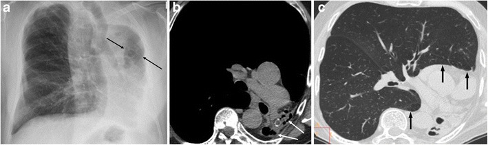
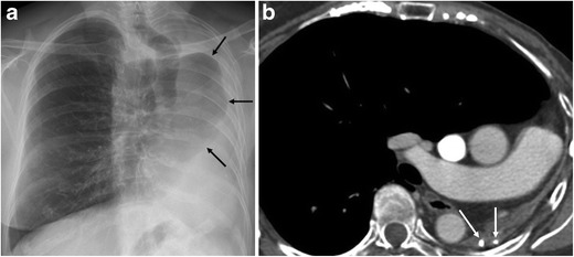
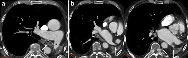
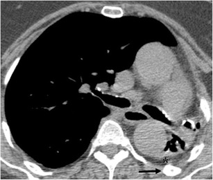
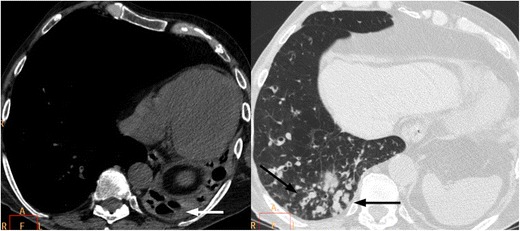
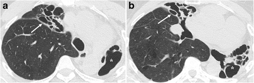
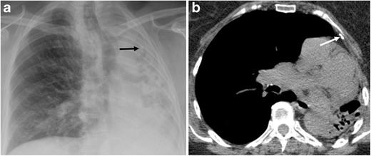
Results: The left lung was more commonly involved (85%). The following radiological findings were found in all cases: a decrease in the diameter of the pulmonary vessels of the affected lung, herniation of the contralateral lung and hypertrophy of the ribs and/or thickening of extrapleural fat. Two radiological patterns were identified: UTLD with cystic bronchiectasis (85%) and UTLD without residual cystic bronchiectasis (15%). Forty-six per cent of cases had respiratory infection symptoms with presence of air-fluid levels in the affected lung as the most common finding in these patients.

Conclusions: Total unilateral post-tuberculous lung destruction is an irreversible complication with the following main radiological features: predominantly left-sided location, decreases in the diameter of the ipsilateral pulmonary vessels, herniation of the contralateral lung and hypertrophy of the ribs and/or thickening of extrapleural fat.

Teaching points: • Unilateral tuberculous lung destruction is an irreversible complication of tuberculosis. • Left-side predominance and herniation of the contralateral lung are characteristic. • Decreased diameter of the ipsilateral pulmonary vessels occurred in all patients. • The pattern with residual cystic bronchiectasis is the most frequent. • Superimposed non-tuberculous infections may affect the destroyed lung.

Keywords: CT scanner, X-ray; Infections, respiratory; Radiography; Thoracic; Tuberculosis, pulmonary.


Figures

Fig. 1
Fig. 1
A 58-year-old male with a history of pulmonary TB 20 years previously presented with dyspnoea on exertion. (a) The chest radiograph shows marked loss of left lung volume, with an isolated hyperlucent lesion (arrows) that reflects herniation of the contralateral lung toward the left hemithorax. (b) Unenhanced multislice CT depicts total destruction of the left lung with residual cystic bronchiectasis (arrows) and herniation of the right lung toward the left hemithorax. (c) Unenhanced multislice CT clearly demonstrates herniation of the right lung toward the left hemithorax, that is, anterior herniation of the upper lobe and posterior herniation of the lower lobe (arrows)
Fig. 2
Fig. 2
A 68-year-old female with a background of pulmonary TB at age 29 was seen at our hospital for a breast cancer follow-up study. (a) Chest radiography shows marked loss of left lung volume and herniation of the contralateral lung (arrows). (b) Contrast-enhanced multislice CT demonstrates total left lung destruction with no residual cystic bronchiectasis. Calcifications are seen in the remnant lung (arrows), and the contralateral lung is herniated
Fig. 3
Fig. 3
An 83-year-old male who had pulmonary TB in infancy came to our hospital for weakness and paresthesia of both hands. (a) On contrast-enhanced multislice CT, total left lung destruction with calcifications (black arrow) in the remnant lung, occupation of the left main bronchus (white arrow) and decreased diameter of the left main pulmonary artery (asterisk) are visualised. (b) The diameter of the ipsilateral superior and inferior pulmonary veins is also decreased (arrows)
Fig. 4
Fig. 4
A 76-year-old female with TB history in infancy presenting with dry cough for 1 month. Non-enhanced CT shows total unilateral left lung destruction with bronchiectasis. Upper ipsilateral ribs are hypertrophic (black arrow) and extrapleural fat proliferation is also evident (asterisk)
Fig. 5
Fig. 5
A 73-year-old male with a history of pulmonary TB as a youth and chronic obstructive pulmonary disease was seen for fever and purulent expectoration. Unenhanced multislice CT depicted complete left pulmonary destruction with residual cystic bronchiectasis associated with air-fluid levels (white arrow in the right image) and airway occupation with mucus impaction in the lower right lobe (black arrows in the left image). Sputum culture was positive for Pseudomona aeruginosa and Streptococcus pneumoniae. Antibiotic treatment was established without success, and the patient died 15 days after hospitalisation
Fig. 6
Fig. 6
A 43-year-old female with TB history in her youth and recurrent infections presenting with fever. Sputum culture was postive for Pseudomona aeruginosa. (a) Non-enhanced CT shows total unilateral left lung destruction with bronchiectasias (asterisk) and lung atelectasis with saccular bronchiectasis in the right upper lobe (white arrow). (b) Right middle lobe atelectasis is also seen along with bronchiectasis (white arrow)
Fig. 7
Fig. 7
Same patient as in Fig. 4. (a) Chest radiograph shows total unilateral left lung destruction and calcification projected on the upper half of left hemithorax. (b) Non-enhanced CT confirms the presence of left upper pleural calcification in correspondence with the calcification seen on chest radiograph

References

    1. World Health Organization (2016) Global tuberculosis report 2015. World Health Organization, Geneva. Available via http://www.who.int/tb/publications/global_report/gtbr15_main_text.pdf?ua=1. Accessed 5 Aug 2016
    1. Fawibe AE, et al. Profile and outcome of unilateral tuberculous lung destruction in Ilorin, Nigeria. West Afr J Med. 2011;30(2):130–135. - PubMed
    1. Palmer PE. Pulmonary tuberculosis—usual and unusual radiographic presentations. Semin Roentgenol. 1979;14(3):204–243. doi: 10.1016/0037-198X(79)90007-5. - DOI - PubMed
    1. Jeong YJ, Lee KS. Pulmonary tuberculosis: up-to-date imaging and management. AJR Am J Roentgenol. 2008;191(3):834–844. doi: 10.2214/AJR.07.3896. - DOI - PubMed
    1. Ashour M, et al. Unilateral post-tuberculous lung destruction: the left bronchus syndrome. Thorax. 1990;45(3):210–212. doi: 10.1136/thx.45.3.210. - DOI - PMC - PubMed

LinkOut - more resources