. 2017 Feb 15:7:41083.
doi: 10.1038/srep41083.

Human IDO-competent, long-lived immunoregulatory dendritic cells induced by intracellular pathogen, and their fate in humanized mice

Affiliations

Human IDO-competent, long-lived immunoregulatory dendritic cells induced by intracellular pathogen, and their fate in humanized mice

Rajeev K Tyagi et al. Sci Rep. .

Abstract

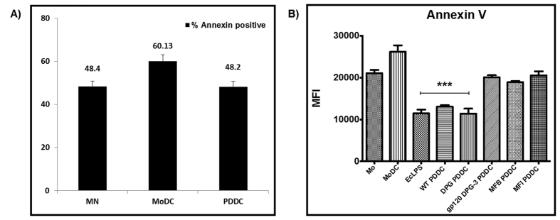
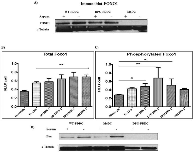
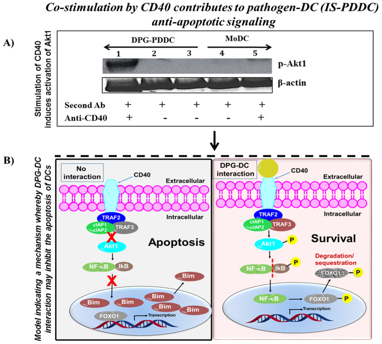
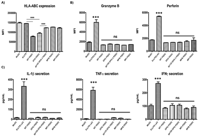
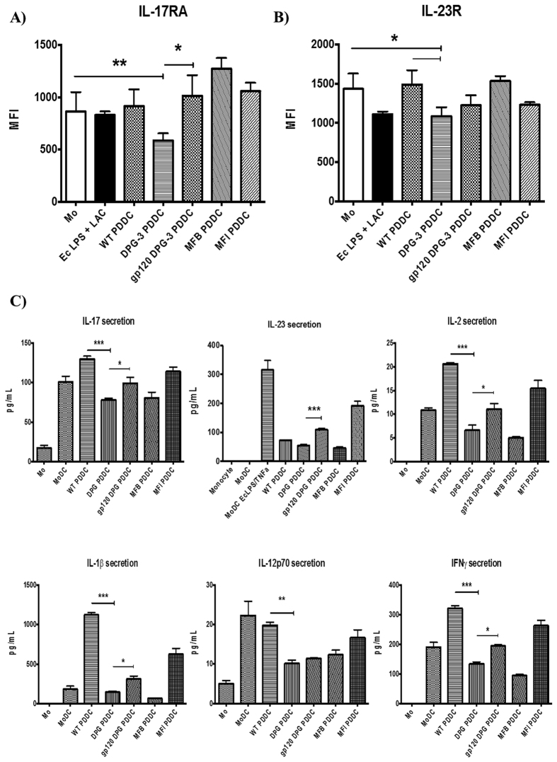
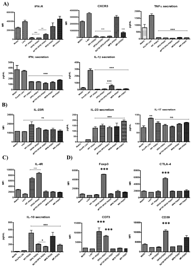
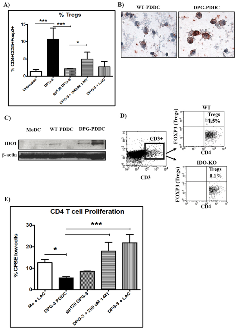
Targeting of myeloid-dendritic cell receptor DC-SIGN by numerous chronic infectious agents, including Porphyromonas gingivalis, is shown to drive-differentiation of monocytes into dysfunctional mDCs. These mDCs exhibit alterations of their fine-tuned homeostatic function and contribute to dysregulated immune-responses. Here, we utilize P. gingivalis mutant strains to show that pathogen-differentiated mDCs from primary human-monocytes display anti-apoptotic profile, exhibited by elevated phosphorylated-Foxo1, phosphorylated-Akt1, and decreased Bim-expression. This results in an overall inhibition of DC-apoptosis. Direct stimulation of complex component CD40 on DCs leads to activation of Akt1, suggesting CD40 involvement in anti-apoptotic effects observed. Further, these DCs drove dampened CD8+ T-cell and Th1/Th17 effector-responses while inducing CD25+Foxp3+CD127- Tregs. In vitro Treg induction was mediated by DC expression of indoleamine 2,3-dioxygenase, and was confirmed in IDO-KO mouse model. Pathogen-infected &CMFDA-labeled MoDCs long-lasting survival was confirmed in a huMoDC reconstituted humanized mice. In conclusion, our data implicate PDDCs as an important target for resolution of chronic infection.


Conflict of interest statement

The authors declare no competing financial interests.

Figures

Figure 1
Figure 1. MoDCs show higher rate of apoptosis than PDDCs.
(A) Unpulsed MoDC and Pg-wt differentiated DC (WT-PDDC) were stained with propidium iodide staining solution using apoptosis detection kit (Annexin V staining kit, eBioscience). Cells were washed in RPMI supplemented with 10% FBS, and cytospin in order to perform immunofluorescence assay. Apoptotic MoDC and PDDC (arrows), with a characteristic condensed, fragmented, brighter nucleus than non-apoptotic DCs. Insets, view of PI staining of representative apoptotic cells. Scale bars: DCs, 35 μm; Monocytes (MN), 15 μm, (B) Unpulsed MoDC and Pg-wt differentiated DC (WT-PDDC) were stained with propidium iodide staining solution using apoptosis detection kit (Annexin V staining kit). Percentage of MN, MoDCs and PDDCs that present condensed or fragmented nuclei (PI) or the outer membrane (annexin+), (C) PDDCs generated from E. Coli LPS treatment or infection with Mfa-1+ strains (WT, DPG) displays a significantly lower level of surface Annexin V staining compared to starved monocytes and MoDC controls. This significant decrease is lost when cells are pretreated with HIV-1gp120 to block DC-SIGN prior to infection or PDDCs are generated with Mfa-1 deficient strains (MFI, MFB). These results are the mean of three independent experiments (n = 3).
Figure 2
Figure 2. Phosphorylated Foxo1 decreases with PDDC apoptosis rate.
(A) FOXO1 expression was observed on protein level in PDDCs (WT & DPG) by carrying out Western blot using Anti-FOXO1 Mab. α-Tubulin, loading control (n = 2). The amount of total PDDC Foxo1 (B) and phosphorylated Foxo1 (C) were measured by whole cell ELISA and normalized to total cell numbers (n = 3) (B) PDDCs generated with E. coli LPS or either of the P. gingivalis strains have significantly higher levels of Foxo1. (C) PDDCs generated by the minor-fimbriae expressing WT and DPG-3 or the non-fimbriated MFB have significantly higher levels of phosphorylated Foxo1 (D) DCs were washed in RPMI and then incubated for 10 hrs in 10% FBS in RPMI. DCs were then lysed and Bim was detected by immunoblot. α-tubulin, loading control (n = 2).
Figure 3
Figure 3. Stimulation of CD40 induces activation of Akt.
(A) MoDC and PDDC were suspended in 0.1% BSA in RPMI and then treated with anti-CD40 antibody. DCs were then treated with secondary antibody (sheep anti-mouse (Fab) 2 fragment antibody) for 15 min to induce clustering of CD40. The DCs were lysed and phosphorylated Akt1 was detected by using anti-Akt1 antibody. Well (1–3) loaded with DPG-PDDC ; well (4–5) loaded with MoDC. β-actin, loading control (n = 3) (B) model indicating a mechanism whereby the DPG-DC interaction may inhibit the apoptosis of DCs. Lower left panel, without DPG-DC interaction, transcription factor NF-kB, associated with its inhibitor IkB, and remains in the cytoplasm. On the contrary, pro-apoptotic factor FOXO1 from nucleus regulates the expression of pro-apoptotic family member Bim. Lower right panel, with DPG-DC interaction, CD40 located at the DPG-DC association induces activation of kinase Akt1. The activated Akt1 leads to, (1) phosphorylation by IkB, which is subsequently degraded, and allowing the translocation of NF-kB to the nucleus. NF-kB may control the transcription of pro-survival genes, and (2) phosphorylates FOXO1 in the nucleus and translocates it to cytoplasm which in turn inhibits the expression of Bim.
Figure 4
Figure 4. P. gingivalis generated PDDCs have inhibited activation of effector CD8 T cell function.
PDDCs were generated for 24 hours and then cultured with autologous naïve CD8 T cells (A) Surface levels of HLA-ABC on the PDDC groups was analyzed to determine stimulatory capacity. WT and DPG-3 PDDC groups had significantly lower levels of HLA-ABC expression. HLA-ABC was significantly increased with pre-treatment of cells with HIV gp120 to prevent DPG-3 uptake. (B) Surface expression of Granzyme B and perforin were analyzed on CD8 T cells after culture. PDDC groups did not drive expression of granzyme B or perforin above baseline levels seen after culture with untreated MoDCs and were much lower than in the presence of E. coli LPS and leukocyte activation cocktail (C) Analysis of cytokine secretion from supernatants of the co-cultures show that inflammatory cytokines IL-1β, TNFα, and IFNγ are only produced in the presence of E. coli LPS and LAC. These results are the mean of three independent experiments (n = 3).
Figure 5
Figure 5. Dampened Th1/Th17 response of PDDCs in absence of P. gingivalis major fimbriae.
PDDCs were analyzed for surface expression (A,B) and cytokine secretion (C) of Th1 or Th17 biased inflammatory mediators. (A) PDDCs express relatively low levels of surface IL-17 receptor, with minor-fimbriated DPG-3 PDDCs showing a significant reduction in IL-17 receptor expression. The expression is significantly restored when cells are pre-treated with HIV gp120 to prevent DPG-3 uptake. (B) PDDCs express relatively low levels of surface IL-23 receptor and DPG-3 generated PDDCs have significantly lower IL-23 receptor than either untreated monocytes or WT generated PDDCs. (C) DPG-3 generated PDDCs show a significant decrease of the IL-17, IL-2, IL-1β, IL-12p70, and IFNγ secretion compared to WT generated PDDCs. Secretion of IL-17, IL-23, IL-2, IL-1β, and IFNγ were all significantly increased when cells were pre-treated with gp120. These results are the mean of three independent experiments (n = 3).
Figure 6
Figure 6. PDDCs are unable to generate robust inflammatory responses from naïve CD4 T cells.
PDDC groups were generated for 24 hours and then cultured with autologous CD4 T cells for 5 days. CD4 T cells were analyzed for expression of selected Th1 inflammatory molecules (A), Th17 inflammatory molecules (B), Th2 molecules (C), or Treg molecules (D). (A) PDDCs generated with P. gingivalis minor-fimbriated strains WT and DPG-3 led to a significant reduction of the Th1-biasing IFNγ receptor and CXCR3 on CD4 T cell surface. Expression of IFNγ receptor was significantly increased when initial DPG-3 uptake was blocked with HIV gp120. TNFα, IFNγ and IL-1β secretion were significantly lower in all PDDC group co-cultures compared to the positive control of E. coli LPS and leukocyte activation cocktail. IL-1β was significantly increased with gp120 treatment. (B) The Th17-biasing IL-23 receptor was significantly increased on CD4 T cells by WT PDDC, but not seen in any of the other PDDC co-cultures. IL-17 secretion was not significantly changed from untreated MoDC co-cultures by any PDDC group co-cultures. IL-23 secretion was significantly increased in all PDDC co-cultures. (C) The expression of Th2-biasing IL-4 receptor was significantly increased in WT and DPG-3 co-cultures and significantly decreased when DPG-3 uptake of PDDCs was blocked by gp120. IL-10 secretion was significantly increased in all PDDC co-cultures compared to LAC-treated controls and was significantly decreased in gp120 treatments prior to DPG-3 infection compared to DPG-3 alone. (D) CD4 T cells cultured with DPG-3 PDDCs showed a significant increase in Foxp3, CTLA-4, CD39, and CD73 expression. Pre-treatment with HIV gp120 to block DPG-3 uptake significantly reduced expression of each marker. These results are the mean of three independent experiments (n = 3).
Figure 7
Figure 7. DPG-3 PDDCs induce IDO-dependent regulatory T cell responses from naïve CD4 T cells.
PDDC groups were generated for 24 hours and then cultured with autologous CD4 T cells for 5 days. (A) Tregs were significantly induced in DPG-3 PDDC co-cultures and this induction was lost with gp120 pre-treatment or in the presence of 1-MT or LAC (n = 3) (B) Immunocytochemistry carried out by IDO specific antibody on cytospins of WT-PDDCs and DPG-PDDCs showed greater expression of IDO in DPG differentiated DCs (n = 3). The ICC of IDO expression is well supported and confirmed by, (C) immunoblot detection of IDO in WT and DPG-PDDCs. MoDCs and PDDCs were cultured in 10% HI-FBS, cells were lysed by cell lysis solution to extract protein. β-actin, loading control (n = 2). (D) IDO plays a crucial role in modulating systemic inflammatory responses by affecting Tregs induction in IDO-KO mouse model of gingivitis. WT (IDO sufficient) mice were able to induce markedly higher level of Tregs (1.5% of CD3+ CD4+ cells) compared to their IDO-KO counterparts (0.1% of CD3+ CD4+ cells) after P. gingivalis LPS injection (n = 3). (E) CD4 T cells were pre-labeled with CFSE to measure proliferation in a parallel experiment. DPG-3 PDDC co-cultures showed a significant reduction in T cell proliferation compared to stimulated MoDC controls. T cell proliferation was significantly increased in DPG-3 PDDC co-cultures in the presence of IDO or LAC (n = 3).

References

    1. Blazquez A. B., Cuesta J. & Mayorga C. Role of dendritic cells in drug allergy. Current opinion in allergy and clinical immunology 11, 279–284, doi: 10.1097/ACI.0b013e3283489bab (2011). - DOI - PubMed
    1. Dejnirattisai W. et al.. A complex interplay among virus, dendritic cells, T cells, and cytokines in dengue virus infections. Journal of immunology (Baltimore, Md.: 1950) 181, 5865–5874 (2008). - PubMed
    1. van Kooten C. et al.. Dendritic cells as a tool to induce transplantation tolerance: obstacles and opportunities. Transplantation 91, 2–7 (2011). - PubMed
    1. Palucka K., Ueno H., Fay J. & Banchereau J. Dendritic cells and immunity against cancer. J Intern Med 269, 64–73 (2011). - PMC - PubMed
    1. Garg N. K., Dwivedi P., Prabha P. & Tyagi R. K. RNA pulsed dendritic cells: an approach for cancer immunotherapy. Vaccine 31, 1141–1156, doi: 10.1016/j.vaccine.2012.12.027 (2013). - DOI - PubMed

Publication types

MeSH terms