. 2017 Apr;29(16):10.1002/adma.201606209.
doi: 10.1002/adma.201606209. Epub 2017 Feb 15.

Erythrocyte-Platelet Hybrid Membrane Coating for Enhanced Nanoparticle Functionalization

Affiliations

Erythrocyte-Platelet Hybrid Membrane Coating for Enhanced Nanoparticle Functionalization

Diana Dehaini et al. Adv Mater. 2017 Apr.

Abstract

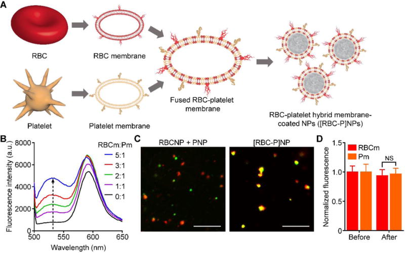
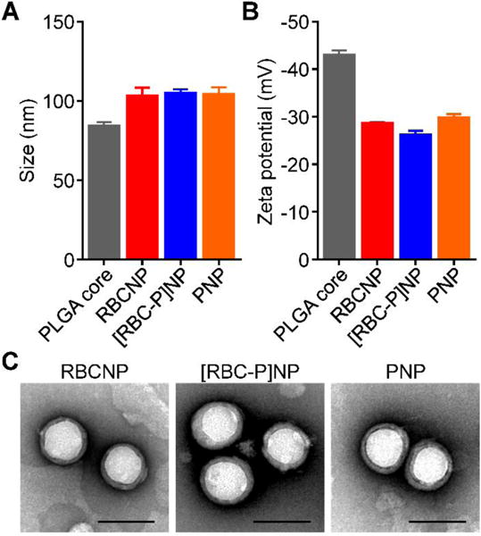
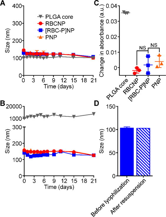
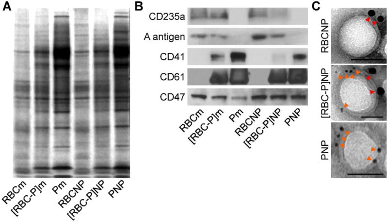
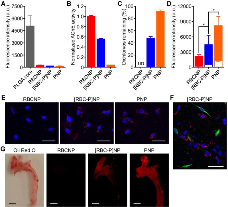
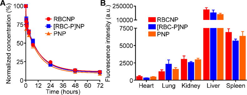
Cell-membrane-coated nanoparticles have recently been studied extensively for their biological compatibility, retention of cellular properties, and adaptability to a variety of therapeutic and imaging applications. This class of nanoparticles, which has been fabricated with a variety of cell membrane coatings, including those derived from red blood cells (RBCs), platelets, white blood cells, cancer cells, and bacteria, exhibit properties that are characteristic of the source cell. In this study, a new type of biological coating is created by fusing membrane material from two different cells, providing a facile method for further enhancing nanoparticle functionality. As a proof of concept, the development of dual-membrane-coated nanoparticles from the fused RBC membrane and platelet membrane is demonstrated. The resulting particles, termed RBC-platelet hybrid membrane-coated nanoparticles ([RBC-P]NPs), are thoroughly characterized, and it is shown that they carry properties of both source cells. Further, the [RBC-P]NP platform exhibits long circulation and suitability for further in vivo exploration. The reported strategy opens the door for the creation of biocompatible, custom-tailored biomimetic nanoparticles with varying hybrid functionalities, which may be used to overcome the limitations of current nanoparticle-based therapeutic and imaging platforms.

Keywords: biomimetic nanoparticles; long circulation; membrane fusion; nanomedicine; targeted delivery.


Figures

Figure 1
Figure 1
Fabrication of RBC-platelet hybrid membrane-coated nanoparticles (denoted [RBC-P]NPs). A) Schematic of membrane fusion and coating. Membrane material is derived from both RBCs and platelets and then fused together. The resulting fused membrane is used to coat poly(lactic-co-glycolic acid) (PLGA) polymeric cores to produce [RBC-P]NPs. B) Platelet membrane was doped with a FRET pair of fluorescent probes and mixed with increasing amounts of RBC membrane. The recovery of the fluorescence emission from the donor at the lower emission peak (534 nm) was monitored (RBCm:Pm = RBC membrane to platelet membrane protein ratio). C) Confocal fluorescent microscopy images of either a mixture of RBCNPs and PNPs or of the [RBC-P]NPs (red = RBC membrane, green = platelet membrane; scale bar = 10 μm). D) Fluorescent quantification of dye-labeled RBCm and Pm inputted to [RBC-P]NPs at a 1:1 protein ratio before the coating process and after purification of the coated nanoparticles (n = 3; mean ± SD). For each type of membrane, the data was normalized to the before values. NS = not significant, Student’s t-test.
Figure 2
Figure 2
Physicochemical characterization. A) Z-average size of bare PLGA cores, RBCNPs, [RBC-P]NPs, and PNPs as measured by DLS (n = 3; mean ± SD). B) Surface zeta potential of bare PLGA cores, RBCNPs, [RBC-P]NPs, and PNPs as measured by DLS (n = 3; mean ± SD). C) Representative TEM images of RBCNPs, [RBC-P]NPs, and PNPs negatively stained with vanadium (scale bar = 100 nm).
Figure 3
Figure 3
Nanoparticle stability. A) Z-average size of bare PLGA cores, RBCNPs, [RBC-P]NPs, and PNPs over 3 weeks in water (n = 3; mean ± SD). B) Z-average size of bare PLGA cores, RBCNPs, [RBC-P]NPs, and PNPs over 3 weeks in PBS (n = 3; mean ± SD). C) Change in absorbance at 560 nm of PLGA cores, RBCNPs, [RBC-P]NPs, and PNPs after transferring into 100% serum from water. An increase in absorbance was used to indicate particle aggregation. NS = not significant, Student’s t-test. D) Z-average size of [RBC-P]NPs before lyophilization in 10 wt% sucrose and after resuspension (n = 3; mean ± SD).
Figure 4
Figure 4
Protein characterization. A) Protein content visualization of RBC membrane (RBCm), fused RBC-platelet membrane ([RBC-P]m), platelet membrane (Pm), RBCNPs, [RBC-P]NPs, and PNPs run on SDS-PAGE at equivalent protein concentrations followed by Coomassie staining. B) Western blot analysis of RBCm, [RBC-P]m, Pm, RBCNPs, [RBC-P]NPs, and PNPs for characteristic RBC markers CD235a (glycophorin A) and A antigen, characteristic platelet markers CD41 (integrin αIIb) and CD61 (integrin β3), and a shared marker CD47. All samples were run at equivalent protein concentrations. C) Immunogold TEM images of RBCNP, [RBC-P]NP, and PNP samples probed for CD235a (red arrows, large gold) and CD61 (orange arrows, small gold) followed by negative staining with vanadium (scale bars = 50 nm).
Figure 5
Figure 5
Membrane biological function assays. A) Uptake of fluorescently labeled bare PLGA cores, RBCNPs, [RBC-P]NPs, and PNPs when incubated with human macrophage-like cells as analyzed by flow cytometry (n = 3; mean ± SD). B) Acetylcholinesterase activity of RBCNPs, [RBC-P]NPs, and PNPs measured by an Amplex acetylcholinesterase assay kit (n = 3; mean ± SD). C) Amount of free dichlorvos, a model organophosphate, remaining in solution after incubation with RBCNPs, [RBC-P]NPs, or PNPs (n = 3; mean ± SD). UD = undetectable. D) Binding of fluorescently labeled RBCNPs, [RBC-P]NPs, and PNPs to human MDA-MB-231 breast cancer cells (solid bars) or human HFF-1 foreskin fibroblasts (open bar overlays) as analyzed by flow cytometry (n = 3; mean ± SD). *P < 0.05, one-tailed Student’s t-test. E) Confocal fluorescence imaging of dye-labeled RBCNPs, [RBC-P]NPs, and PNPs after incubation with MDA-MB-231 cells (red = nanoparticles, blue = nuclei; scale bars = 50 μm). F) Confocal fluorescence imaging of dye-labeled [RBC-P]NPs after incubation with a co-culture of MDA-MB-231 and HFF-1 cells (red = nanoparticles, green = HFF-1 membrane, blue = nuclei; scale bar = 50 μm). G) Imaging of aortas from ApoE knockout mice fed with a high fat western diet after intravenous administration with dye-labeled RBCNPs, [RBC-P]NPs, and PNPs (red = nanoparticles; scale bars = 1 mm). Oil Red O staining was used to confirm the presence of atherosclerotic plaque.
Figure 6
Figure 6
In vivo characterization. A) Circulation time of fluorescently labeled RBCNPs, [RBC-P]NPs, and PNPs after intravenous administration to mice via the tail vein (n = 4; mean ± SEM; lines represent two-phase decay model). B) Biodistribution of fluorescently labeled RBCNPs, [RBC-P]NPs, and PNPs 24 hours after intravenous administration to mice via the tail vein (n = 4; mean ± SEM).

References

    1. Hu CMJ, Fang RH, Luk BT, Zhang L. Nanoscale. 2014;6:65. - PubMed
    1. Farokhzad OC, Langer R. ACS Nano. 2009;3:16. - PubMed
    1. Allen TM, Cullis PR. Adv Drug Deliv Rev. 2013;65:36. - PubMed
    1. Gupta AK, Naregalkar RR, Vaidya VD, Gupta M. Nanomedicine. 2007;2:23. - PubMed
    1. Brannon-Peppas L, Blanchette JO. Adv Drug Deliv Rev. 2004;56:1649. - PubMed