Diagnostic performance of T1 and T2 mapping to detect intramyocardial hemorrhage in reperfused ST-segment elevation myocardial infarction (STEMI) patients
- PMID: 28199043
- PMCID: PMC5573941
- DOI: 10.1002/jmri.25638
Diagnostic performance of T1 and T2 mapping to detect intramyocardial hemorrhage in reperfused ST-segment elevation myocardial infarction (STEMI) patients
Abstract
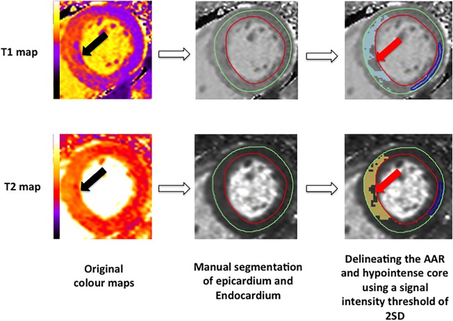
Purpose: To investigate the performance of T1 and T2 mapping to detect intramyocardial hemorrhage (IMH) in ST-segment elevation myocardial infarction (STEMI) patients treated by primary percutaneous coronary intervention (PPCI).
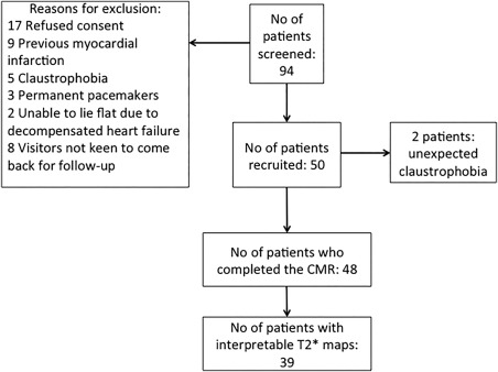
Materials and methods: Fifty STEMI patients were prospectively recruited between August 2013 and July 2014 following informed consent. Forty-eight patients completed a 1.5T cardiac magnetic resonance imaging (MRI) with native T1 , T2 , and T2* maps at 4 ± 2 days. Receiver operating characteristic (ROC) analyses were performed to assess the performance of T1 and T2 to detect IMH.
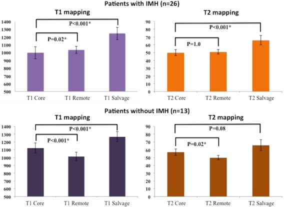
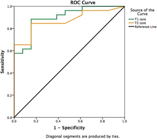
Results: The mean age was 59 ± 13 years old and 88% (24/48) were male. In all, 39 patients had interpretable T2* maps and 26/39 (67%) of the patients had IMH ( T2* <20 msec on T2* maps). Both T1 and T2 values of the hypointense core within the area-at-risk (AAR) performed equally well to detect IMH (T1 maps AUC 0.86 [95% confidence interval [CI] 0.72-0.99] versus T2 maps AUC 0.86 [95% CI 0.74-0.99]; P = 0.94). Using the binary assessment of presence or absence of a hypointense core on the maps, the diagnostic performance of T1 and T2 remained equally good (T1 AUC 0.87 [95% CI 0.73-1.00] versus T2 AUC 0.85 [95% CI 0.71-0.99]; P = 0.90) with good sensitivity and specificity (T1 : 88% and 85% and T2 : 85% and 85%, respectively).
Conclusion: The presence of a hypointense core on the T1 and T2 maps can detect IMH equally well and with good sensitivity and specificity in reperfused STEMI patients and could be used as an alternative when T2* images are not acquired or are not interpretable.
Level of evidence: 2 Technical Efficacy: Stage 2 J. MAGN. RESON. IMAGING 2017;46:877-886.
Keywords: T2* mapping; ST-segment elevation myocardial infarction; T1 mapping; T2 mapping; intramyocardial hemorrhage; microvascular obstruction.
© 2017 The Authors Journal of Magnetic Resonance Imaging published by Wiley Periodicals, Inc. on behalf of International Society for Magnetic Resonance in Medicine.
Figures


Similar articles
-
Residual Myocardial Iron Following Intramyocardial Hemorrhage During the Convalescent Phase of Reperfused ST-Segment-Elevation Myocardial Infarction and Adverse Left Ventricular Remodeling.Circ Cardiovasc Imaging. 2016 Oct;9(10):e004940. doi: 10.1161/CIRCIMAGING.116.004940. Circ Cardiovasc Imaging. 2016. PMID: 27894068 Free PMC article.
-
Cardiac magnetic resonance imaging detection of intramyocardial hemorrhage in patients with ST-elevated myocardial infarction: comparison between susceptibility-weighted imaging and T1/T2 mapping techniques.Quant Imaging Med Surg. 2024 Jan 3;14(1):476-488. doi: 10.21037/qims-23-591. Epub 2023 Nov 28. Quant Imaging Med Surg. 2024. PMID: 38223054 Free PMC article.
-
Detection of Intramyocardial Iron in Patients Following ST-Elevation Myocardial Infarction Using Cardiac Diffusion Tensor Imaging.J Magn Reson Imaging. 2022 Oct;56(4):1171-1181. doi: 10.1002/jmri.28063. Epub 2022 Jan 12. J Magn Reson Imaging. 2022. PMID: 35019174 Free PMC article.
-
Cardiac MRI to Visualize Myocardial Damage after ST-Segment Elevation Myocardial Infarction: A Review of Its Histologic Validation.Radiology. 2021 Oct;301(1):4-18. doi: 10.1148/radiol.2021204265. Epub 2021 Aug 24. Radiology. 2021. PMID: 34427461 Review.
-
Impact of Intramyocardial Hemorrhage on Clinical Outcomes in ST-Elevation Myocardial Infarction: A Systematic Review and Meta-analysis.J Soc Cardiovasc Angiogr Interv. 2022 Aug 26;1(6):100444. doi: 10.1016/j.jscai.2022.100444. eCollection 2022 Nov-Dec. J Soc Cardiovasc Angiogr Interv. 2022. PMID: 39132339 Free PMC article. Review.
Cited by
-
CMR for myocardial characterization in ischemic heart disease: state-of-the-art and future developments.Eur Radiol Exp. 2021 Mar 25;5(1):14. doi: 10.1186/s41747-021-00208-2. Eur Radiol Exp. 2021. PMID: 33763757 Free PMC article. Review.
-
Quantitative T1 Mapping for Detecting Microvascular Obstruction in Reperfused Acute Myocardial Infarction: Comparison with Late Gadolinium Enhancement Imaging.Korean J Radiol. 2020 Aug;21(8):978-986. doi: 10.3348/kjr.2019.0736. Korean J Radiol. 2020. PMID: 32677382 Free PMC article.
-
The role of cardiac magnetic resonance non-contrast T1 mapping in differentiation between injured and salvaged myocardium in acute and chronic myocardial infarction.Postepy Kardiol Interwencyjnej. 2022 Dec;18(4):472-475. doi: 10.5114/aic.2022.121032. Epub 2022 Nov 14. Postepy Kardiol Interwencyjnej. 2022. PMID: 36967854 Free PMC article. No abstract available.
-
Quantification of both the area-at-risk and acute myocardial infarct size in ST-segment elevation myocardial infarction using T1-mapping.J Cardiovasc Magn Reson. 2017 Aug 1;19(1):57. doi: 10.1186/s12968-017-0370-6. J Cardiovasc Magn Reson. 2017. PMID: 28764773 Free PMC article.
-
Head-to-head comparison of multiple cardiovascular magnetic resonance techniques for the detection and quantification of intramyocardial haemorrhage in patients with ST-elevation myocardial infarction.Eur Radiol. 2021 Mar;31(3):1245-1256. doi: 10.1007/s00330-020-07254-1. Epub 2020 Sep 14. Eur Radiol. 2021. PMID: 32929640 Free PMC article.
References
-
- Bulluck H, Hausenloy DJ. Microvascular obstruction: the bane of myocardial reperfusion. Revista espanola de cardiologia 2015. - PubMed
-
- O'Regan DP, Ahmed R, Karunanithy N, et al. Reperfusion hemorrhage following acute myocardial infarction: assessment with T2* mapping and effect on measuring the area at risk. Radiology 2009;250:916–922. - PubMed
-
- van Kranenburg M, Magro M, Thiele H, et al. Prognostic value of microvascular obstruction and infarct size, as measured by CMR in STEMI patients. JACC Cardiovasc Imaging 2014;7:930–939. - PubMed
Publication types
MeSH terms
Grants and funding
LinkOut - more resources
Full Text Sources
Other Literature Sources
Medical
Research Materials