Review
. 2017 Mar 1;22(8):1330-1343.
doi: 10.2741/4546.

The mucolipin-1 (TRPML1) ion channel, transmembrane-163 (TMEM163) protein, and lysosomal zinc handling

Affiliations
Review

The mucolipin-1 (TRPML1) ion channel, transmembrane-163 (TMEM163) protein, and lysosomal zinc handling

Math P Cuajungco et al. Front Biosci (Landmark Ed). .

Abstract

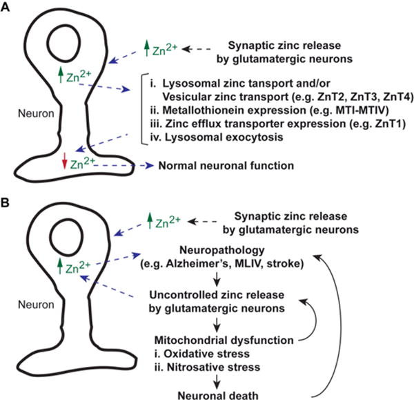
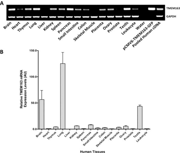
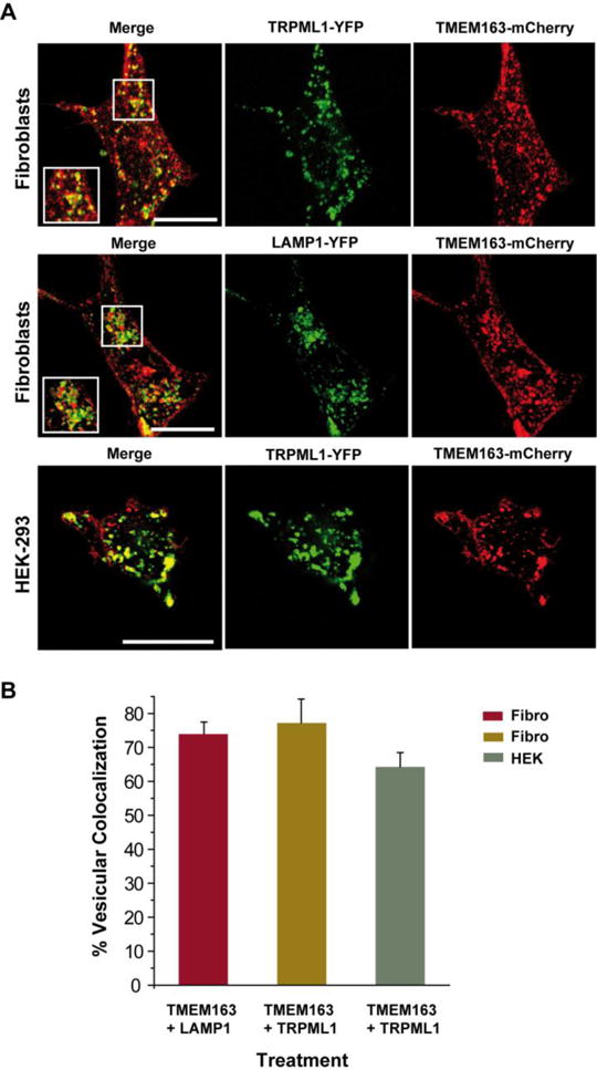
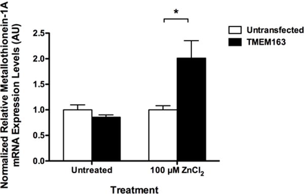
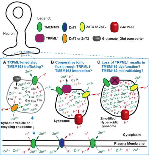
Lysosomes are emerging as important players in cellular zinc ion (Zn2+) homeostasis. The series of work on Zn2+ accumulation in the neuronal lysosomes and the mounting evidence on the role of lysosomal Zn2+ in cell death during mammary gland involution set a biological precedent for the central role of the lysosomes in cellular Zn2+ handling. Such a role appears to involve cytoprotection on the one hand, and cell death on the other. The recent series of work began to identify the molecular determinants of the lysosomal Zn2+ handling. In addition to zinc transporters (ZnT) of the solute-carrier family type 30A (SLC30A), the lysosomal ion channel TRPML1 and the poorly understood novel transporter TMEM163 have been shown to play a role in the Zn2+ uptake by the lysosomes. In this review, we summarize the current knowledge on molecular determinants of the lysosomal Zn2+ handling, uptake, and release pathways, as well as discuss their possible roles in health and disease.


Conflict of interest statement

The authors declare no conflicts of interest.

Figures

Fig. 1
Fig. 1. Cellular zinc status
A) Normal zinc release by glutamatergic neurons results in extracellular increase of Zn2+. The ions are taken up by cells, which elevate intracellular Zn2+ levels. The ions are taken up by lysosomes or vesicular compartments until increases in the expression levels of Metallothionein and efflux zinc transporter have occurred. Lysosomal exocytosis is also a mechanism to reduce excess intracellular Zn2+. B) In pathological conditions caused by stroke or neurodegenerative diseases, glutamatergic neurons that release Zn2+ becomes a vicious cycle. Extracellular Zn2+ elevation perpetuates intracellular Zn2+ accumulation. The flood of Zn2+ results in oxidative and nitrosative stresses in mitochondria. Failure of lysosomes to buffer Zn2+ increase contributes to cellular stress, which subsequently results in cell death.
Fig. 2
Fig. 2. Analyses of TMEM163 gene expression in human tissues
A) Standard PCR analysis of human TMEM163 transcripts using normalized multiple tissue cDNA (MTC) panel commercially purchased from Clontech. No template control (H2O) represented the negative control, while pCMV6-GFP-TMEM163 and non-normalized pooled cDNA were used as positive controls. The housekeeping gene, GAPDH, was used as an internal loading control. B) Real-time quantitative reverse-transcription polymerase reaction (RT-PCR) analysis of TMEM163 using the same MTC panel used in A. The samples were analyzed using the Livak method (ΔΔCq). The housekeeping gene, 18s rRNA, was used as a reference (normalizer). The leukocyte sample was used as the calibrator (value = 1), which makes the tissue mRNA levels all relative to leukocyte. Data are represented as mean ± SEM (n = 3). AU, arbitrary units; bp, basepair. Reprinted with permission from Cuajungco et al. (2014), Traffic, 15, 1247-1265. Copyright 2014 Wiley.
Fig. 3
Fig. 3. Subcellular distribution of heterologously co-expressed TRPML1 and TMEM163 proteins
A) Representative laser scanning micrographs showing subcellular co-localization of TRPML1-YFP and TMEM163-mCherry upon heterologous expression in human primary fibroblast cells (top panel) and HEK-293 cells (bottom panel). TMEM163-mCherry partially co-localized with TRPML1-YFP and LAMP1-YFP (a marker for late endosomes and lysosomes). TMEM163 localized on the plasma membrane, but also exhibited a punctate distribution pattern with either TRPML1 or LAMP1. In HEK-293 cells, co-expression of TMEM163-mCherry with the TRPML1-YFP showed similar a subcellular distribution pattern to the fibroblast cells. Scale bar = 20 μm. B) Cell count showing the percentage of vesicular co-localization pattern between co-expressed TMEM163 plus LAMP1, and TMEM163 plus TRPML1 (for both human fibroblast and HEK-293 cells). The data showed that 70–80% of TMEM163 co-localized with LAMP1 and TRPML1 in late endosomes and lysosomes of fibroblast cells, while 60–70% of TMEM163 co-localized with TRPML1 in late endosomes and lysosomes of HEK-293 cells (n = 50 cells). Reprinted with permission from Cuajungco et al. (2014), Traffic, 15, 1247–1265. Copyright 2014 Wiley.
Fig. 4
Fig. 4. Amino acid sequence map of TMEM163 protein
The map indicates H and D amino acid residues that could potentially bind zinc (red line). The predicted TM domains are shown as solid green bar, while both N- and C-termini are demarcated by a solid orange bar. The putative lysosomal targeting sequence (LTS) is indicated by a red line. The LTS contains a consensus sequence motif of [D/E]XXXL[L/I] residues where X is any amino acid.
Fig. 5
Fig. 5. Cultured SH-SY5Y neuroblastoma cells heterologously expressing human TMEM163 increases Metallothionein-1A expression levels upon exogenous zinc exposure
Real-time quantitative RT-PCR of Metallothionein-1A (MT1A) transcripts at 24 hours following transient ZnCl2 exposure (100 μM, 1 h) of TMEM163-expressing SH-SY5Y neuroblastoma and untransfected control cells. Significant up-regulation of MT1A transcripts is evident in the TMEM163-expressing cells exposed to zinc compared to untreated cells. This result suggests that TMEM163 mediates intracellular zinc flux upon exogenous zinc exposure. Data are represented as mean ± SEM (n = 3, Student’s t-test, paired, two-tailed, *p < 0.05). AU, arbitrary units.
Fig. 6
Fig. 6. Proposed cellular function of TMEM163 protein in zinc-rich cells or neurons
TMEM163 has been observed to localize in the plasma membrane, lysosomes, and vesicular compartments. A) The schematic model depicts that TMEM163 is a zinc transporter that is similar to the ZnT proteins in that it is a zinc (Zn2+)/proton (H+) exchanger. It is proposed that TRPML1 may be responsible to the subcellular trafficking of TMEM163 from the plasma membrane to endocytic compartments, synaptic vesicles, or lysosomes; and vice versa. B) The illustration shows that TRPML1 is a release channel that controls the flux of ions (H+, Ca2+, Zn2+, Fe2+, Mn2+) within the lysosomes. The physical interaction between TMEM163 and TRPML1 is hypothesized to result in cooperative release of Zn2+ and possibly other cations, in order to prevent pathological buildup. C) The loss of TRPML1 function produces hyperacidic lysosomes that is also filled with Zn2+ through the activity of ZnT4 proteins (ZnT2 in other cell types or ZnT3 in neurons). Consequently, the loss of TMEM163 and TRPML1 interaction prevents the cooperative release and exacerbates Zn2+ accumulation.

References

    1. Altarescu G, Sun M, Moore DF, Smith JA, Wiggs EA, Solomon BI, Patronas NJ, Frei KP, Gupta S, Kaneski CR, Quarrell OW, Slaugenhaupt SA, Goldin E, Schiffmann R. The neurogenetics of mucolipidosis type IV. Neurology. 2002;59:306–313. - PubMed
    1. Balaji RV, Colvin RA. A proton-dependent zinc uptake in PC12 cells. Neurochemical research. 2005;30:171–176. - PubMed
    1. Bargal R, Avidan N, Ben-Asher E, Olender Z, Zeigler M, Frumkin A, Raas-Rothschild A, Glusman G, Lancet D, Bach G. Identification of the gene causing mucolipidosis type IV. Nat Genet. 2000;26:118–123. - PubMed
    1. Barth J, Zimmermann H, Volknandt W. SV31 is a Zn2+-binding synaptic vesicle protein. J Neurochem. 2011;118:558–570. - PubMed
    1. Bassi MT, Manzoni M, Monti E, Pizzo MT, Ballabio A, Borsani G. Cloning of the gene encoding a novel integral membrane protein, mucolipidin-and identification of the two major founder mutations causing mucolipidosis type IV. Am J Hum Genet. 2000;67:1110–1120. - PMC - PubMed

Publication types

LinkOut - more resources