Review
. 2017 Mar 1;22(9):1439-1457.
doi: 10.2741/4552.

Endocytosis and membrane receptor internalization: implication of F-BAR protein Carom

Affiliations
Review

Endocytosis and membrane receptor internalization: implication of F-BAR protein Carom

Yanjie Xu et al. Front Biosci (Landmark Ed). .

Abstract

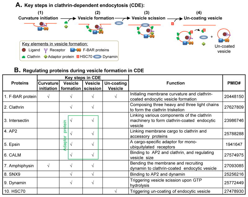
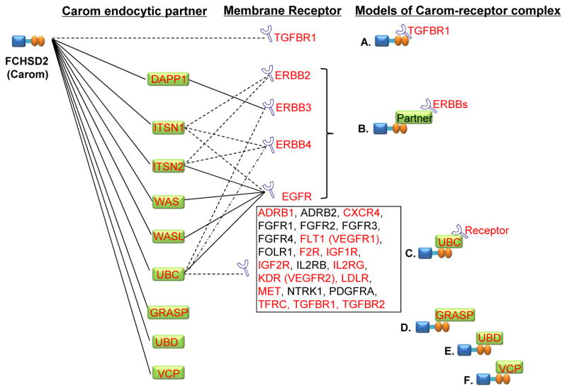
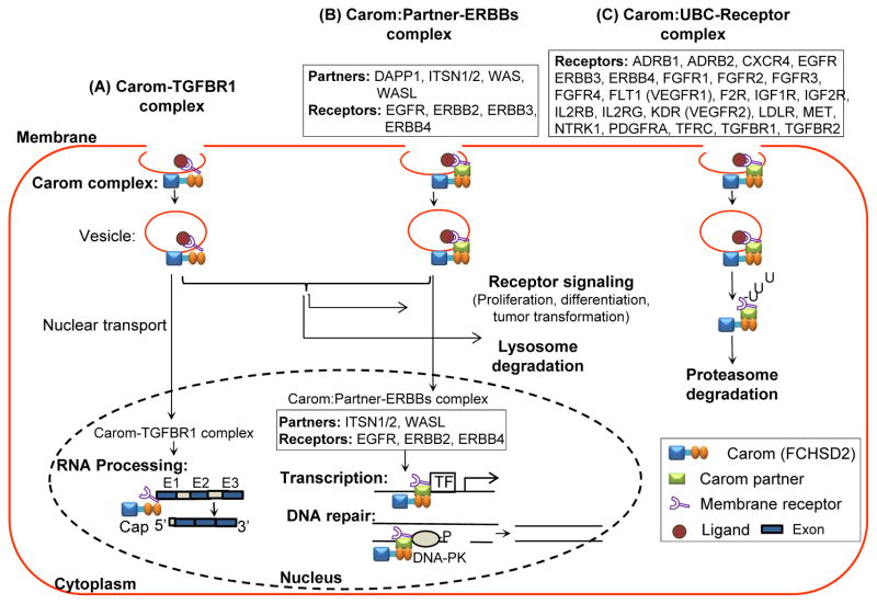
Endocytosis is a cellular process mostly responsible for membrane receptor internalization. Cell membrane receptors bind to their ligands and form a complex which can be internalized. We previously proposed that F-BAR protein initiates membrane curvature and mediates endocytosis via its binding partners. However, F-BAR protein partners involved in membrane receptor endocytosis and the regulatory mechanism remain unknown. In this study, we established database mining strategies to explore mechanisms underlying receptor-related endocytosis. We identified 34 endocytic membrane receptors and 10 regulating proteins in clathrin-dependent endocytosis (CDE), a major process of membrane receptor internalization. We found that F-BAR protein FCHSD2 (Carom) may facilitate endocytosis via 9 endocytic partners. Carom is highly expressed, along with highly expressed endocytic membrane receptors and partners, in endothelial cells and macrophages. We established 3 models of Carom-receptor complexes and their intracellular trafficking based on protein interaction and subcellular localization. We conclude that Carom may mediate receptor endocytosis and transport endocytic receptors to the cytoplasm for receptor signaling and lysosome/proteasome degradation, or to the nucleus for RNA processing, gene transcription and DNA repair.


Figures

Figure 1
Figure 1. Overview of endocytosis
Endocytosis is a cellular process by which molecules or substances are transported into the cell via cell membrane engulfment. A. Classification of endocytosis Endocytosis is generally classified as phagocytosis and pinocytosis. Pinocytosis can be further divided into 4 subtypes; macropinocytosis, clathrin-dependent, caveolae-dependent, and clathrin/caveolae independent endocytosis based on clatherin or caveolae involvement. Most of the receptor-mediated endocytosis (REM) are processed via clathrin-dependent mechanism. B. Features of endocytosis. Features of endocytosis are summarized for the size of internalized particle, membrane domain localization and cargo content. C. Schematic diagram of endocytosis process. Pathogens and ligands induce endocytosis by binding to the cell membrane via receptor-dependent or -independent mechanism, and then form phagosome or endocytic vesicle which may be coated with clathrin/caveolae or regulated by flotillin, GRAF1, Arf6 and RhoA. Membrane–bounded F-BAR protein are linked to actin-associated proteins, can cause cell membrane curvature and facilitate clathrin-meditaed or caveolae-dependent endocytosis. During phagocytosis, cells bring in solid particles into phagosomes and then fuse with lysosomes (marked by LAMP1). During pinocytosis, internalized vesicles are transported to early endosome (marked by Rab5). The early endosome can send the cargoes to three locations: 1) late endosome (marked by Rab7) then lysosome for degradation, 2) recycling endosome (marked by Rab11) for signal transduction or recycling to plasma membrane, and 3)nucleus for transcription factor regulation or chromatin remolding machinery. Abbreviation: RTK, receptor tyrosine kinase; GPCR, G protein-coupled receptor; TFR, transferrin receptor; LDLR, low-density lipoprotein receptor; GPI, glycosylphosphatidylinositol; TGF-βR, transforming growth factor-beta receptor; IGF-IR, insulin-like growth factor I receptor; IL-2RB, interleukin 2 receptor beta; Rab, Ras associated protein; EEA1, Early Endosome Antigene 1; LAMP1, Lysosomal associated membrane protein 1;GRAF1, Rho GTPase Activating Protein 26; Arf6, ADP-Ribosylation Factor 6; RhoA, Ras Homolog Family Member A;
Figure 2
Figure 2. Key steps in clathrin-dependent endocytosis and its regulating proteins
A. Key steps in vesicle formation in clathrin-dependent endocytosis (CDE). There are four steps during vesicle formation: (1) curvature initiation, (2) vesicle formation, (3) vesicle scission, and (4) un-coating vesicle. . At first, F-BAR protein binds to plasma membrane and initiates membrane curvature. F-BAR protein can recruit adaptor protein via its SH3 domain during vesicle formation. Clathrin are recruited directly from the cytosol to the site of adaptor-concentrated membrane to help the formation of coated vesicle. GTPase dynamin can then bind to the membrane and cause vesicle constriction, scission, and release. HSC70 binds to Clathrin, disassociates Clathrin, Intersectin and Dynamin from the vesicle and produces an un-coated endocytic vesicle containing the cargo molecules. B. Regulating proteins during vesicle formation in CDE. A group of proteins are involved in endocytic vesicle formation. F-BAR protein initiates membrane curvature and clathrin-coated endocytic vesicle formation. Adaptor proteins (Intersectin, AP2, Epsin, CALM) links various components of the clathrin machinery to the membrane and helps the formation of adaptor-concentrated clathrin-coated vesicle. Dynamin triggers vesicle scission upon GTP hydrolysis. HSC70 triggers un-coating of endocytic vesicle. Abbreviation: AP2, adaptor protein 2; SNX9, sorting nexin 9; HSC70, ATPase heat shock cognate 70; CALM, clathrin assembly lymphoid myeloid leukaemia; (N-)WASP/WAVE, Wiskott-Aldrich Syndrome Like.
Figure 3
Figure 3. Endocytic membrane receptor, Carom and Carom endocytic partner expression profile in human primary cells
A. Heat map of membrane receptor, Carom and Carom partner expression in human primary cells. mRNA levels are obtained from microarray data available in the web site (https://www.genevestigator.com/gv/) and expressed as heat map. Gradient bars indicate percent of expression potential. The dark and light color shadings represent relatively high and low expression levels, respectively. Dashed frames indicate body system and cells with relative high gene expression. B. Relationship of Carom, endocytic membrane receptor and partner expression in human circulatory and immune system cells. Noted that in aortic ECs, membrane receptor CXCR4, F2R, FLT1 (VEGFR1), KDR (VEGFR2), MET, IGF2R, LDLR and TFRC, and Carom endocytic partner GRASP, ITSN1-2, UBC, VCP and WASL are highly expressed. Carom is highly expressed in EC, LYM, MC, Mϕ and myoblast. Abbreviation: CMC, Cardiomyocyte; VSMC, vascular smooth muscle cell; EC, endothelial cell. LYM, lymphocyte; MC, Monocyte; Mϕ, Macrophage; GPCR, G-protein coupled receptor; RTK, Receptor tyrosine kinase; TRM, Transmembrane receptor; other refer to Table 1 and 3.
Figure 4
Figure 4. Models of Carom, endocytic partner and membrane receptor complexes
Interaction of membrane receptor with Carom and Carom partner were identified via NCBI and String databases. Solid lines indicate known interaction deposited in NCBI Gene database which was established from affinity capture-MS, affinity capture-RNA, affinity capture-western, reconstituted complex and two-hybrid experimental data. Dashed lines indicate computational-predicted interaction in String database based on analyzing genomic information (‘genomic context’-methods) or from transferring associations/interactions between organisms (‘interolog’-transfer). Letters in red indicate genes with comparable high level of expression paralleled with high levels Carom in human cells identified in Figure 3B. Noted that Carom may directly bind to TGBR1 (model A), indirectly interact with EGFR, ERBB2, ERBB3 and ERBB4 via Carom partner DAPP1, ITSN1, ITSN2, WAS, and WASL (model B), bind to receptors through the partner UBC (model C), and directly interact with partner GRASP, UBD and VCP (model D, E and F). Abbreviation: refer to Table 1 and 3.
Figure 5
Figure 5. Hypothetic working model of Carom-mediated membrane receptor trafficking and endocytosis
Carom can form 3 types of receptor complexes and mediate membrane receptor trafficking and endocytosis. A) Carom-TGFBR1 complex can enter nucleus via nuclear localization signal and facilitate RNA processing. B) Carom:Partner-ERBBs complexes that Carom associate with receptor via its partner, such as Carom:DAPP1-ERBB3; Carom:ITSN1-ERBB2, RBB4, EGFR; Carom:ITSN2-ERBB2, ERBB4, EGFR; Carom:WAS-EGFR; Carom:WASL-EGFR (details in table 5). Carom:Partner-ERBBs complex (Carom:ITSN1-ERBB2, RBB4, EGFR; Carom:ITSN2-ERBB2, ERBB4, EGFR; Carom:WASL-EGFR) can be transported into nucleus, bind to transcriptional factor and promote transcription, or bind to damaged DNA to facilitate DNA repair via activating DNA-PK. Both Carom-TGFBR1 and Carom:Partner-ERBBs complexes can facilitate receptor signaling in the cytoplasm leading to proliferation, differentiation and tumor transformation, and can be degradated in lysosome. C) Carom:UBC-Receptor complexes may bind to its partner UBC which further bind to 21 membrane receptor and facilitate ubiquitination and proteasome degradation. Abbreviation: DNA-PK, DNA-protein kinase, others refer to Table 1 and 3.

References

    1. Du Toit A. Endocytosis. A new gateway into cells. Nat Rev Mol Cell Biol. 2015;16(2):68. doi: 10.1038/nrm3939. - DOI - PubMed
    1. McMahon HT, Boucrot E. Molecular mechanism and physiological functions of clathrin-mediated endocytosis. Nat Rev Mol Cell Biol. 2011;12(8):517–33. doi: 10.1038/nrm3151. - DOI - PubMed
    1. Cheng ZJ, Singh RD, Marks DL, Pagano RE. Membrane microdomains, caveolae, and caveolar endocytosis of sphingolipids. Mol Membr Biol. 2006;23(1):101–10. doi: 10.1080/09687860500460041. - DOI - PubMed
    1. Aguilar RC, Wendland B. Endocytosis of membrane receptors: two pathways are better than one. Proc Natl Acad Sci U S A. 2005;102(8):2679–80. doi: 10.1073/pnas.0500213102. - DOI - PMC - PubMed
    1. Kourouniotis G, Wang Y, Pennock S, Chen X, Wang Z. Non-Ligand-Induced Dimerization is Sufficient to Initiate the Signalling and Endocytosis of EGF Receptor. Int J Mol Sci. 2016;17(8) doi: 10.3390/ijms17081200. - DOI - PMC - PubMed

Publication types