Review
. 2017 Mar 1;22(9):1493-1522.
doi: 10.2741/4555.

Base excision repair of oxidative DNA damage: from mechanism to disease

Affiliations
Review

Base excision repair of oxidative DNA damage: from mechanism to disease

Amy M Whitaker et al. Front Biosci (Landmark Ed). .

Abstract

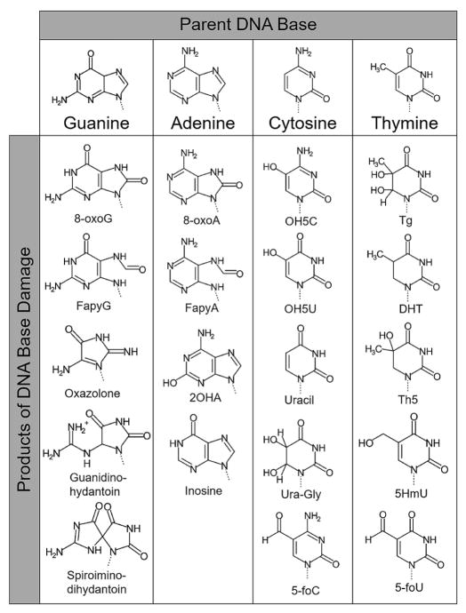
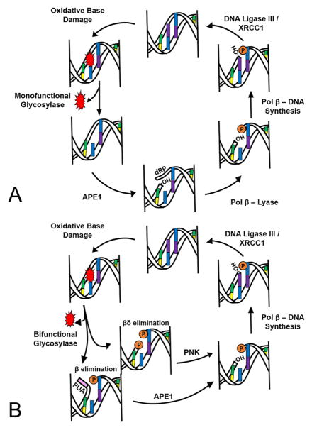
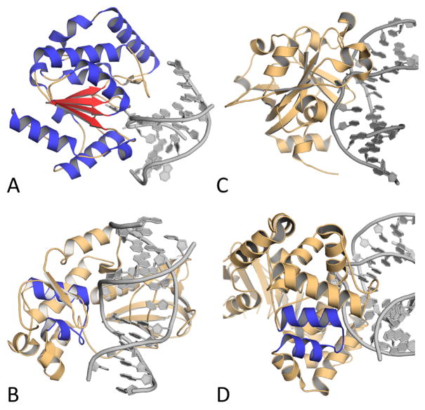
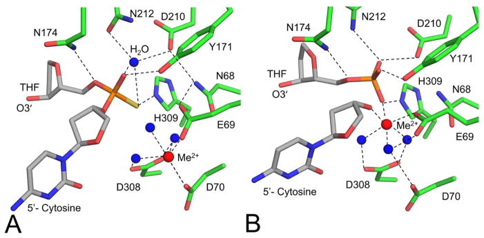
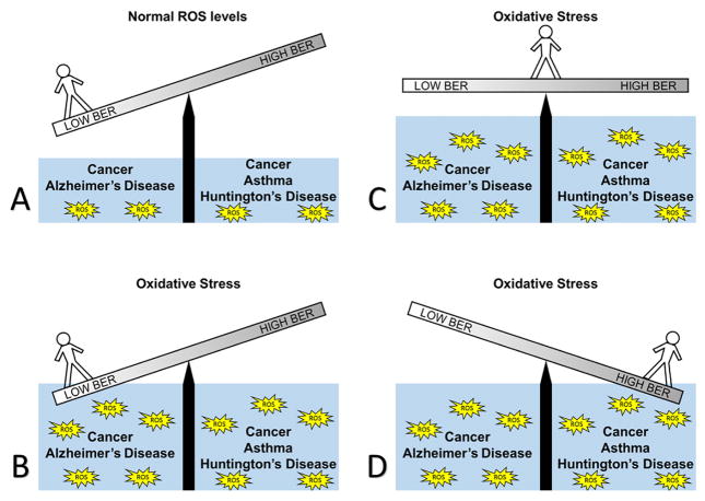
Reactive oxygen species continuously assault the structure of DNA resulting in oxidation and fragmentation of the nucleobases. Both oxidative DNA damage itself and its repair mediate the progression of many prevalent human maladies. The major pathway tasked with removal of oxidative DNA damage, and hence maintaining genomic integrity, is base excision repair (BER). The aphorism that structure often dictates function has proven true, as numerous recent structural biology studies have aided in clarifying the molecular mechanisms used by key BER enzymes during the repair of damaged DNA. This review focuses on the mechanistic details of the individual BER enzymes and the association of these enzymes during the development and progression of human diseases, including cancer and neurological diseases. Expanding on these structural and biochemical studies to further clarify still elusive BER mechanisms, and focusing our efforts toward gaining an improved appreciation of how these enzymes form co-complexes to facilitate DNA repair is a crucial next step toward understanding how BER contributes to human maladies and how it can be manipulated to alter patient outcomes.


Figures

Figure 1
Figure 1
A compilation of biologically significant base lesions organized by parent base. All lesions or several ROS mediated attacks, and/or other damage related to oxidative stress. Specifically, cytosine can give rise to uracil via deamination by N2O3, an oxidized nitric oxide, and a hydrolysis reaction (27).
Figure 2
Figure 2
A representation of the base excision repair (BER) pathway for removing oxidative damage. (A) Classical BER cycle initiated by a monofunctional glycosylase and (B) a BER cycle started by a bifunctional glycosylase. The orange “P” represents a phosphate, -OH represents a 3′-hydroxyl and a 5′-2-deoxyribose-5-phosphate is represented by dRP. The 3′-phospho-α,β-unsaturated aldehyde (PUA) is in pink.
Figure 3
Figure 3
Images of representative members of each structural fold family, with the structural motifs highlighted. (A) Alpha-beta fold of UNG with alpha helices in blue and beta sheets in red. (B) Helix-two-turn-helix (H2TH) motif of NEIL1 colored in blue. (C) MPG fold containing none of the other motifs from MPG. (D) Helix-hairpin-helix (HhH) fold of OGG1 in blue. PDB accession codes are 1EMG, 5ITR, 1F6O, and 1EBM, respectively.
Figure 4
Figure 4
High resolution substrate and product structures of APE1 led to a refined mechanism of strand cleavage. (A) Active site of APE1:DNA substrate complex (PDB code 5DG0). (B) Active site of APE1:DNA product complex (PDB code 5DFF). Waters are shown as blue spheres and metals are shown as red spheres. Tetrahydrofuran, a stable AP site analog, is labeled as THF.
Figure 5
Figure 5
Structural overview of pol β. (A) A ribbon representation of the pol β binary complex (PDB code 3ISB) highlighting the polymerase domain in gray, lyase domain in magenta, and helix-N in blue. (B) Pol β open binary complex (PDB code 3ISB; blue helix-N) superimposed with the ternary complex (PDB code 2FMS; green helix-N). The DNA is truncated in panel B for clarity.
Figure 6
Figure 6
Pol β pre-catalytic complexes demonstrating varying strategies used to accommodate the oxidized DNA lesion, 8-oxoG. (A) 8-oxoG(anti) in the templating positon opposite cytosine (PDB code 3RJI, gray carbons) overlaid with the guanine(anti):cytosine (PDB code 2FMP, yellow carbons). (B) 8-oxoG(syn):adenine with 8-oxoG in the templating position (PDB code 3RJF). (C) Incoming 8-oxodGTP(anti) opposite cytosine (PDB code 4UBC). (D) Incoming 8-oxodGTP(syn) opposite adenine (PDB code 4UAW). Metals are shown in red.
Figure 7
Figure 7
An illustration of the importance of balance in BER. (A) In conditions of low oxidative stress, a person with low BER could still be disease free, as would a person with normal or high BER. (B) When oxidative stress rises, a person with low BER is at risk for disease; in this case, AD or cancer could result. (C) In conditions with high oxidative stress, a person with balanced BER can results in no disease. (D) In conditions with high oxidative stress and overactive BER, the balance shifts, and a person would have an increased risk of HD, cancer, and asthma.

References

    1. Lindahl T, Nyberg B. Rate of depurination of native deoxyribonucleic acid. Biochemistry. 1972;11(19):3610–8. doi: 10.1021/bi00769a018. - DOI - PubMed
    1. Cadet J, Wagner JR. DNA base damage by reactive oxygen species, oxidizing agents, and UV radiation. Cold Spring Harb Perspect Biol. 2013;5(2) doi: 10.1101/cshperspect.a012559. - DOI - PMC - PubMed
    1. Jovanovic SV, Simic MG. One-Electron Redox Potentials of Purines and Pyrimidines. Journal of Physical Chemistry. 1986;90(5):974–978. doi: 10.1021/j100277a053. - DOI
    1. Zhang Y, Yuan F, Wu X, Taylor JS, Wang Z. Response of human DNA polymerase iota to DNA lesions. Nucleic Acids Res. 2001;29(4):928–35. doi: 10.1093/nar/29.4.928. - DOI - PMC - PubMed
    1. Oda Y, Uesugi S, Ikehara M, Nishimura S, Kawase Y, Ishikawa H, Inoue H, Ohtsuka E. NMR studies of a DNA containing 8-hydroxydeoxyguanosine. Nucleic Acids Res. 1991;19(7):1407–12. doi: 10.1093/nar/19.7.1407. - DOI - PMC - PubMed

MeSH terms

LinkOut - more resources