. 2017 Mar 2;543(7643):118-121.
doi: 10.1038/nature21407. Epub 2017 Feb 15.

Mechanical stretch triggers rapid epithelial cell division through Piezo1

Affiliations

Mechanical stretch triggers rapid epithelial cell division through Piezo1

S A Gudipaty et al. Nature. .

Abstract

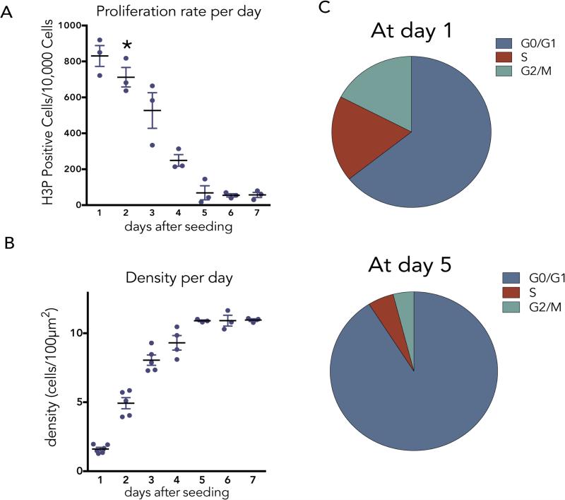
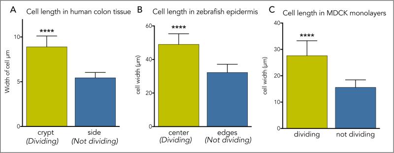
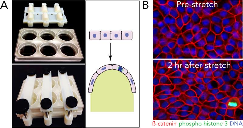
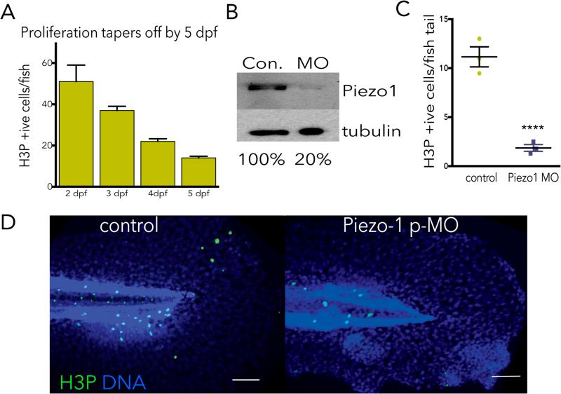
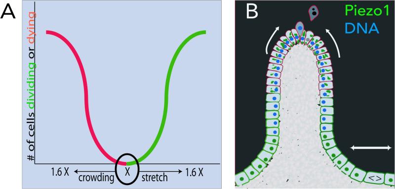
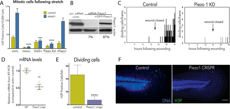
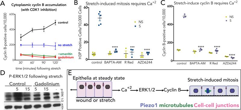
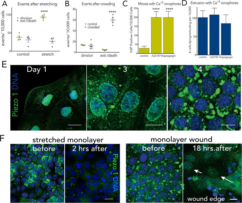
Despite acting as a barrier for the organs they encase, epithelial cells turn over at some of the fastest rates in the body. However, epithelial cell division must be tightly linked to cell death to preserve barrier function and prevent tumour formation. How does the number of dying cells match those dividing to maintain constant numbers? When epithelial cells become too crowded, they activate the stretch-activated channel Piezo1 to trigger extrusion of cells that later die. However, it is unclear how epithelial cell division is controlled to balance cell death at the steady state. Here we show that mammalian epithelial cell division occurs in regions of low cell density where cells are stretched. By experimentally stretching epithelia, we find that mechanical stretch itself rapidly stimulates cell division through activation of the Piezo1 channel. To stimulate cell division, stretch triggers cells that are paused in early G2 phase to activate calcium-dependent phosphorylation of ERK1/2, thereby activating the cyclin B transcription that is necessary to drive cells into mitosis. Although both epithelial cell division and cell extrusion require Piezo1 at the steady state, the type of mechanical force controls the outcome: stretch induces cell division, whereas crowding induces extrusion. How Piezo1-dependent calcium transients activate two opposing processes may depend on where and how Piezo1 is activated, as it accumulates in different subcellular sites with increasing cell density. In sparse epithelial regions in which cells divide, Piezo1 localizes to the plasma membrane and cytoplasm, whereas in dense regions in which cells extrude, it forms large cytoplasmic aggregates. Because Piezo1 senses both mechanical crowding and stretch, it may act as a homeostatic sensor to control epithelial cell numbers, triggering extrusion and apoptosis in crowded regions and cell division in sparse regions.


Figures

EDFigure 1
EDFigure 1. Profile of epithelial cell proliferation and cell density over time
(A) Daily proliferation rates of MDCK cells, as measured by phospho-Histone H3 (H3P) immunostaining, where asterisk indicates when confluence is reached (n=3). Cell division slows but does not stop at day 5. (B), Cell density plateaus by 5 days of growth when cells reach ~11/1000μm2 (n=3). All values are the mean of ten fields from 3 independent experiments with error bars as s.e.m. (C) Cell cycle profiles by FACS at day 1 and day 5 after plating at high density.
EDFigure 2
EDFigure 2. Epithelial cells divide in sparsest regions of epithelia
Density of cells in dividing versus non-dividing regions of epithelia, as measured by cell length through longest axis in 4 videos (except fixed colon sections) in (A) human colon tissue n=8 (B) developing zebrafish epidermis, n=50, and (C) MDCK cells in culture, n=50, where p-value of unpaired t-test is 0.0001 for **** and error bars=s.e.m for all graphs.
EDFigure 3
EDFigure 3. Stretching steady state monolayers
(A) New printable device used for stretching cells uniaxially on Flexcell plates, unassembled (top) and assembled in stretched state (bottom) and schematic of uniaxial stretch on an epithelium in the unassembled (top) and assembled (bottom) states (photo-credit, Jody Rosenblatt). (B) Immunostained MDCK monolayers before (top) and after (bottom) stretching, representative of >200 images captured each. Bar=10μm
EDFigure 4
EDFigure 4. Piezo1 morphants have reduced cell division in zebrafish epidermis at steady state
(A) Cell division in zebrafish reaches a low steady state at day 4-5 post-fertilization, where n=50 fish each day and error bars=s.e.m. (B) Photo-activation of zebrafish injected with Piezo1 translation-blocking morpholino results in knockdown of Piezo1 protein, as shown by an immunoblot. Please see SI Figure 1 for scan of full blots. (C, D) Zebrafish Piezo1 morphants have dramatically reduced epidermal mitoses at 5 days post-fertilisation when cells homeostatic growth rate, where values are the averages of the means from 3 separate experiments, error bars are the s.e.m. of the mean, and the P-value is from an unpaired t-test is <0.005. Each micrograph is representative of ~75 samples, bar=100μm.
EDFigure 5
EDFigure 5. A single calcium spark occurs around an hour before cells divide
(A) Blocking stretch-induced proliferation with gadolinium at two hours post-stretch does not affect the percentages of cells in S phase, as measured by EdU incorporation from the mean of six independent experiments, where error bars are s.e.m. and P values from a t-test compared to the non-stretched control show no significance. (B) Inhibition of Piezo1 with Gd3+ or transcription with alpha-amanitin blocks stretch-induced cytoplasmic cyclin B accumulation and mitosis (H3P), where each micrograph is representative of >100 samples except for alpha-amanitin (40 samples) and bar=10μm. (C) Quantification of time from calcium spark to cell rounding measured from 10 mitotic events in 8 videos. Error bars=s.e.m. (D) Sample graph measuring the time (minutes) from calcium spark (arrow) to cell rounding (arrowhead) in MDCK cells expressing the calcium indicator, CMV-R-GECO1 using Nikon Elements.
EDFigure 6
EDFigure 6. Models for how Piezo1 controls cell division in response to stretch and cell extrusion in response to crowding
(A) Theoretical graph of density-dependent Piezo1 function for cell division and cell death: Epithelia trend to a steady-state density, X. If density is reduced, stretching causes Piezo1 to activate cell division, if it increases, crowding causes Piezo1 to activate cells to extrude and die. (B) Schematic showing how Piezo1 (green) localizes to plasma membrane in sub-regions of epithelia that are sparser and divide and accumulate into cytoplasmic aggregates in sub-regions that are crowded and extrude.
EDFigure 7
EDFigure 7. Stretch affects cells at steady state to enter mitosis
(A) Characterization of Piezo1 antibody using an shRNA to Piezo1 tagged with mCherry indicates that cells expressing red mCherry lack Piezo1 (green). Micrographs are representative of ~50 samples and bar=10μm. (B) High-density monolayers (>500 cells/40X field) where intrinsic proliferation rate is low (<50 H3P-positive cells/10,000 cells) proliferate in response to mechanical stretch (with P value= 0.0007 from a unpaired t-test with error bars=s.e.m.) or wounding. (C) Low density MDCK monolayers just reaching confluence with a higher intrinsic rate of proliferation (>50 H3P-positive cells/10,000 cells) do not significantly proliferate in response to stretch or wounding (P value from a t-test is not significant). N=12 experiments for each case.
Figure 1
Figure 1. Mechanical stretch induces epithelial monolayers to rapidly divide
(A) Proliferation rates (A) and cell lengths (B) at various times following stretch show that stretch-induced cell divisions return cell densities to control levels, where values are the averages of 3 experiments measuring the mean of 6 areas, error bars = s.e.m. P-values from unpaired T-tests compared to control are ***<0.0005, **<0.005, *<0.01. (C) Stills showing cumulatively where and when cells divide (red dots) during wound healing of an MDCK monolayer, where wound edge (highlighted with white line) with time in hours. (D) Graph (one of 14 similar) of cell divisions after monolayer wounding, with arrow indicating wound closure.
Figure 2
Figure 2. Stretch-induced mitosis requires the stretch-activated channel Piezo1
(A) Inhibition or siRNA-mediated knockdown of Piezo1 blocks stretch-induced mitosis, and is rescued with Piezo1-GFP, where values are means of 6 measurements averaged from 5 experiments. (B) Immunoblot confirming knockdown and rescue (SI Fig.1 for full blots). (C) Piezo1 knockdown blocks cell division following wound closure, where graphs are representative of 20 similar videos. (D) CRISPR-mediated Piezo1 knockout reduces Piezo1 mRNA by 50% (n=3) and dramatically decreases mitosis rates, measured in 26 wild type and 53 knockout 4-day old larvae (E, F). Bar = 100μm (images represent 12 total for each). For all graphs, except (C), error bars = s.e.m. P values from an unpaired t-test are **<0.005, ***<0.0005, ****<0.0001.
Figure 3
Figure 3. Stretch-activation of Piezo1 triggers calcium and ERK1/2-dependent cyclin B transcription
(A) Stretch induces cyclin B accumulation (with CDK1 inhibition), which is blocked with Gd3+ or alpha-amanitin, where values are the means from 6 experiments, with error bars = s.e.m. P-values <0.0001 from two-way Anova. Inhibiting intracellular calcium (BAPTA-AM, Ruthenium Red) or MEK1/2 (AZD6244) blocks stretch-induced mitosis (B) and cytoplasmic cyclin B (with CDK1 inhibition) (C). Data points represent averaged means of 6 areas from 3 experiments. Error bars = s.e.m. P-values from an unpaired T-test are ****<0.0001 and ***<0.002. (D) Stretch rapidly activates ERK1/2 phosphorylation in a Piezo1-dependent manner (see SI Fig. 1 for full scans). (E) Model: stretch triggers cells in early G2 to activate Piezo1-dependent calcium influx, which stimulates ERK1/2-dependent transcription of cyclin B and cell division.
Figure 4
Figure 4. Piezo1 controls proliferation in response to stretch and extrusion and death in response to crowding
Stretch triggers only mitosis (A) whereas crowding induces only cell extrusion/death (B), where values are averages of the means of 4 experiments from 6 fields each. Experimental induction of calcium influx induces proliferation, n=8 (C) but not extrusion/death, n=9 (D) within two hours. For all graphs, error bars = s.e.m. and P-values from an unpaired T-test are ****<0.0001 and ***<0.002. (E) Piezo1 (green) shifts from the nuclear envelope in cells at low density to the cytoplasm/plasma membrane to large cytoplasmic formations in cells at high density. (F) Piezo1 disappears after stretch or wounding, eventually localizing to nuclear envelope (arrows, wound). All images representative of 10 others, with scale bars=10μm.

Comment in

Similar articles

Cited by

References

    1. Eisenhoffer GT, et al. Crowding induces live cell extrusion to maintain homeostatic cell numbers in epithelia. Nature. 2012;484:546–549. doi:10.1038/nature10999. - PMC - PubMed
    1. Puliafito A, et al. Collective and single cell behavior in epithelial contact inhibition. Proc Natl Acad Sci U S A. 2012;109:739–744. doi:10.1073/pnas.1007809109. - PMC - PubMed
    1. Coste B, et al. Piezo1 and Piezo2 are essential components of distinct mechanically activated cation channels. Science. 2010;330:55–60. doi:10.1126/science.1193270. - PMC - PubMed
    1. Shah AN, Davey CF, Whitebirch AC, Miller AC, Moens CB. Rapid reverse genetic screening using CRISPR in zebrafish. Nat Methods. 2015;12:535–540. doi:10.1038/nmeth.3360. - PMC - PubMed
    1. Shah AN, Moens CB, Miller AC. Targeted candidate gene screens using CRISPR/Cas9 technology. Methods Cell Biol. 2016;135:89–106. doi:10.1016/bs.mcb.2016.01.008. - PubMed

Publication types

MeSH terms