. 2017 Feb 1;22(2):24002.
doi: 10.1117/1.JBO.22.2.024002.

Implementation of fluorescence confocal mosaicking microscopy by "early adopter" Mohs surgeons and dermatologists: recent progress

Affiliations

Implementation of fluorescence confocal mosaicking microscopy by "early adopter" Mohs surgeons and dermatologists: recent progress

Manu Jain et al. J Biomed Opt. .

Abstract

Confocal mosaicking microscopy (CMM) enables rapid imaging of large areas of fresh tissue ex vivo without the processing that is necessary for conventional histology. When performed in fluorescence mode using acridine orange (nuclear specific dye), it enhances nuclei-to-dermis contrast that enables detection of all types of basal cell carcinomas (BCCs), including micronodular and thin strands of infiltrative types. So far, this technique has been mostly validated in research settings for the detection of residual BCC tumor margins with high sensitivity of 89% to 96% and specificity of 99% to 89%. Recently, CMM has advanced to implementation and testing in clinical settings by “early adopter” Mohs surgeons, as an adjunct to frozen section during Mohs surgery. We summarize the development of CMM guided imaging of ex vivo skin tissues from bench to bedside. We also present its current state of application in routine clinical workflow not only for the assessment of residual BCC margins in the Mohs surgical setting but also for some melanocytic lesions and other skin conditions in clinical dermatology settings. Last, we also discuss the potential limitations of this technology as well as future developments. As this technology advances further, it may serve as an adjunct to standard histology and enable rapid surgical pathology of skin cancers at the bedside.


Figures

Fig. 1
Fig. 1
Acetic acid stained reflectance confocal mosaic (a) shows nodular BCCs (red arrows) that can be readily identified at 2× magnification and compares well to (b) the corresponding H&E frozen histopathology. Nests of tumor (red arrows) within the dermis, the epidermal margin (green arrows) and hair follicles (yellow arrows) are seen. Scale bars: A=1  mm. Figure reproduced with permission, courtesy of Wiley.
Fig. 2
Fig. 2
Acetic acid stained reflectance confocal mosaic (a) shows nodular and infiltrative BCCs and (b) corresponding H&E stained frozen histology. Tiny strands of infiltrative BCC (red boxed area) are difficult to identify and differentiate from thin bright strands of collagen fibres (yellow dotted circles) at low magnification in contrast to the nodular BCC (red arrow) that appears distinct (a). (c) Digital zooming to 30× magnifications is often required to identify the tiny strands of infiltrative BCC. The epidermal margin (green arrows) is seen. Scale bars: A=1  mm; C=150  μm. Figure reproduced with permission, courtesy of Wiley.
Fig. 3
Fig. 3
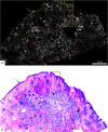
Low tumor volume of infiltrative BCC can be readily detected on a wide-field fluorescent confocal mosaic. Acridine orange stained fluorescence confocal mosaic (a) shows a small focus of residual infiltrative BCC (dashed red box) as bright strands that can be identified within the reticular dermis even at this low magnification, corresponding to 2× magnification on (b) H&E-stained frozen histopathology. However, it can sometimes be challenging to differentiate these particularly thin tumor strands (four to eight cells thick; dashed red box) from the surrounding inflammatory infiltrate (green dotted circles) and may require digital zooming to higher magnification (4× to 10×). (c) Submosaic shows digital zooming of the area under the red dashed box from mosaic “A” necessary to appreciate pleomorphic nuclei and enlarged nuclear cytoplasmic ratio of the tumor strands (red arrowheads) and differentiate them from bright smaller monomorphic inflammatory cells (green dotted circles). Epidermis (green arrow) and hair follicle (yellow arrow) are seen. On a separate note, the dark bands that are seen in this particular mosaic (but not the others shown in this paper) are due to incomplete correction for the illumination fall-off (vignetting) that occurs across each image and incomplete stitching along the edges of images. This was merely due to the developmental state of the stitching algorithm and software at the time. (Further development led to a second algorithm that produces much more seamless appearing mosaics, as seen in the other figures.) Scale bars: A, C=1  mm.
Fig. 4
Fig. 4
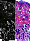
Acridine orange stained fluorescence confocal mosaic of a micronodular BCC (a) that equates well to 2× view on standard H&E stained histology section (b). Small and tiny nodules or nests of tumor (red dotted area) can be identified at this magnification. Epidermis (green arrow), hair follicle (yellow arrow), sebaceous glands (green asterisk), dermal collagen (yellow asterisk) are seen. Scale bar: A = 2mm. Figure reproduced from Gareau et al. “Confocal mosaicing microscopy in Mohs skin excisions: feasibility of rapid surgical pathology.”
Fig. 5
Fig. 5
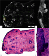
Acridine orange stained fluorescence confocal submosaic (a) obtained by digitally zooming (6× magnification) in the mosaic (Fig. 4(a), dashed boxed area) to appreciate morphological features of micronodular BCC tumor (red dotted area) such as nuclear pleomorphism, increased nuclear density, and clefting. Epidermis (green arrow), hair follicle (yellow arrow), sebaceous glands (green asterisk), dermal collagen (yellow asterisk) are seen. This corresponds well with H&E stained frozen histopathology (b). A = 0.5 mm. Figure reproduced from Gareau et al. “Confocal mosaicing microscopy in Mohs skin excisions: feasibility of rapid surgical pathology.”
Fig. 6
Fig. 6
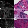
(a) Acridine orange stained fluorescence confocal submosaic shows a small bright focus (dotted circle) of superficial BCC along the epidermal margin (green arrow) raising suspicion for residual tumor and (b) corresponding H&E-stained frozen histopathology at 4× magnification. Digital zooming to higher magnification (30×) reveals the subtle but characteristic features of (c) superficial BCC on confocal image as well as on (d) H&E-stained frozen histopathology. This figure demonstrates that small foci of superficial BCC may be sometimes missed on confocal imaging either due to their subtle morphological features or uneven flattening of the tissue. Scale bar: A=1  mm. Figure reproduced with permission, courtesy of Wiley.
Fig. 7
Fig. 7
Fluorescent confocal microscopy criteria for various subtypes of BCCs. (a, c, e) Submosaics show characteristic morphological features of BCCs: presence of fluorescence, nuclear pleomorphism, increased N/C ratio, and palisading in (a) superficial BCC, (c) nodular BCC, and (e) infiltrative BCC. The tumor islands (red arrows) are well demarcated in (c) nodular BCC and (e) infiltrative BCC than the (a) superficial BCC, whereas clefting (red arrowheads) appears more prominent in superficial BCC (a) and nodular BCC (c). (e) “Starry sky” (yellow asterisk) appearance of the tumor stroma is pronounced in infiltrative BCC. (b, d, f) And corresponding H&E-stained frozen histopathology (10×). Scale bars: A=200  μm; C & E=600  μm.
Fig. 8
Fig. 8
DSCMs of normal skin tissue closely mimicking H&E stained tissue histology. Mosaic of normal skin tissue in (a) fluorescence mode, (b) in reflectance mode, and (c) digital H&E (DHE) image created by overlapping fluorescence and reflectance channels. Nucleated structures, such as epidermis (red arrows) and hair follicle (yellow arrow), appear bright in fluorescence mode (a) and dull-grey on reflectance mode (b). Adipocytes (blue asterixis) appear darker and well defined in the fluorescence mode than in reflectance mode. By contrast, dermal collagen (green asterixis) appears bright in both the modes. (d) The H&E image (2×) compares well with the DHE image (c). Scale bars: A, B=2  mm.
Fig. 9
Fig. 9
DSCMs of BCCs closely mimicking H&E stained tissue histology. Submosaics shows (a, c) infiltrative BCC and nodular BCC in fluorescence mode, (b, d) in reflectance mode, and (e, g) digital H&E (DHE) images created by overlapping fluorescence and reflectance channels, respectively. (a, e) Nucleated structures, such as hair follicle (yellow arrows) and tumor cords and nodules (red arrows), appear bright in fluorescence mode and (b, f) are not visible (yellow and red asterixis) on reflectance mode. By contrast, dermal collagen (green arrows) appears more pronounced in the reflectance mode bright in (b, f; green asterixis). Corresponding H&E images of (f) infiltrative and (h) nodular BCC at 10× magnification compares well with the DHE images (d and g, respectively). Scale bars: A, B=0.3  mm; C, D=0.15  mm.

Similar articles

Cited by

References

    1. Rogers H. W., et al. , “Incidence estimate of nonmelanoma skin cancer (keratinocyte carcinomas) in the U.S. population, 2012,” JAMA Dermatol. 151(10), 1081–1086 (2015).10.1001/jamadermatol.2015.1187 - DOI - PubMed
    1. Mohan S. V., Chang A. L., “Advanced basal cell carcinoma: epidemiology and therapeutic innovations,” Curr. Dermatol. Rep. 3, 40–45 (2014).10.1007/s13671-014-0069-y - DOI - PMC - PubMed
    1. Karia P. S., Han J., Schmults C. D., “Cutaneous squamous cell carcinoma: estimated incidence of disease, nodal metastasis, and deaths from disease in the United States, 2012,” J. Am. Acad. Dermatol. 68(6), 957–966 (2013).JAADDB10.1016/j.jaad.2012.11.037 - DOI - PubMed
    1. Chen J. T., Kempton S. J., Rao V. K., “The economics of skin cancer: an analysis of medicare payment data,” Plast. Reconstructive Surg. Global Open 4(9), e868 (2016).10.1097/GOX.0000000000000826 - DOI - PMC - PubMed
    1. Donaldson M. R., Coldiron B. M., “No end in sight: the skin cancer epidemic continues,” Semin. Cutaneous Med. Surg. 30(1), 3–5 (2011).10.1016/j.sder.2011.01.002 - DOI - PubMed

MeSH terms