. 2017 Mar 1;38(3):281-292.
doi: 10.1093/carcin/bgw121.

Six1 promotes colorectal cancer growth and metastasis by stimulating angiogenesis and recruiting tumor-associated macrophages

Affiliations

Six1 promotes colorectal cancer growth and metastasis by stimulating angiogenesis and recruiting tumor-associated macrophages

Hanwen Xu et al. Carcinogenesis. .

Abstract

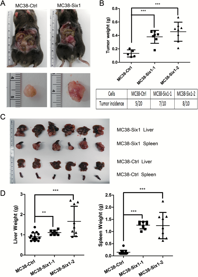
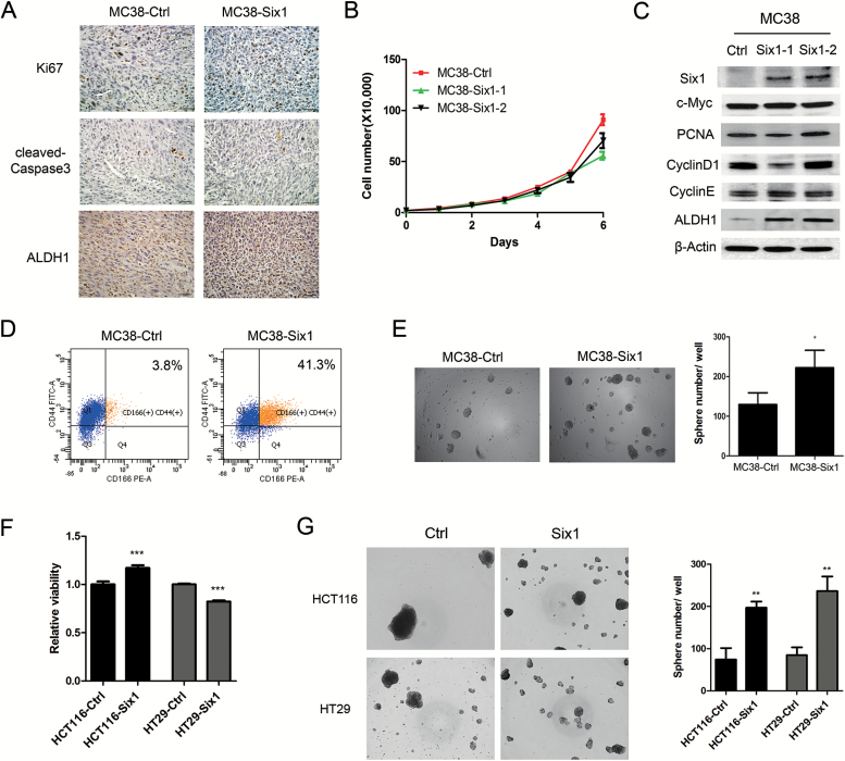
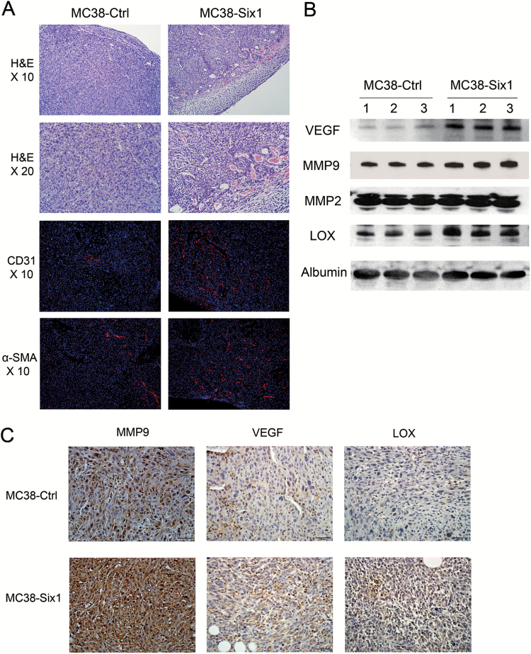
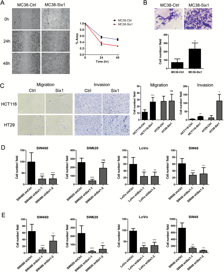
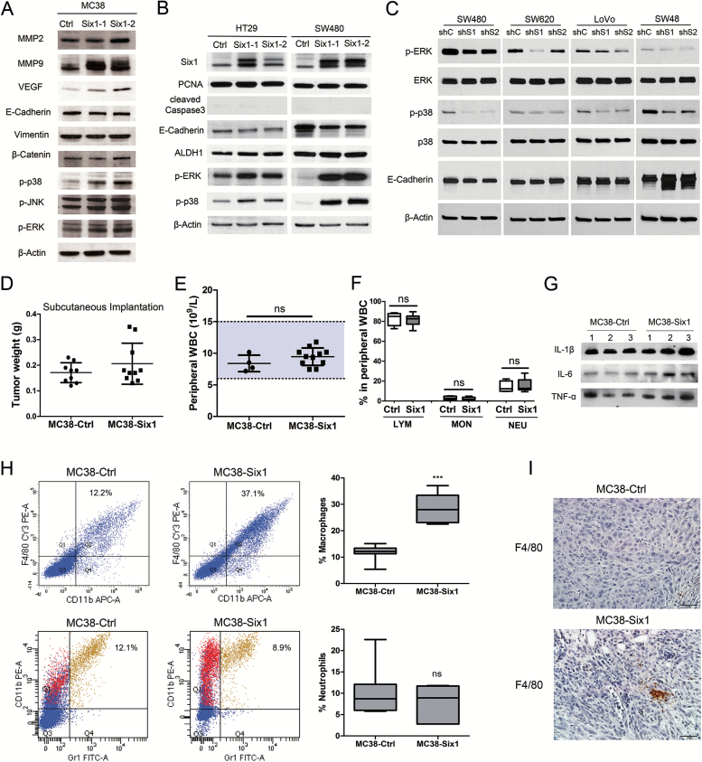
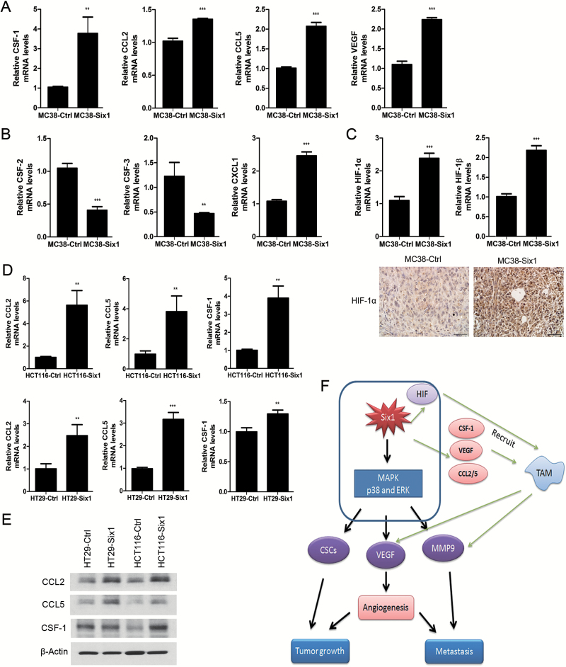
The homeoprotein Six1 is overexpressed in many human cancers and is associated with increased tumor progression and metastasis. Recent studies have shown that Six1 is associated with poorer overall survival in advanced-stage colorectal cancer (CRC). In the current study, we explored the functional changes and molecular events associated with Six1 overexpression in a mouse model of CRC. An orthotopic model and a splenic injection metastasis model were used to investigate the role of Six1 in CRC tumor growth and metastasis using mouse colon adenocarcinoma MC38 cells overexpressing Six1. We found that overexpression of Six1 dramatically promotes CRC tumor growth and metastasis in vivo. Six1 overexpression in MC38 increased protein levels of aldehyde dehydrogenase-1 and expanded CD44+/CD166+ populations, indicating Six1 increased features of cancer stem cells. In addition, Six1 overexpression stimulated angiogenesis by upregulating the expression of vascular endothelial growth factor (VEGF). Six1-overexpressing tumor cells recruited tumor-associated macrophages (TAM) by increasing the expression of macrophage-specific colony stimulating factor, chemokine (C-C motif) ligand 2/5 and VEGF, further facilitating CRC tumor growth and metastasis. Furthermore, we determined that Six1 activated mitogen-activated protein kinase (MAPK) signaling in CRC cells. In summary, our studies strongly suggest that Six1 overexpression promotes CRC growth and metastasis and remodels tumor stroma by stimulating angiogenesis and recruiting TAM. MAPK activation may be a pivotal event in Six1-associated tumor progression, which may provide opportunities for pharmacologic intervention.


Figures

Figure 1.
Figure 1.
Six1 overexpression increases CRC tumor growth and metastasis in vivo. (A) MC38-Six1 or MC38-Ctrl were orthotopically injected into the cecum subserosa of C57BL/6 mice and mice were killed after 6 weeks. Left panels: tumors from MC38-Ctrl. Right panels: tumors from MC38-Six1. (B) The weight of tumors and tumor incidence in mice injected in the cecum with MC38-Ctrl or isolates of MC38-Six1 cells. (C) Upper panel: livers and spleens from MC38-Six1 tumor-bearing mice. Lower panel: livers and spleens from MC38-Ctrl tumor-bearing mice. (D) The weight of livers (left panel) and spleens (right panel) from MC38-Six1 or MC38-Ctrl tumor-bearing mice. Bars indicate SD, and ** and *** indicate P values <0.01 and <0.001, respectively.
Figure 2.
Figure 2.
Six1 overexpression in MC38 cells increases CSC characteristics. (A) The expression of Ki67, cleaved caspase-3 and ALDH1 in tumors arising from mice injected in the cecum with MC38-Ctrl and MC38-Six1 as detected by immunohistochemistry. (X400) (B) Cell proliferation was determined by directly counting cells at the days indicated in two isolates of MC38-Six1 and MC38-Ctrl. (C) Western blots for Six1, c-Myc, PCNA, cyclin D1, cyclin E and ALDH1 in MC38-Ctrl and two isolates of MC38 overexpressing Six1 (MC38-Six1). β-Actin was used as a loading control. (D) Analysis by flow cytometry of CD44 and CD166 expression in MC38-Ctrl or MC38-Six1. (E) Tumorsphere formation ability in MC38-Ctrl and MC38-Six1. (F) Cell proliferation was measured by WST-1 in HCT116-Ctrl, HCT116-Six1, HT29-Ctrl and HT29-Six1. (G) Tumorsphere formation ability in HCT116-Ctrl, HCT116-Six1, HT29-Ctrl and HT29-Six1. Bars indicate SD and *, ** and *** indicate P values <0.05, <0.01 and <0.001, respectively.
Figure 3.
Figure 3.
Six1 overexpression promotes angiogenesis. (A) Upper panels: H&E staining of primary tumors formed in the cecum of mice from MC38-Ctrl and MC38-Six1. Lower panels: immunofluorescent staining of CD31 (red) and α-SMA (red) merged with nuclei (blue) in MC38-Ctrl and MC38-Six1 tumors. (B) Western blots for VEGF, MMP9, MMP2 and LOX in sera from MC38-Six1 tumor-bearing mice and MC38-Ctrl mice. Albumin was used as a loading control. (C) The expression of MMP9, VEGF and LOX in MC38-Six1 and MC38-Ctrl tumors as assessed by immunohistochemistry (×400).
Figure 4.
Figure 4.
Six1 promotes migration and invasion in CRC cells in vitro. (A) Cell migration was determined in MC38-Ctrl and MC38-Six1 using a wound healing assay. Images are at ×100 magnification. The area of the wound is quantified to determine the extent of wound repair. (B) Cell invasion in MC38-Ctrl and MC38-Six1 was determined using a Matrigel transwell invasion assay. The number of invading cells was quantified after crystal violet staining. Images are shown at ×400 magnification. (C) Cell invasion and migration in HCT116-Ctrl, HCT116-Six1, HT29-Ctrl and HT29-Six1 were determined using a transwell assay with or without Matrigel. Images are shown at ×100 magnification. (D and E) The quantification of numbers of migrating (D) or invading (E) cells after crystal violet staining in non-target shRNA control (shCtrl) and Six1 knockdown (shSix1-1 and shSix1-2) of SW480, SW620, LoVo and SW48. Each column represents the mean of five different fields. Bars indicate SD and *, ** and *** indicate p values < 0.05, 0.01 and 0.001, respectively.
Figure 5.
Figure 5.
(AC) Six1 actives MAPK in vitro. (A) Western blots for MMP2, MMP9, VEGF, E-cadherin, vimentin, β-catenin, p-p38, p-JNK and p-ERK in two different isolates of MC38-Six1 and a MC38-Ctrl line. (B) Western blots for Six1, PCNA, cleaved caspase-3, E-cadherin, ALDH1, p-ERK and p-p38 in two different isolates of Six1-overexpressing HT29 (HT29-Six1) and SW480 (SW480-Six1) and corresponding controls (HT29-Ctrl and SW480-Ctrl). (C) Western blots for p-ERK, ERK, p-p38, p38 and E-cadherin in Six1-knockdown SW480 (SW480-shS1 and SW480-shS2), SW620 (SW620-shS1 and SW620-shS2), LoVo (LoVo-shS1 and LoVo-shS2) and SW48 (SW48-shS1 and SW48-shS2) as well as corresponding controls (SW480-shC, SW620-shC, LoVo-shC and SW48-shC). β-Actin was used as a loading control. (DI) MC38 cells overexpressing Six1 recruit macrophages to tumors. (D) MC38-Six1 or MC38-Ctrl were injected into the flank of C57BL/6 mice and the mice killed 6 weeks later (n = 9–10). The weight of tumors formed from MC38-Ctrl and MC38-Six1 is shown. (E) Peripheral white blood cell (WBC) count from MC38-Ctrl and MC38-Six1 tumor-bearing mice (cecal implantation). (F) Percentage of lymphocytes (LYM), monocytes (MON) and neutrophils (NEU) in peripheral WBC from MC38-Ctrl and MC38-Six1 tumor-bearing mice (cecal implantation). (G) Western blot of IL-1β, IL-6 and TNF-α in sera from MC38-Six1or MC38-Ctrl tumor-bearing mice (cecal implantation). (H) Left panels: Flow cytometry analysis of the expression of CD11b, F4/80 and Gr1 in MC38-Ctrl and MC38-Six1 tumors from the cecum. Right panels: the percentage of total cells positive for either CD11b+/F4/80+ (a marker for macrophages) or CD11b+/Gr1+ (a marker for neutrophils) cells. Results are representative of seven independent experiments. Bars indicate SD and *** indicates P < 0.001. (I) Immunohistochemical analysis of the expression of F4/80 in MC38-Ctrl and MC38-Six1 derived tumors (×400).
Figure 6.
Figure 6.
Six1 overexpression in MC38 increases the expression of molecules that induce chemotaxis of TAM. mRNA expression of CSF-1, CCL2, CCL5 and VEGF (A), CSF-2, CSF-3 and CXCL1 (B) and HIF-1α and HIF-1β (C, upper panel) was determined by real-time PCR in MC38-Ctrl and MC38-Six1. Bars indicate SD, and ** and *** indicate P values <0.01 and <0.001, respectively. (C, lower panel) Protein levels of HIF-1α were examined by immunohistochemistry (×400) in MC38-Ctrl and MC38-Six1 derived tumors. (D) mRNA expression of CCL2, CCL5 and CSF-1 in Six1-overexpressing HCT116 (HCT116-Six1) and HT29 (HT29-Six1) and their corresponding controls (HCT116-Ctrl and HT29-Ctrl). (E) Protein levels by Western blot of CCL2, CCL5 and CSF-1 in HT29-Ctrl, HT29-Six1, HCT116-Ctrl and HCT116-Six1. β-Actin was used as a loading control (F) Model illustrating how Six1 overexpression promotes CRC tumor growth and metastasis.

References

    1. Siegel R., et al. (2014) Colorectal cancer statistics, 2014. CA Cancer J. Clin., 64, 104–117. - PubMed
    1. Zheng W., et al. (2003) The role of Six1 in mammalian auditory system development. Development, 130, 3989–4000. - PMC - PubMed
    1. Ikeda K., et al. (2010) Six1 is indispensable for production of functional progenitor cells during olfactory epithelial development. Int. J. Dev. Biol., 54, 1453–1464. - PubMed
    1. Xu P.X., et al. (2003) Six1 is required for the early organogenesis of mammalian kidney. Development, 130, 3085–3094. - PMC - PubMed
    1. Christensen K.L., et al. (2008) The six family of homeobox genes in development and cancer. Adv. Cancer Res., 101, 93–126. - PubMed

Publication types

MeSH terms