. 2017 May 5;45(8):4550-4563.
doi: 10.1093/nar/gkx096.

Human CTF18-RFC clamp-loader complexed with non-synthesising DNA polymerase ε efficiently loads the PCNA sliding clamp

Affiliations

Human CTF18-RFC clamp-loader complexed with non-synthesising DNA polymerase ε efficiently loads the PCNA sliding clamp

Ryo Fujisawa et al. Nucleic Acids Res. .

Abstract

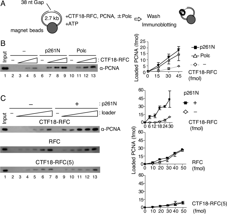
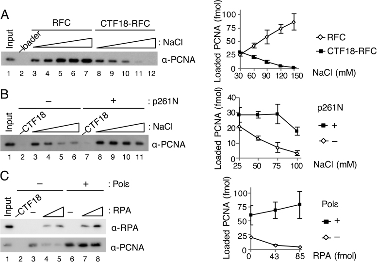
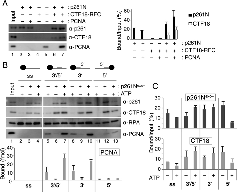
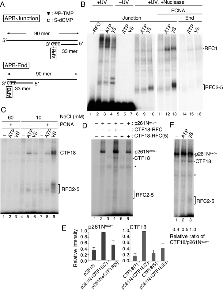
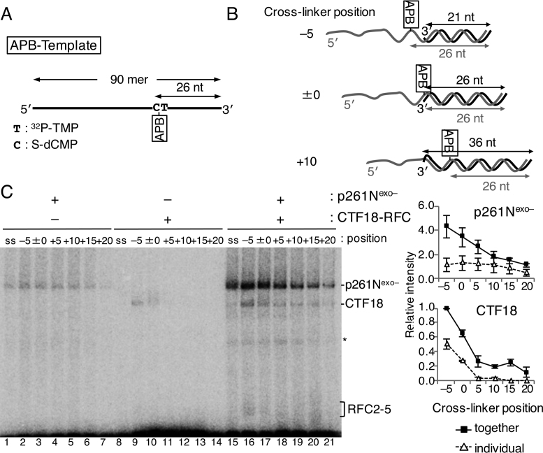
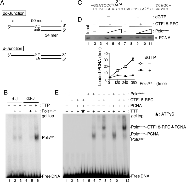
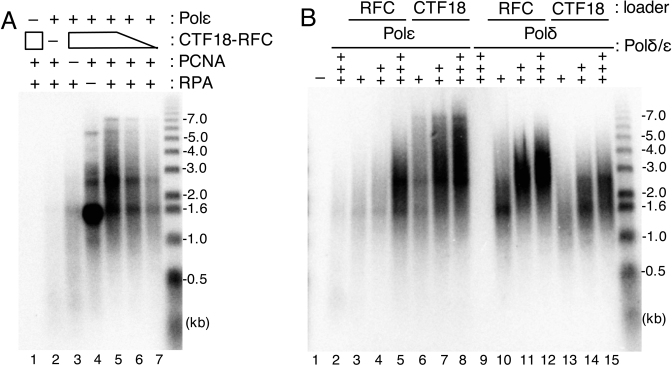
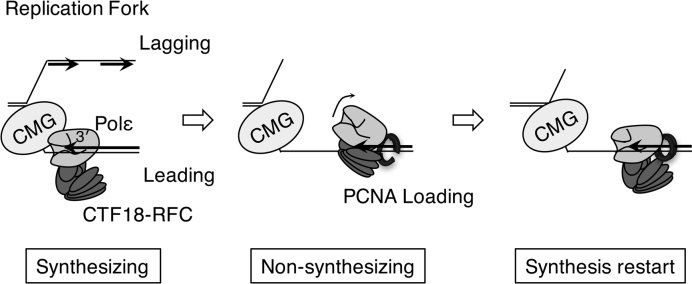
The alternative proliferating-cell nuclear antigen (PCNA)-loader CTF18-RFC forms a stable complex with DNA polymerase ε (Polε). We observed that, under near-physiological conditions, CTF18-RFC alone loaded PCNA inefficiently, but loaded it efficiently when complexed with Polε. During efficient PCNA loading, CTF18-RFC and Polε assembled at a 3΄ primer-template junction cooperatively, and directed PCNA to the loading site. Site-specific photo-crosslinking of directly interacting proteins at the primer-template junction showed similar cooperative binding, in which the catalytic N-terminal portion of Polε acted as the major docking protein. In the PCNA-loading intermediate with ATPγS, binding of CTF18 to the DNA structures increased, suggesting transient access of CTF18-RFC to the primer terminus. Polε placed in DNA synthesis mode using a substrate DNA with a deoxidised 3΄ primer end did not stimulate PCNA loading, suggesting that DNA synthesis and PCNA loading are mutually exclusive at the 3΄ primer-template junction. Furthermore, PCNA and CTF18-RFC-Polε complex engaged in stable trimeric assembly on the template DNA and synthesised DNA efficiently. Thus, CTF18-RFC appears to be involved in leading-strand DNA synthesis through its interaction with Polε, and can load PCNA onto DNA when Polε is not in DNA synthesis mode to restore DNA synthesis.


Figures

Figure 1.
Figure 1.
PCNA loading by CTF18-RFC in the presence of Polε. (A) Schematic diagram of the PCNA loading assay with a gapped DNA attached to magnetic beads. A linearised 2.7 kb plasmid DNA harbouring a 38 nt gap was used. After incubation of the DNA beads with purified proteins and ATP, the loaded PCNA was recovered from the bead-bound fraction. (B) PCNA loading by 15–45 fmol of CTF18-RFC with either 100 fmol of p261N (lanes 6–9) or 100 fmol of Polε (lanes 10–13), or with neither (lanes 2–5). The 10 μl reaction mixture contained 30 mM NaCl, 40 mM creatine phosphate and 25 ng/μl creatine-phosphate kinase. Input control (17 fmol of trimeric PCNA) (lane 1) and 50% of bound samples (lanes 2–13) were analysed by immunoblotting with anti-PCNA antibody. The loaded PCNA was quantified and graphed with mean ± S.E. of two experimental replicates (right). (C) PCNA loading by 6–30 fmol of CTF18-RFC (top), 10–50 fmol of RFC (middle) or 10–50 fmol of CTF18-RFC(5) (bottom) in the same reaction mixture with (+) or without (−) 200 fmol of p261N. Input (lane 1 of each) used 17 fmol (top and middle) or 12 fmol (bottom) PCNA. The bound fractions (50%) were analysed for loaded PCNA (lanes 2–13), which was quantified and graphed as indicated at the right with mean ± S.E. of two experimental replicates.
Figure 2.
Figure 2.
Effect of salt concentrations and RPA on PCNA loading by CTF18-RFC. (A) Titration of NaCl concentration from 30 mM to 150 mM for PCNA loading by 50 fmol each of RFC (lanes 3–7) or CTF18-RFC (lanes 8–12). PCNA in 50% bound fractions (lanes 2–12) and input control (12 fmol) were detected by immunoblotting (left). The band intensities were quantified and graphed with mean ± S.E. of two experimental replicates (right). (B) Titration of NaCl (25–100 mM) for PCNA loading by 30 fmol of CTF18-RFC with (lanes 7–11) or without (lanes 2–6) 200 fmol of p261N. PCNA in 50% bound fractions (lanes 2–11) and input control (12 fmol) were detected by immunoblotting (left), and their band intensities were quantified and graphed with mean ± S.E. of two experimental replicates (right). (C) Effects of RPA (42, 85 fmol) on PCNA loading with 30 ng gapped-DNA beads at 60 mM NaCl using 100 fmol CTF18-RFC with (lanes 6–8) or without (lanes 3–5) 200 fmol Polε. The total bound fractions (lanes 2–8) and 42 fmol RPA and 12 fmol PCNA as the input control (lane 1) were analysed by immunoblotting with the indicated antibodies (left). Band intensities of PCNA were quantified and graphed with the mean ± S.E. of three experimental replicates (right).
Figure 3.
Figure 3.
Analyses of loaded PCNA and bound CTF18-RFC and p261N on various structures of DNA. (A) Pull-down assay with 30 ng of gapped-DNA beads at 100 mM NaCl using 100 fmol of p261N, 100 fmol of CTF18-RFC and 6.2 pmol of PCNA, as indicated. Input (10%; p261N, CTF18-RFC) and 12 fmol of PCNA (lane 1), and 50% bound fractions (lanes 2–7), were analyzed by immunoblotting with the indicated antibodies (left). Bound p261N and CTF18 from two experimental replicates are graphed with mean ± S.E. (right). (B) PCNA-loading assay with oligo-DNA beads containing conjugated ssDNA (ss), a 3΄ and 5΄ recessed primer–template DNA (3΄/5΄), a 3΄ recessed primer–template DNA (3΄), or a 5΄ recessed primer–template DNA (5΄). Reactions were performed at 120 mM NaCl with 6.2 pmol of PCNA, 4.2 pmol of RPA, 200 fmol of CTF18-RFC and the indicated combination of 2 mM ATP and 1 pmol of p261Nexo–. Input bands (lane 1) indicate 60 fmol of p261Nexo–, 10 fmol of CTF18-RFC, 340 fmol of RPA and 12 fmol of PCNA. Bound proteins were analysed with 50% samples (lanes 2–13). Band intensities of PCNA were quantified and graphed with the mean ± S.E. from two experimental replicates (bottom). (C) The amounts of p261Nexo– and CTF18 that bound to four different DNA bead substrates were compared with or without ATP by quantification of their bound % versus input as indicated by the graph, with mean ± S.E. of two experimental replicates.
Figure 4.
Figure 4.
Analyses of proteins directly bound to the 3΄ primer end at a primer–template junction during PCNA loading with CTF18-RFC–p261N complex by photo-crosslinking. (A) Substrate DNA “APB-Junction” for photo-crosslinking analyses has azidophenacyl bromide (APB) at the 3΄ primer end of the primer–template junction. Two 32P-TMP, and one S-dCMP, were incorporated at the 3΄ primer end of RF30 annealed to TEMP90-R, and a photoreactive crosslinker APB was conjugated to S-dCMP. The control DNA “APB-End” has APB at the blunted end. (B) Photo-crosslinking of 25 fmol APB-Junction (Junction; lanes 1–13) or APB-End (End; lanes 14–16) at 60 mM NaCl by 150 fmol of RFC (lanes 2–16) and 500 fmol of PCNA (lanes 11–16). A set of results without nucleotide (−) or with 2 mM ATP or 250 μM ATPγS (γS) is shown for each condition. Samples with (lanes 1–4, 8–16) or without (lanes 5–7) UV irradiation and with (lanes 8–16) or without (lanes 1–7) nuclease treatment were separated by SDS-PAGE and visualised. (C) Photo-crosslinking of 25 fmol of APB-Junction at 60 mM (lanes 1–3) or 10 mM (lanes 4–9) NaCl by 150 fmol of CTF18-RFC with or without 500 fmol of PCNA, as indicated. A set of results without nucleotides (−) or with 2 mM ATP or 250 μM ATPγS is shown for each condition. (D) Photo-crosslinking of 25 fmol of APB-Junction with combinations of 150 fmol of CTF18-RFC, 250 fmol of CTF18-RFC(5) and 150 fmol of p261Nexo–, as indicated, in the presence of 500 fmol of PCNA and 250 μM ATPγS. (E) Crosslinked bands in (D) corresponding to p261Nexo– and CTF18 were quantified, and their relative intensities were graphed on the right using the highest intensity bands (lane 4) as reference (1.0), with mean ± S.E. of three experimental replicates. “p261N”, “CTF18(7)” and “CTF18(5)” represent p261Nexo–, CTF18-RFC and CTF18-RFC(5), respectively. (F) Photo-crosslinking of the 25 fmol APB-Junction, with 150 fmol of p261Nexo–, 150 fmol of CTF18-RFC and 500 fmol of PCNA in the presence or absence of 2 mM ATP or 250 μM ATPγS. Crosslinked bands corresponding to p261Nexo– and CTF18 were quantified, and the relative values of the CTF18:p261Nexo– ratios are indicated below, with the ratio with ATPγS as 1.0.
Figure 5.
Figure 5.
Photo-crosslinking analyses of proteins directly bound to the template strand of a primer–template junction during PCNA loading by the CTF18-RFC–p261N complex. (A) The template DNA, “APB-Template”, had 32P-TMP and S-dCMP 25 nt and 26 nt from the 3΄ end on a 90-mer oligonucleotide. (B) Three representatives of the six primer–template junction substrates with differently positioned crosslinkers (azidophenacyl bromide; APB) relative to the 3΄ primer end are indicated. In ‘–5’, the APB is located in single-stranded DNA 5 nt from the junction, in ‘±0’ APB is at the junction and in ‘+10’ APB is in double-stranded DNA 10 nt from the junction. (C) Photo-crosslinked bands from 25 fmol of indicated substrate DNA and 150 fmol of p261Nexo– (lanes 1–7, 15–21), or 150 fmol of CTF18-RFC (lanes 8–21) as indicated in the presence of 500 fmol of PCNA and 250 μM ATPγS. Protein bands corresponding to p261Nexo–, CTF18 and RFC2–5 are indicated. Some degraded proteins are indicated with an asterisk. Results with APB-Template without primers are indicated as the controls (‘ss’; lanes 1, 8, 15). Relative band intensities of p261Nexo– and CTF18 using that of CTF18 in lane 16 as a reference (1.0) were measured and graphed on the right, with mean ± S.E. of three experimental replicates.
Figure 6.
Figure 6.
Analyses of PCNA loading in the presence of Polε in synthesis mode. (A) Two 3΄ primer–template junction substrates with ddAMP (“dd-Junction”) or dAMP (“d-Junction”) at their 3΄ primer ends. (B) 60 fmol of Polεexo– was mixed with 25 fmol of d-Junction (“d-J”; lanes 2, 3) or dd-Junction (“dd-J”; lanes 5, 6) at 60 mM NaCl in the presence (+) or absence (−) of 100 μM TTP, and binding was analysed by EMSA after glutaraldehyde fixation. Lanes 1 and 4 were controls without Polεexo–. Bands produced by binding of Polεexo– to DNA are indicated. (C) To study PCNA loading in the presence of Polεexo– in synthesis mode, a gapped DNA with ddAMP at the 3΄ primer end was prepared. The sequence shows a 51 bp region with the 35 nt gap on the substrate DNA. The nucleotides shown in bold represent sequence extension to prepare the 3΄ primer end with ddAMP. (D) Comparison of PCNA loading with the gapped-DNA beads with Polεexo– in non-synthesising (–dGTP) and synthesising (+dGTP) modes. The indicated DNA beads (15 ng) were incubated with 100 fmol of CTF18-RFC and 6.2 pmol of PCNA in the presence of 0, 120, 240 and 360 fmol of Polεexo– in a 10 μl reaction mixture at 60 mM NaCl. dGTP (100 μM) was added in lanes 7–11. Input control of 12 fmol of PCNA (lane 1) and 100% bound fractions (lanes 2–11) were applied to immunoblotting with anti-PCNA antibody. Lanes 2 and 7 were the negative controls without CTF18-RFC and Polεexo–. Bound PCNA was quantified and graphed below with mean ± S.E. of two experimental replicates. (E) Assembly of 60 fmol each of CTF18-RFC and Polεexo– and 500 fmol of PCNA to 25 fmol of dd-Junction substrate was analysed by EMSA as above (B) using indicated combinations of proteins at 60 mM NaCl. Additions of 100 μM TTP and 250 μM ATPγS are indicated by ‘+’ and an asterisk (lane 3), respectively. DNA bands shifted at positions by added proteins are indicated at the right.
Figure 7.
Figure 7.
Analysis of DNA synthesis with CTF18-RFC–Polε–PCNA. Product DNA profiles after electrophoresis in alkaline agarose gels are indicated together with DNA size markers (right). (A) Holoenzyme assay was done with indicated combinations of 200 fmol Polε, 600 fmol CTF18-RFC, 2 pmol PCNA and 6 pmol RPA. Lower amounts of CTF18-RFC (400 or 200 fmol) were used in lanes 6 and 7. (B) Titration of Polε (+, ++, +++ for 147, 293, 440 fmol, respectively; lanes 2–8) or Polδ (+, ++, +++ for 53, 107, 160 fmol, respectively; lanes 9–15) in the holoenzyme assay with 600 fmols RFC (lanes 3–5, 10–12) or CTF18-RFC (lanes 6–8, 13–15) or without (lanes 2 and 9) in the presence of 6 pmol RPA and 2 pmol PCNA.
Figure 8.
Figure 8.
A possible PCNA-loading mechanism by CTF18-RFC–Polε complex. Synthesising: In the normal replication fork, CMG helicase and Polε form a processive DNA polymerase complex for leading-strand synthesis. CTF18-RFC will associate with Polε in the complex, but PCNA loading will be minimised because Polε is in synthesising mode. Non-synthesising: Upon arrest of Polε by an obstacle on the chromosome, Polε is decoupled from CMG and shifts to non-synthesising mode. Under this condition, CTF18-RFC is able to access the 3΄ primer end and load PCNA at the site. Synthesis restart: Participation of PCNA in the CTF18-RFC–Polε complex at the 3΄ primer end will stabilise the complex and facilitate restart of the DNA synthesis by forming a secondary processive DNA polymerase complex lacking CMG.

Similar articles

Cited by

References

    1. Bloom L.B. Loading clamps for DNA replication and repair. DNA Repair. 2009; 8:570–578. - PMC - PubMed
    1. Hedglin M., Kumar R., Benkovic S.J.. Replication clamps and clamp loaders. Cold Spring Harb. Perspect. Biol. 2013; 5:a010165. - PMC - PubMed
    1. Moldovan G.L., Pfander B., Jentsch S.. PCNA, the maestro of the replication fork. Cell. 2007; 129:665–679. - PubMed
    1. Kelch B.A. Review: The lord of the rings: Structure and mechanism of the sliding clamp loader. Biopolymers. 2016; 105:532–546. - PubMed
    1. Bermudez V.P., Maniwa Y., Tappin I., Ozato K., Yokomori K., Hurwitz J.. The alternative Ctf18-Dcc1-Ctf8-replication factor C complex required for sister chromatid cohesion loads proliferating cell nuclear antigen onto DNA. Proc. Natl. Acad. Sci. U.S.A. 2003; 100:10237–10242. - PMC - PubMed

MeSH terms

LinkOut - more resources