. 2017 Feb 14;8(2):235-248.
doi: 10.1016/j.stemcr.2016.12.019.

HuCNS-SC Human NSCs Fail to Differentiate, Form Ectopic Clusters, and Provide No Cognitive Benefits in a Transgenic Model of Alzheimer's Disease

Affiliations

HuCNS-SC Human NSCs Fail to Differentiate, Form Ectopic Clusters, and Provide No Cognitive Benefits in a Transgenic Model of Alzheimer's Disease

Samuel E Marsh et al. Stem Cell Reports. .

Abstract

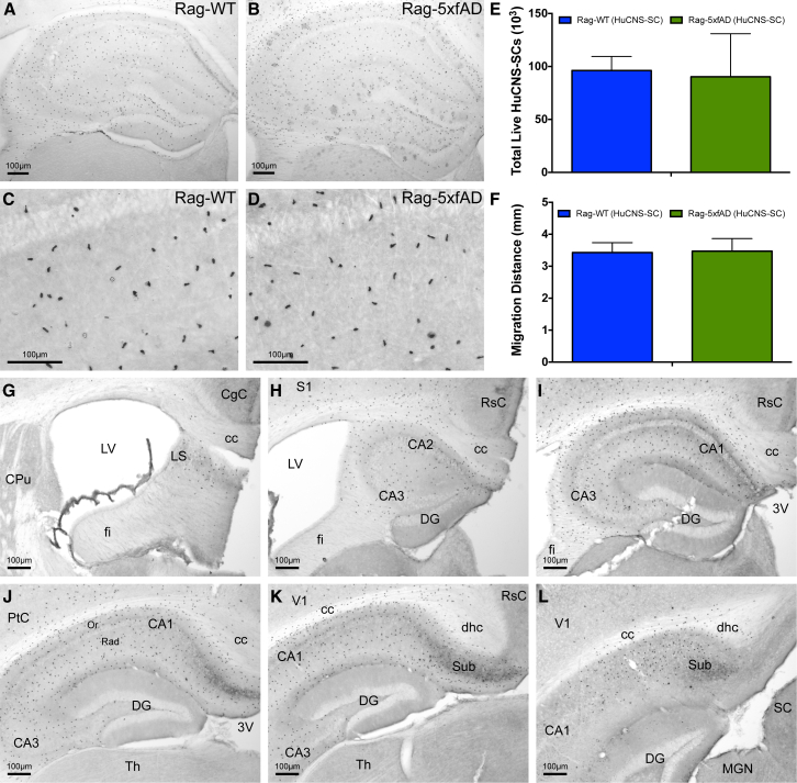
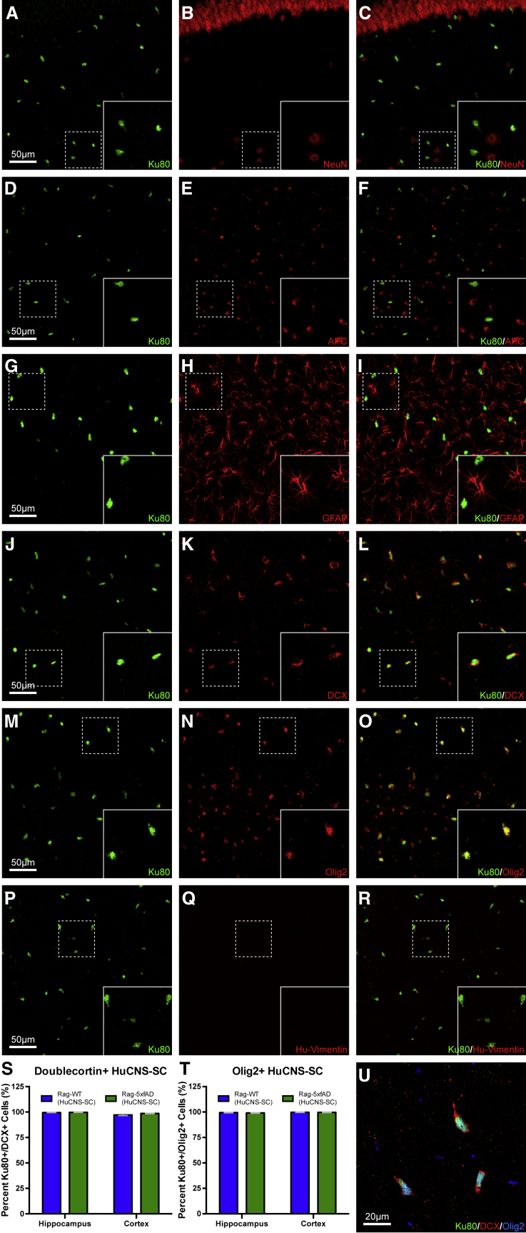
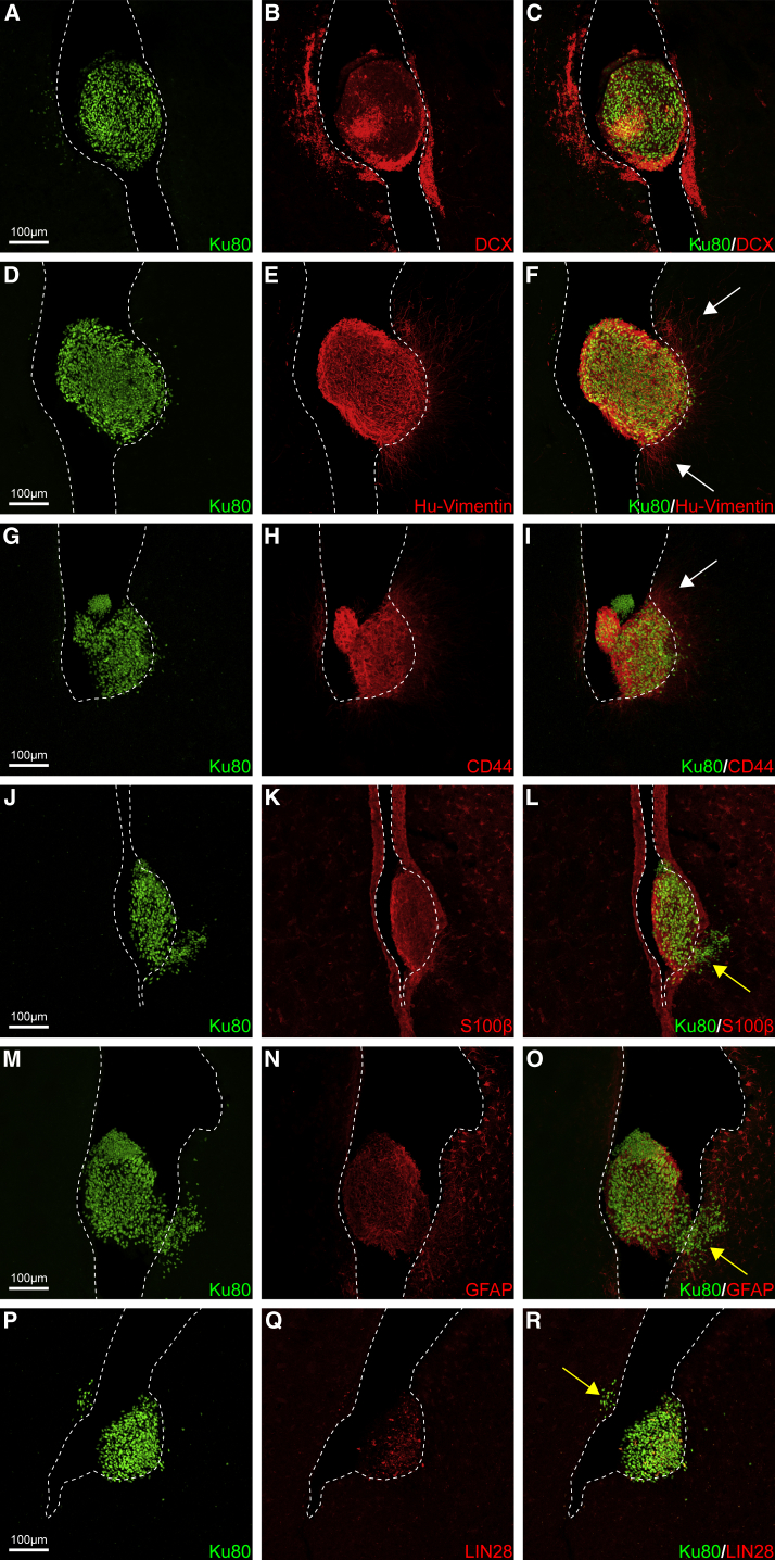
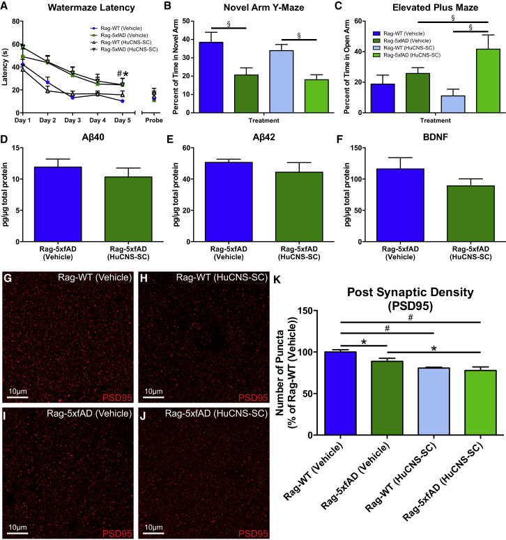
Transplantation of neural stem cells (NSCs) can improve cognition in animal models of Alzheimer's disease (AD). However, AD is a protracted disorder, and prior studies have examined only short-term effects. We therefore used an immune-deficient model of AD (Rag-5xfAD mice) to examine long-term transplantation of human NSCs (StemCells Inc.; HuCNS-SCs). Five months after transplantation, HuCNS-SCs had engrafted and migrated throughout the hippocampus and exhibited no differences in survival or migration in response to β-amyloid pathology. Despite robust engraftment, HuCNS-SCs failed to terminally differentiate and over a quarter of the animals exhibited ectopic human cell clusters within the lateral ventricle. Unlike prior short-term experiments with research-grade HuCNS-SCs, we also found no evidence of improved cognition, no changes in brain-derived neurotrophic factor, and no increase in synaptic density. These data, while disappointing, reinforce the notion that individual human NSC lines need to be carefully assessed for efficacy and safety in appropriate long-term models.

Keywords: Alzheimer's disease; HuCNS-SC; NSC; cognition; dementia; hippocampus; stem cells; translation; transplantation; β-amyloid.


Figures

Figure 1
Figure 1
HuCNS-SCs Engraft and Migrate Equally Well in AD and Wild-Type Immune-Deficient Mice (A–D) HuCNS-SCs (Ku80: human-specific nuclear marker) exhibit robust survival and migration throughout the rostral-caudal extent of the hippocampus in Rag-WT (A, C) and Rag-5xfAD mice (B, D). (E and F) Unbiased stereological analysis of HuCNS-SC engraftment reveals no significant differences in total engrafted cells (E) or anterior-posterior (A/P) migration distance (F). Data presented as means ± SEM. N ≥ 8 mice/group. (G–L) Representative images illustrating A/P migration of HuCNS-SC throughout hippocampal formation. 3V, third ventricle; CA1, field CA1 of the hippocampus; CA2, field CA2 of the hippocampus; CA3, field CA3 of the hippocampus; cc, corpus callosum; CgC, cingulate cortex; CPu, caudate putamen (striatum); DG, dentate gyrus; dhc, dorsal hippocampal commissure; fi, fimbria; LS, lateral septal nucleus; LV, lateral ventricle; MGN, medial geniculate nucleus; Or, oriens layer of the hippocampus; PtC, parietal cortex; Rad, radiatum layer of the hippocampus; RSC, retrosplenial cortex; S1, primary somatosensory cortex; SC, superior colliculus; Sub, subiculum; Th, thalamus; V1, primary visual cortex. Scale bar, 100 μm. See also Movie S1.
Figure 2
Figure 2
Five Months Post-Transplantation HuCNS-SCs Show No Evidence of Terminal Neuronal or Glial Differentiation Sections from all animals, both Rag-5xfAD (shown) and Rag-WT, probed with human-specific nuclei marker (Ku80, green) to identify engrafted HuCNS-SCs and co-labeled with mature (A–I) and immature (J–R) markers of neurons, oligodendrocytes, and astrocytes. Co-labeling of HuCNS-SCs with mature neuronal marker NeuN (A–C), mature oligodendroglial marker APC (D–F), and mature astrocytic marker GFAP (G–I) revealed no expression of any mature markers in HuCNS-SCs. Co-labeling of HuCNS-SCs with immature neuronal marker doublecortin (J–L, U) revealed that greater than 97% of engrafted HuCNS-SCs expressed DCX (S). Intriguingly, co-labeling of HuCNS-SCs with immature oligodendrocyte marker Olig2 (M–O, U) revealed that greater than 99% of engrafted HuCNS-SCs also expressed Olig2 (T). However, none of the engrafted HuCNS-SCs expressed immature astrocytic marker vimentin (P–R). Thus, taken together, it appears that HuCNS-SCs fail to terminally differentiate in immune-deficient mouse brains. Data presented as means ± SEM. Two-way ANOVA p < 0.05 and Fisher's PLSD post hoc. N ≥ 6 animals/group and one section/region of interest was analyzed for differentiation. The total number of cells counted was equivalent between genotypes. Scale bar, 50 μm.
Figure 3
Figure 3
HuCNS-SCs Form Ectopic Clusters within the Lateral Ventricle Over a quarter of animals that received HuCNS-SCs exhibited ectopic clusters within the anterior lateral ventricle (∼+1.5 mm relative to bregma, 3.5 mm from site of injection, +1.5 mm from furthest anterior parenchymal cell migration). Interestingly, HuCNS-SCs within the ventricle were immunoreactive for several immature glial markers that failed to label intraparenchymally engrafted cells. Instead, these HuCNS-SC ventricular clusters (Ku80, green) were immunoreactive for (A–C) DCX (immature neurons, red), (D–F) vimentin (immature glia/neurons, red), (G–I) CD44 (immature glia, red), (J–L) S100β (glia, red), and (M–O) GFAP (astrocytes, red). In some clusters (G and M), examples of what appears to be varying Ku80+ nuclei size or pleomorphism can also be observed. (P–R) Some cells within the ventricles as well infiltrating HuCNS-SCs (arrow in R) were also positive for LIN28, a marker of one type of PNET that exhibits similar characteristic uneven immunoreactivity (Picard et al., 2012). Ventricular clusters also exhibit growth into the surrounding tissue of both fibers (white arrows) and infiltration of cells through the ventricle wall into the adjacent striatum or septum (yellow arrows, see also Movie S2). Dotted lines indicate boundary of ventricle walls. Scale bar, 100 μm. See also Figures S1 and S2 and Movie S2.
Figure 4
Figure 4
HuCNS-SCs Fail to Improve Learning and Memory in Rag-5xfAD Mice (A) Analysis of Morris water maze (MWM) learning curve demonstrates that vehicle-injected Rag-5xfAD mice (green squares) take significantly longer to find the platform compared with vehicle-injected Rag-WT mice (blue circles). Thus, Rag-5xfAD mice exhibit characteristic AD-associated impairments in MWM performance. Transplantation of HuCNS-SCs provides no improvement in MWM performance as HuCNS-SC-injected Rag-5xfAD mice (green triangles) perform no better than vehicle-injected Rag-5xfAD mice (green squares). (B) Rag-5xfAD mice regardless of treatment were also significantly impaired in a novel arm Y-maze test of spatial working memory, spending a smaller percentage of their time in the novel arm. (C) Analysis of anxiety in elevated plus maze (EPM) revealed no differences between Rag-5xfAD and Rag-WT animals receiving vehicle injections. However, Rag-5xfAD (HuCNS-SC) animals were significantly less anxious than Rag-WT (HuCNS-SC) animals, suggesting that HuCNS-SCs may promote impaired EPM performance. (D and E) ELISA analysis of hippocampal lysates revealed no difference in Aβ40 or Aβ42 levels regardless of treatment in Rag-5xfAD mice. (F) HuCNS-SC transplantation also failed to elevate hippocampal BDNF protein levels and surprisingly produced a non-significant trend toward decreased BDNF (p = 0.21). (G–J) Representative high-power confocal images of presynaptic PSD-95 labeling in the stratum oriens of the hippocampus. (K) As previously reported in AD mouse models, quantification of PSD-95 density revealed a significant reduction in postsynaptic terminals in vehicle-treated Rag-5xfAD versus vehicle-treated Rag-WT mice. However, no increase in PSD95 density was detected after HuCNS-SC transplantation. Instead, delivery of HuCNS-SCs led to a significant further reduction of PSD95 in Rag5xfAD mice and reduced postsynaptic puncta in Rag-WT mice. ANOVA p < 0.05 and Fisher's PLSD post hoc p < 0.05, #p < 0.001. Aβ and BDNF; PSD95 N ≥ 6 animals/group. Data presented as means ± SEM. ANOVA p < 0.05 and Fisher's PLSD post hoc. N ≥ 6 animals/group (B–C), N ≥ 4 animals/group (D–F), (K) N ≥ 6 animals/group. Value for each animal was an average of five randomly selected regions of interest within images of the stratum oriens of two different brain sections (total of ten measurements). (A) p < 0.05 between Rag-WT (Vehicle) and Rag-5xfAD (Vehicle) and #p < 0.05 between Rag-WT (Vehicle) and Rag-5xfAD (HuCNS-SC). (B, C, and K) p < 0.05, §p < 0.01, #p < 0.001. Scale bar, 10 μm.

Comment in

  • Lessons Learned from Pioneering Neural Stem Cell Studies.
    Temple S, Studer L. Temple S, et al. Stem Cell Reports. 2017 Feb 14;8(2):191-193. doi: 10.1016/j.stemcr.2017.01.024. Stem Cell Reports. 2017. PMID: 28199825 Free PMC article.
  • Response to StemCells Inc.
    Monuki ES, Anderson AJ, Blurton-Jones M, Cummings BJ. Monuki ES, et al. Stem Cell Reports. 2017 Feb 14;8(2):195-197. doi: 10.1016/j.stemcr.2017.02.002. Stem Cell Reports. 2017. PMID: 28199827 Free PMC article. No abstract available.

References

    1. Ager R.R., Davis J.L., Agazaryan A., Benavente F., Poon W.W., LaFerla F.M., Blurton-Jones M. Human neural stem cells improve cognition and promote synaptic growth in two complementary transgenic models of Alzheimer’s disease and neuronal loss. Hippocampus. 2015;25:813–826. - PMC - PubMed
    1. Alzheimer’s Association 2016 Alzheimer’s disease facts and figures. Alzheimers Dement. 2016;12:459–509. - PubMed
    1. Anderson A.J., Haus D.L., Hooshmand M.J., Perez H., Sontag C.J., Cummings B.J. Achieving stable human stem cell engraftment and survival in the CNS: is the future of regenerative medicine immunodeficient. Regen. Med. 2011;6:367–406. - PMC - PubMed
    1. Anderson A.J., Piltti P.M., Hooshmand M.J., Nishi R.A., Cummings B.J. Preclinical Efficacy Failure of Human CNS-Derived Stem Cells for Use in the Pathway Study of Cervical Spinal Cord Injury. Stem Cell Rep. 2017;8 in this issue. - PMC - PubMed
    1. Berkowitz A.L., Miller M.B., Mir S.A., Cagney D., Chavakula V., Guleria I., Aizer A., Ligon K.L., Chi J.H. Glioproliferative lesion of the spinal cord as a complication of “Stem-Cell Tourism”. N. Engl. J. Med. 2016;375:196–198. - PubMed

Publication types

MeSH terms