. 2017 Feb 14;8(2):249-263.
doi: 10.1016/j.stemcr.2016.12.018.

Preclinical Efficacy Failure of Human CNS-Derived Stem Cells for Use in the Pathway Study of Cervical Spinal Cord Injury

Affiliations

Preclinical Efficacy Failure of Human CNS-Derived Stem Cells for Use in the Pathway Study of Cervical Spinal Cord Injury

Aileen J Anderson et al. Stem Cell Reports. .

Abstract

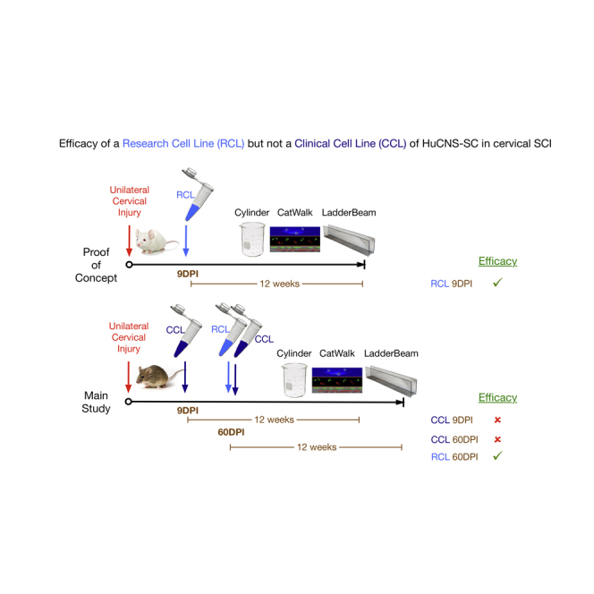
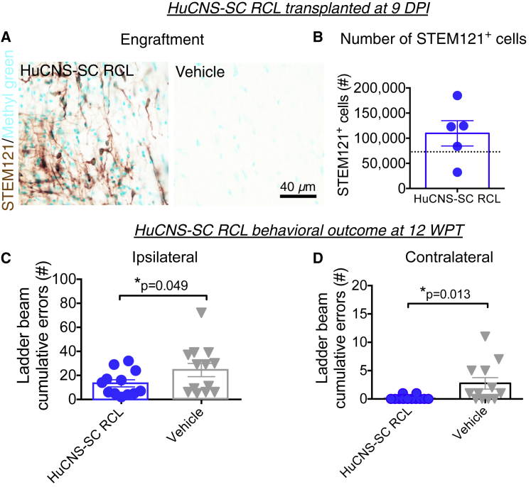
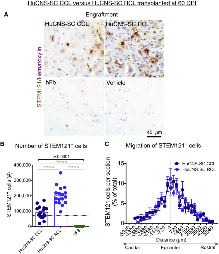
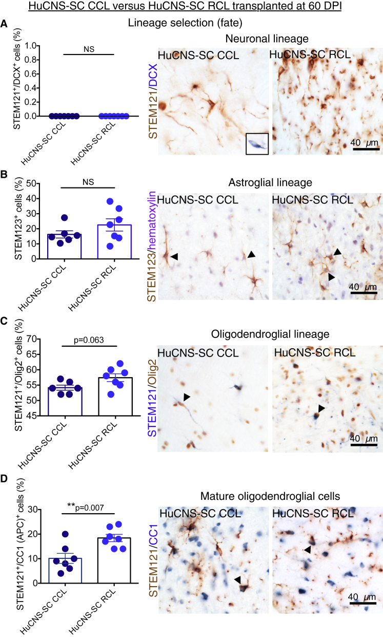
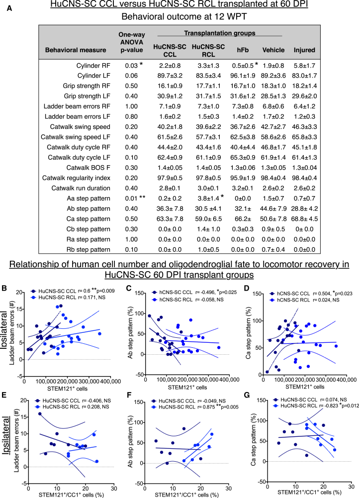
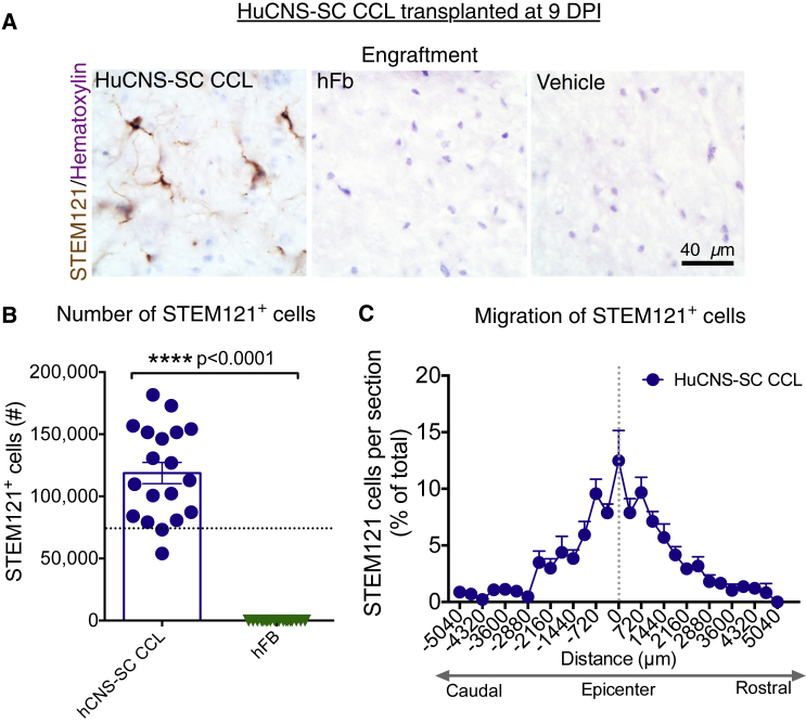
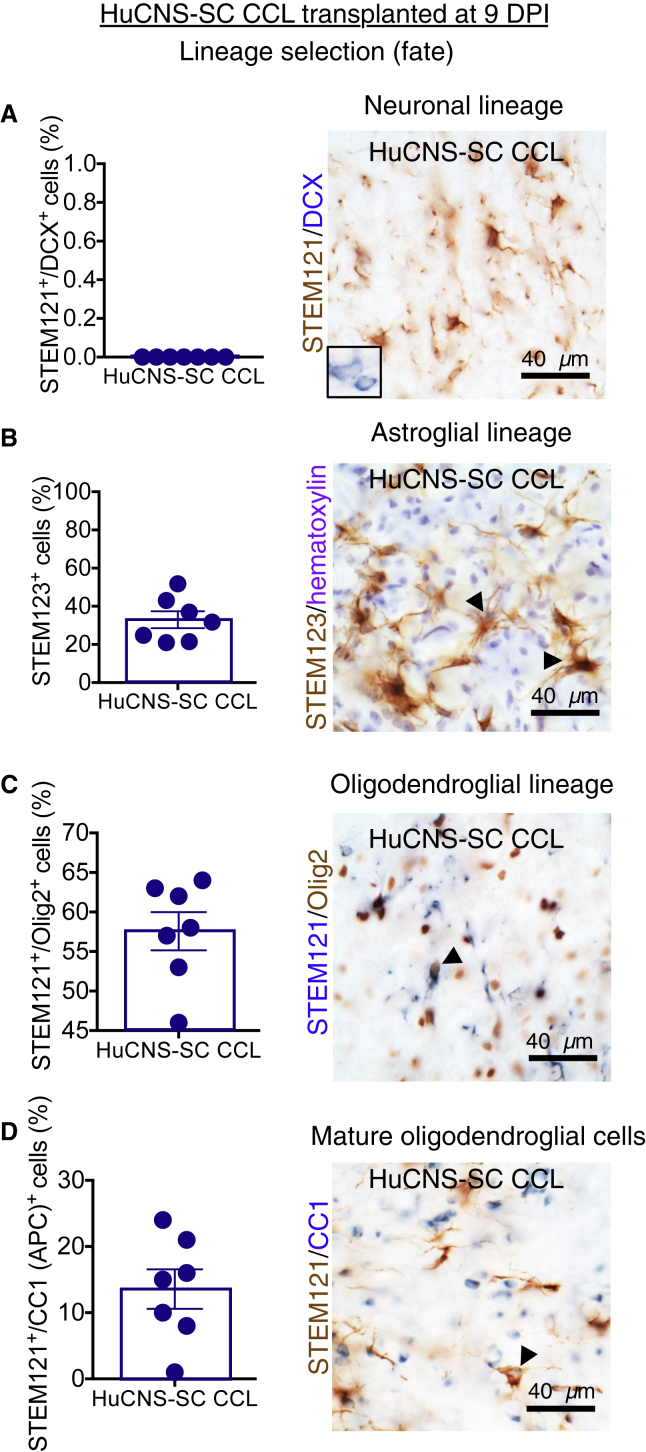
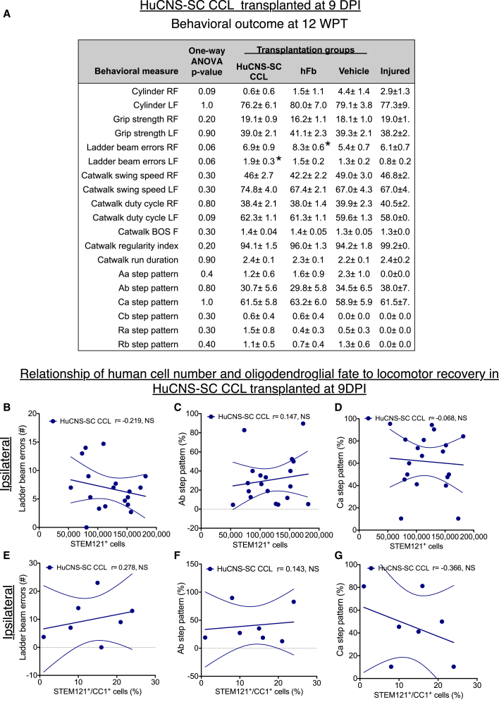
We previously showed the efficacy of multiple research cell lines (RCLs) of human CNS neural stem cells (HuCNS-SCs) in mouse and rat models of thoracic spinal cord injury (SCI), supporting a thoracic SCI clinical trial. Experts recommend in vivo preclinical testing of the intended clinical cell lot/line (CCL) in models with validity for the planned clinical target. We therefore tested the efficacy of two HuCNS-SC lines in cervical SCI: one RCL, and one CCL intended for use in the Pathway Study of cervical SCI in man. We assessed locomotor recovery and sensory function, as well as engraftment, migration, and fate. No evidence of efficacy of the CCL was observed; some data suggested a negative impact of the CCL on outcomes. These data raise questions about the development and validation of potency/comparability assays for clinical testing of cell products, and lack of US Food and Drug Administration requirements for in vivo testing of intended clinical cell lines.

Keywords: FDA guidelines for cell therapies; RIGOR; cervical; efficacy; good manufacturing practices (GMP); human neural stem cells (hNSC); spinal cord injury (SCI).


Figures

None
Graphical abstract
Figure 1
Figure 1
HuCNS-SC RCLs Exhibit Engraftment and Improve Locomotor Recovery in a 9 DPI Transplantation Paradigm (A) Representative images from cervical SCI groups receiving HuCNS-SC RCL transplant (left) or vehicle control (right) 9 DPI. Animals were sacrificed 12 WPT. Sections were immunostained with a human-specific cytoplasmic marker STEM121 (brown) and counterstained with methyl green. (B) Blinded, unbiased stereological quantification 12 WPT revealed an average of 109,695 ± 25,197 human cells in the RCL group (n = 9). Dashed line denotes transplant dose of 75,000 cells. (C and D) Animals receiving RCL 9 DPI (n = 12) exhibited a significant decrease in horizontal ladder beam errors at 12 WPT for both ipsilateral (13.4 ± 3.0 versus 24.5 ± 5.5 for controls) and contralateral (0.2 ± 0.1 versus 3.8 ± 1.4 for controls) forepaws (Student's one-tailed t test, p < 0.05) compared with controls (n = 13 ipsilateral, 12 contralateral). Data shown as means ± SEM.
Figure 2
Figure 2
HuCNS-SC CCLs Exhibit Reduced Engraftment Compared with RCLs in a 60 DPI Paradigm (A) Representative images from cervical SCI groups that received either a CCL, RCL, or hFb transplant, or vehicle control 60 DPI. Animals were sacrificed 12 WPT. Sections were immunostained with STEM121 (brown) and counterstained with hematoxylin (purple). (B) Blinded quantification 12 WPT revealed an average of 91,701 human cells for the CCL group (n = 16), 200,754 for RCL group (n = 17), and no surviving human cells for the hFB group (n = 12) A one-way ANOVA revealed significant differences in survival (solid bar p < 0.0001; post hoc Tukey's test of CCL versus RCL and hFb, ∗∗∗∗p < 0.0001). Dashed line denotes transplant dose of 75,000 cells. (C) No difference was found in the rostral-caudal extent of human cell migration between the CCL (n = 16) and RCL (n = 17) at 12 WPT (unpaired t tests with Holm-Sidak multiple comparison correction, p > 0.05). Dashed vertical line indicates injury epicenter. Data shown as means ± SEM.
Figure 3
Figure 3
Lineage Analysis of HuCNS-SC CCL and RCL in the 60 DPI Paradigm Data for proportional human cell fate were collected by blinded, unbiased stereology. (A) Co-immunostaining for STEM121 (brown) and the early neuronal marker DCX (blue) revealed no STEM121+/DCX+ cells in either the CCL (n = 7) or RCL (n = 7) groups (Student's two-tailed t test, p > 0.05, n.s.). Inset shows positive control for DCX in the hippocampus. (B) Immunostaining for human-specific GFAP (STEM123, brown) with hematoxylin counterstaining (purple) revealed no significant difference in STEM123 proportion between the CCL (n = 6) and RCL (n = 7) groups (Student's two-tailed t test, p > 0.2). (C) Immunostaining for STEM121 (blue) and the oligodendroglial nuclear marker Olig2 (brown) revealed that the largest proportion of STEM121+ cells were also nuclear Olig2+ and there was a trend for a decrease in Olgi2+ cells in CCL (n = 5) versus RCL (n = 7) transplants (Student's two-tailed t test, p = 0.06). (D) Immunostaining for STEM121 (brown) and the mature oligodendroglial marker CC1 (blue) revealed a significant decrease in the proportion of STEM121+/CC1+ cells in CCL (n = 7) versus RCL (n = 7) transplants (Student's two-tailed t test, p < 0.007). Arrowheads indicate double-positive cells. Data shown as means ± SEM.
Figure 4
Figure 4
Analysis of Locomotor Recovery in HuCNS-SC 60 DPI Cohort Transplantation Groups versus Vehicle Controls 12 WPT (A) Table showing average ± SEM values for cylinder reaching (percentage paw placement), grip strength, horizontal ladder beam errors, and CatWalk step kinematic analysis for each experimental group. RF, ipsilateral forelimb; LF, contralateral forelimb. Where one-way ANOVA reached significance (p ≤ 0.05, ∗∗p ≤ 0.01), Tukey's multiple comparison t tests were conducted. Significant differences were found in ipsilateral cylinder reaching between the hFB and Injured only groups (p ≤ 0.05), and in Aa step pattern between the RCL, CCL, and hFB groups (p ≤ 0.05). n = 15–16 for CCL, n = 16–17 for RCL, n = 11–12 for hFB, n = 15–17 for Vehicle, and n = 9–10 for Injured only (see Figure S1D for exact numbers). BOS, base of support. (B–G) Pearson correlations were conducted between the number of human STEM121+ cells and (B) ipsilateral forelimb ladder beam errors, (C) Ab step pattern, and (D) Ca step pattern. Pearson correlation were also conducted between STEM121+/CC1+ human oligodendroglial cell proportion and (E) ipsilateral forelimb ladder beam errors, (F) Ab step pattern, and (G) Ca step pattern. Dashed lines indicate confidence intervals of 95%. n = 7 for CCL and n = 7 for RCL in CC1+ correlational analyses.
Figure 5
Figure 5
HuCNS-SC CCL Exhibit Engraftment and Rostral-Caudal Migration in a 9 DPI Paradigm (A) Representative images from cervical SCI groups that received CCL or hFb transplant, or vehicle control 9 DPI. Animals were sacrificed 12 WPT. Sections were immunostained with STEM121 (brown) and counterstained with hematoxylin. (B) Blinded, unbiased quantification 12 WPT revealed an average of 118,757 human cells for the CCL group (n = 19) and no human cells for the hFB group (n = 18) (Student's two-tailed t test, p < 0.0001). Dashed line indicates the transplant dose of 75,000 cells. (C) Rostral-caudal extent of human cell migration for the CCL at 12 WPT. Dashed vertical line indicates injury epicenter (n = 19). Data shown as means ± SEM.
Figure 6
Figure 6
Lineage Analysis of HuCNS-SC CCL in the 9 DPI Paradigm Data for proportional human cell fate were collected by blinded, unbiased stereology (A) Immunostaining for STEM121 (brown) and DCX (blue) revealed no STEM121+/DCX+ cells in CCL 9 DPI animals. Inset shows positive control for DCX in cortex. (B) Immunostaining for human-specific GFAP (STEM123, brown) with hematoxylin counterstaining (purple) revealed 33% ± 4.4% of CCL 9 DPI were positive for this astroglial marker. (C) Immunostaining for STEM121 (blue) and Olig2 (brown) revealed that the largest proportion of STEM121+ cells in the 9 DPI transplant group were positive for this oligodendroglial marker (57.6% ± 2.4%). (D) Immunostaining for STEM121 (brown) and CC1 (blue) revealed 13.6% ± 3% of CCL 9 DPI were positive for this mature oligodendroglial marker. Arrowheads indicate double-positive cells. n = 7 for all lineage tests. Data shown as means ± SEM.
Figure 7
Figure 7
Locomotor Recovery in HuCNS-SC 9 DPI Cohort Transplantation Groups versus Vehicle Controls 12 WPT Data for proportional human cell fate were collected by blinded, unbiased stereology. (A) Average ± SEM values for cylinder reaching (percentage paw placement), grip strength, horizontal ladder beam errors, and CatWalk kinematic analysis for each group. RF, ipsilateral forelimb; LF, contralateral forelimb. No significant differences were found between groups using one-way ANOVA p = 0.06; however Tukey's multiple comparison revealed significant an increase in ipsilateral forelimb ladder beam errors in the hFB group compared with vehicle group (p < 0.05) and in contralateral forelimb ladder beam errors in CCL 9 DPI group compared with injured only group (p < 0.05), denoted by . n = 18–19 for CCL, n = 17–18 for hFB, n = 18–20 for Vehicle, and n = 9–10 for Injured only (see Figure S1D for exact numbers). (B–G) Pearson correlations were conducted between the number of human STEM121+ cells and (B) ipsilateral forelimb ladder beam error number, (C) Ab step pattern, and (D) Ca step pattern (n = 18). Pearson correlations were also run between STEM121+/CC1+ human oligodendroglial cell proportion and (E) ipsilateral forelimb ladder beam errors, (F) Ab step pattern, and (G) Ca step pattern (n = 7). No significant differences were observed, suggesting neither improvements nor decrements in function at 9 DPI. Dashed lines indicate confidence intervals of 95%.

Comment in

  • Lessons Learned from Pioneering Neural Stem Cell Studies.
    Temple S, Studer L. Temple S, et al. Stem Cell Reports. 2017 Feb 14;8(2):191-193. doi: 10.1016/j.stemcr.2017.01.024. Stem Cell Reports. 2017. PMID: 28199825 Free PMC article.
  • Response to StemCells Inc.
    Monuki ES, Anderson AJ, Blurton-Jones M, Cummings BJ. Monuki ES, et al. Stem Cell Reports. 2017 Feb 14;8(2):195-197. doi: 10.1016/j.stemcr.2017.02.002. Stem Cell Reports. 2017. PMID: 28199827 Free PMC article. No abstract available.

References

    1. Anderson A.J., Cummings B.J. Achieving informed consent for cellular therapies: a preclinical translational research perspective on regulations versus a dose of reality. J. Law Med. Ethics. 2016;44:394–401. - PMC - PubMed
    1. Anderson A.J., Haus D.L., Hooshmand M.J., Perez H., Sontag C.J., Cummings B.J. Achieving stable human stem cell engraftment and survival in the CNS: is the future of regenerative medicine immunodeficient? Regen. Med. 2011;6:367–406. - PMC - PubMed
    1. Arvanian V.L., Schnell L., Lou L., Golshani R., Hunanyan A., Ghosh A., Pearse D.D., Robinson J.K., Schwab M.E., Fawcett J.W., Mendell L.M. Chronic spinal hemisection in rats induces a progressive decline in transmission in uninjured fibers to motoneurons. Exp. Neurol. 2009;216:471–480. - PMC - PubMed
    1. Center for Biologics Evaluation and Research . US Department of Health and Huma Services, US Food and Drug Administration and Center for Biologics Evaluation and Research, Office of Communication, Outreach, and Development (OCOD), US Government; 2013. Guidance for Industry - Preclinical Assessment of Investigational Cellular and Gene Therapy Products; p. 35.
    1. Chang J., Phelan M., Cummings B.J. A meta-analysis of efficacy in pre-clinical human stem cell therapies for traumatic brain injury. Exp. Neurol. 2015;273:225–233. - PubMed

Publication types

MeSH terms