. 2017 Feb 14;18(7):1713-1726.
doi: 10.1016/j.celrep.2017.01.055.

The SIN3A/HDAC Corepressor Complex Functionally Cooperates with NANOG to Promote Pluripotency

Affiliations

The SIN3A/HDAC Corepressor Complex Functionally Cooperates with NANOG to Promote Pluripotency

Arven Saunders et al. Cell Rep. .

Abstract

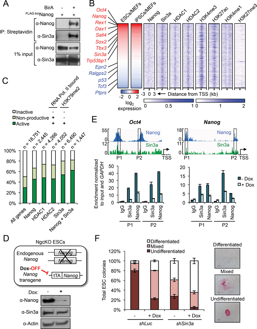
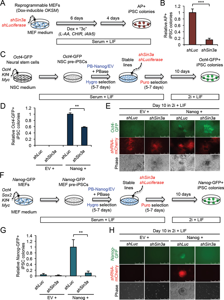
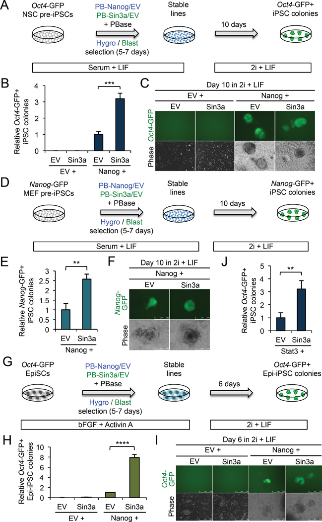
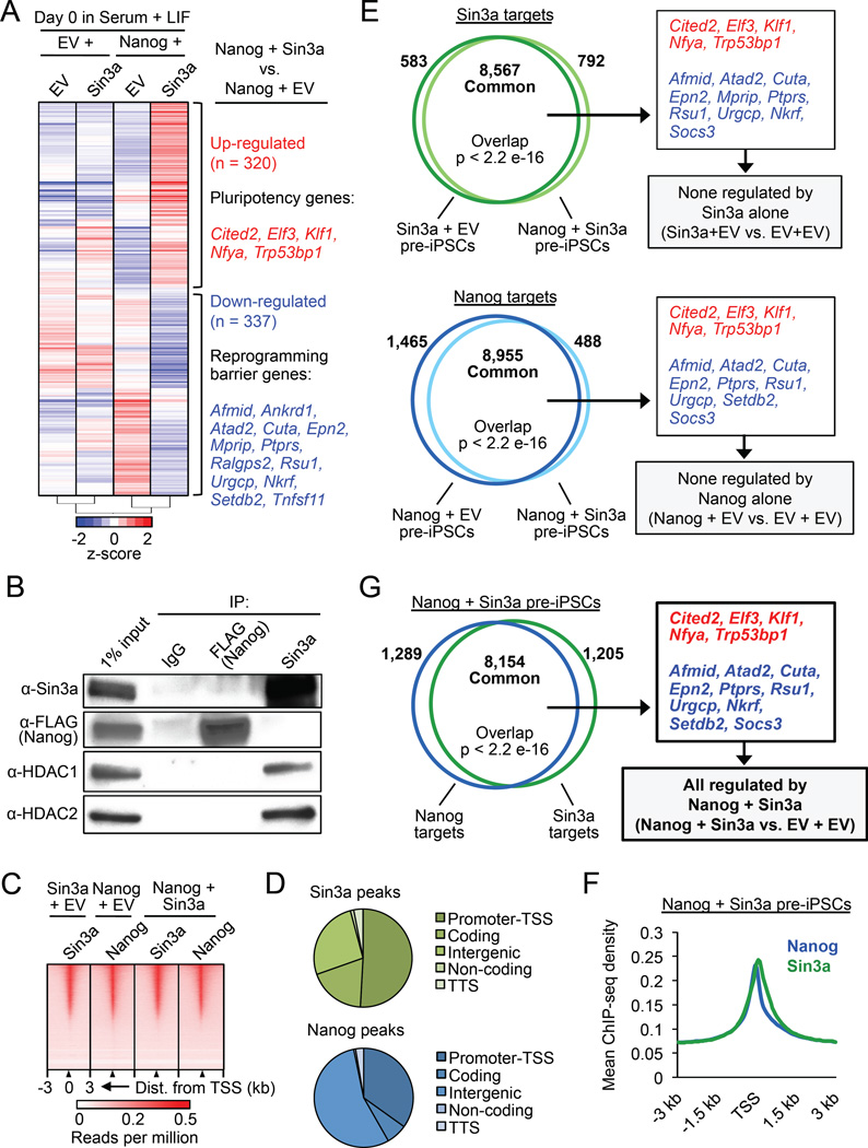
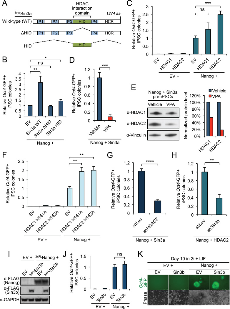
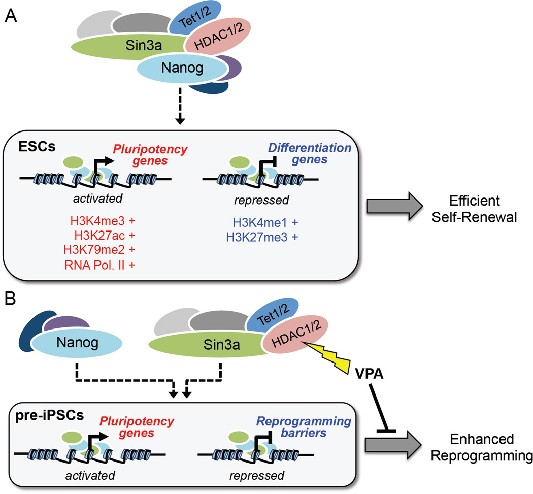
Although SIN3A is required for the survival of early embryos and embryonic stem cells (ESCs), the role of SIN3A in the maintenance and establishment of pluripotency remains unclear. Here, we find that the SIN3A/HDAC corepressor complex maintains ESC pluripotency and promotes the generation of induced pluripotent stem cells (iPSCs). Members of the SIN3A/HDAC corepressor complex are enriched in an extended NANOG interactome and function in transcriptional coactivation in ESCs. We also identified a critical role for SIN3A and HDAC2 in efficient reprogramming of somatic cells. Mechanistically, NANOG and SIN3A co-occupy transcriptionally active pluripotency genes in ESCs and also co-localize extensively at their genome-wide targets in pre-iPSCs. Additionally, both factors are required to directly induce a synergistic transcriptional program wherein pluripotency genes are activated and reprogramming barrier genes are repressed. Our findings indicate a transcriptional regulatory role for a major HDAC-containing complex in promoting pluripotency.

Keywords: ESCs; EpiSCs; HDAC1; HDAC2; SIN3A; SIN3B; TET1/2; VPA; iPSCs; pre-iPSCs.


Figures

Figure 1
Figure 1. Nanog and Sin3a co-occupy active promoters in ESCs. See also Figure S1
(A) IP/co-IP between FLAG-bioNanog and endogenous Sin3a in mouse ESCs. BirA = biotin ligase. (B) Sin3a and Nanog co-bind highly expressed pluripotency genes in ESCs. Shown are all Nanog and Sin3a common target genes in ESCs (n = 1,447) based on ChIP-seq data, sorted by high (red) to low (blue) ESC/MEF expression ratios based on RNA-seq data. Pluripotency genes and reprogramming barrier genes are listed in red and blue text, respectively. Blue heat maps on the right show enrichment of Nanog, the Sin3a/HDAC complex, and four chromatin marks centered on the TSS. (C) Percentages of active (RNA Pol. II bound + H3K79me2), non-productive (RNA Pol. II bound, no H3K79me2), and inactive (no RNA Pol. II bound, no H3K79me2) genes bound by each factor in ESCs, based on ChIP-seq data. “All genes” = genome-wide average of all genes, and numbers above chart indicate the number of target genes identified and analyzed for each individual factor or combination. (D) Illustration of Nanog conditional knockout (NgcKO) ESCs, wherein endogenous Nanog is deleted and cells are maintained by a doxycycline (Dox)-suppressible Nanog transgene. Protein expression upon 8-hour Dox treatment (1 µg/mL) is shown. (E) Nanog-dependent Sin3a binding to common target genes. ChIP-qPCR was performed for Sin3a and Nanog at two different peaks (P1, P2) in the Oct4 and Nanog promoters in NgcKO ESCs. Dox treatment (1 µg/mL) lasted for 8 hours. (F) Colony formation assay after Sin3a knockdown, individually or combined with Nanog conditional knockout (+Dox), in NgcKO ESCs.
Figure 2
Figure 2. Sin3a is required for efficient somatic cell reprogramming. See also Figure S2
(A) The procedure for assessing Sin3a knockdown in doxycycline (Dox)-inducible MEF reprogramming. (B) Sin3a knockdown significantly decreases MEF reprogramming efficiency. Data are presented as average fold change of AP+ iPSC colonies ± SD (n = 3; *** p < 0.001). (C) The procedure for assessing Sin3a knockdown in neural stem cell (NSC)-derived pre-iPSC reprogramming. (D) Sin3a knockdown significantly decreases Nanog-mediated NSC pre-iPSC reprogramming efficiency. Data are presented as average fold change of Oct4-GFP+ iPSC colonies ± SD (n = 3; ** p < 0.01). (E) Representative images of Oct4-GFP+ iPSC colonies. (F) The procedure for assessing Sin3a knockdown in MEF-derived pre-iPSC reprogramming. (G) Sin3a knockdown significantly decreases Nanog-mediated MEF pre-iPSC reprogramming efficiency. Data are presented as average fold change of Nanog-GFP+ iPSC colonies ± SD (n = 3; ** p < 0.01). (H) Representative images of Nanog-GFP+ iPSC colonies.
Figure 3
Figure 3. Sin3a over-expression promotes pre-iPSC and EpiSC reprogramming. See also Figure S3
(A) The procedure for assessing Sin3a over-expression in NSC-derived pre-iPSC reprogramming. (B) Sin3a can synergize with Nanog to significantly increase NSC-derived pre-iPSC reprogramming efficiency. Data are presented as average fold change of Oct4-GFP+ iPSC colonies ± SD (n = 3; *** p < 0.001). (C) Representative images of Oct4-GFP+ iPSC colonies. (D) The procedure for assessing Sin3a over-expression in MEF-derived pre-iPSC reprogramming. (E) Sin3a can synergize with Nanog to significantly increase MEF pre-iPSC reprogramming efficiency. Data are presented as average fold change of Nanog-GFP+ iPSC colonies ± SD (n = 3; ** p < 0.01) (F) Representative images of Nanog-GFP+ iPSC colonies. (G) The procedure for assessing Sin3a over-expression in EpiSC reprogramming. (H) Sin3a can synergize with Nanog to significantly increase EpiSC reprogramming efficiency. Data are presented as average fold change of Oct4-GFP+ Epi-iPSC colonies ± SD (n = 3; **** p < 0.0001). (I) Representative images of Oct4-GFP+ Epi-iPSC colonies. (J) Sin3a can synergize with Stat3 to significantly increase NSC-derived pre-iPSC reprogramming efficiency. Data are presented as average fold change of Oct4-GFP+ iPSC colonies ± SD (n = 3; ** p < 0.01)
Figure 4
Figure 4. Nanog and Sin3a directly activate pluripotency genes and repress reprogramming barrier genes in pre-iPSCs. See also Figure S4
(A) Microarray heat maps of significantly differentially expressed genes (p < 0.05, fold change ≥ 1.5) in NSC pre-iPSCs. Genes in red and blue are up- and down-regulated, respectively, in Nanog + Sin3a pre-iPSCs relative to Nanog + EV pre-iPSCs (right). (B) IP/co-IP for 3×FLAGNanog, Sin3a, HDAC1, and HDAC2 in Nanog + Sin3a NSC pre-iPSCs. (C) Heat maps for genome-wide binding of Nanog and/or Sin3a in indicated pre-iPSC populations. (D) Genome-wide distribution of Nanog and Sin3a peaks in Nanog + Sin3a pre-iPSCs. Promoter-TSS = −1 kb to +100 bp from transcription start site (TSS), coding = exons, TTS = −100 bp to +1 kb from transcription termination site (TTS). (E) Venn diagrams showing significantly overlapping Sin3a targets (top) and Nanog targets (bottom) in indicated pre-iPSC populations, as well as microarray genes of interest contained within common target sets (right). (F) Average ChIP-seq read density for Nanog and Sin3a in Nanog + Sin3a pre-iPSCs. (G) Venn diagram showing significant overlap of Nanog and Sin3a targets in Nanog + Sin3a pre-iPSCs (left), as well as microarray genes that are directly regulated by Nanog + Sin3a (right).
Figure 5
Figure 5. HDAC2 is critical for Nanog/Sin3a functional cooperation in NSC-derived pre-iPSC reprogramming. See also Figure S5
(A) Myc-tagged Sin3a constructs used for assessing the requirement of HDAC1/2 association for Sin3a function in reprogramming. (B) The Sin3a HID domain is critical for Sin3a function in synergizing with Nanog during pre-iPSC reprogramming. Data are presented as average fold change of Oct4-GFP+ iPSC colonies ± SD (n = 3; * p < 0.05, ** p < 0.01, ns = not significant). (C) HDAC2, and to a minimal extent HDAC1, can synergize with Nanog during pre-iPSC reprogramming. Data are presented as average fold change of Oct4-GFP+ iPSC colonies ± SD (n = 3; *** p < 0.001, ns = not significant). (D) VPA treatment (2 mM) completely abrogates the Nanog/Sin3a reprogramming synergy compared to vehicle treated control. Data are presented as average fold change of Oct4-GFP+ iPSC colonies ± SD (n = 3; *** p < 0.001). (E) VPA treatment (2 mM; 24 hours) causes degradation of HDAC1/2 proteins. Western blot band intensities were quantified using ImageJ software and are relative to Vinculin loading control. HDAC1/2 protein levels were normalized to a vehicle treated sample. (F) HDAC1/2 catalytic mutants can synergize with Nanog during pre-iPSC reprogramming. Data are presented as average fold change of Oct4-GFP+ iPSC colonies ± SD (n = 3; ** p < 0.01). (G) HDAC2 is required for efficient pre-iPSC reprogramming by Nanog + Sin3a. Data are presented as average fold change of Oct4-GFP+ iPSC colonies ± SD (n = 3; **** p < 0.0001). (H) Sin3a is required for efficient pre-iPSC reprogramming by Nanog + HDAC2. Data are presented as average fold change of Oct4-GFP+ iPSC colonies ± SD (n = 3; ** p < 0.01). (I) Western blot for pre-iPSCs over-expressing 3×FLSin3b and 3×FLNanog in serum + LIF conditions. (J) Co-expression of Sin3b and Nanog has no effect on pre-iPSC reprogramming efficiency compared to Nanog alone. Data are presented as average fold change of Oct4-GFP+iPSC colonies ± SD (n = 3; ns = not significant). (K) Representative images of Oct4-GFP+ iPSC colonies.
Figure 6
Figure 6. Proposed model of the Sin3a/HDAC complex in maintaining ESC self-renewal and promoting somatic cell reprogramming through functional cooperation with Nanog
(A) Nanog and the Sin3a/HDAC complex co-occupy promoter regions of highly expressed pluripotency genes in ESCs and cooperate to promote ESC self-renewal. (B) Nanog and the Sin3a/HDAC complex functionally cooperate in pre-iPSCs by directly activating pluripotency genes and repressing reprogramming barrier genes resulting in significantly enhanced reprogramming efficiency.

References

    1. Anokye-Danso F, Trivedi CM, Juhr D, Gupta M, Cui Z, Tian Y, Zhang Y, Yang W, Gruber PJ, Epstein JA, Morrisey EE. Highly efficient miRNA-mediated reprogramming of mouse and human somatic cells to pluripotency. Cell Stem Cell. 2011;8:376–388. - PMC - PubMed
    1. Baltus GA, Kowalski MP, Tutter AV, Kadam S. A positive regulatory role for the mSin3A–HDAC complex in pluripotency through Nanog and Sox2. J. Biol. Chem. 2009;284:6998–7006. - PMC - PubMed
    1. Costa Y, Ding J, Theunissen TW, Faiola F, Hore TA, Shliaha PV, Fidalgo M, Saunders A, Lawrence M, Dietmann S, Das S, Levasseur DN, Li Z, Xu M, Reik W, Silva JCR, Wang J. NANOG-dependent function of TET1 and TET2 in establishment of pluripotency. Nature. 2013;495:370–374. - PMC - PubMed
    1. Cowley SM, Iritani BM, Mendrysa SM, Xu T, Cheng PF, Yada J, Liggitt HD, Eisenman RN. The mSin3A chromatin-modifying complex is essential for embryogenesis and T-cell development. Molecular and Cellular Biology. 2005;25:6990–7004. - PMC - PubMed
    1. Das S, Jena S, Levasseur DN. Alternative splicing produces Nanog protein variants with different capacities for self-renewal and pluripotency in embryonic stem cells. Journal of Biological Chemistry. 2011;286:42690–42703. - PMC - PubMed

MeSH terms