Review
. 2016 Nov 1;40(6):875-893.
doi: 10.1093/femsre/fuw027.

Sensing the enemy, containing the threat: cell-autonomous immunity to Chlamydia trachomatis

Affiliations
Review

Sensing the enemy, containing the threat: cell-autonomous immunity to Chlamydia trachomatis

Ryan Finethy et al. FEMS Microbiol Rev. .

Abstract

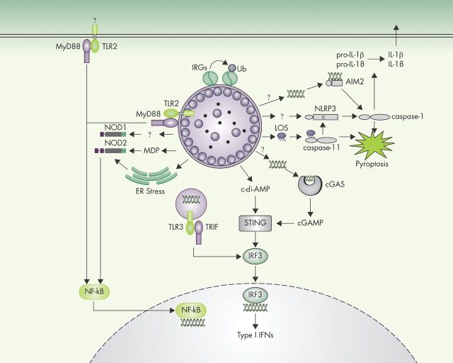
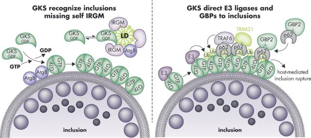
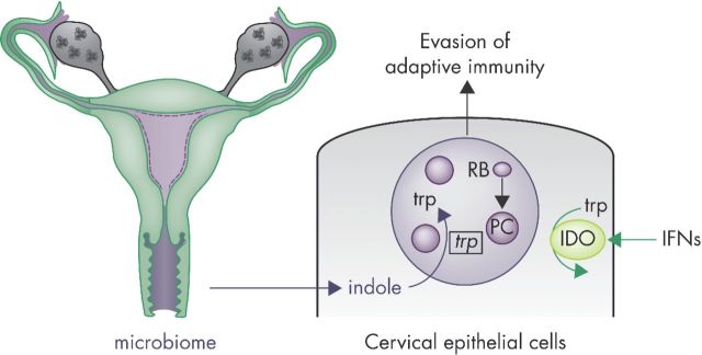
The bacterium Chlamydia trachomatis is the etiological agent of the most common sexually transmitted infection in North America and Europe. Medical complications resulting from genital C. trachomatis infections arise predominantly in women where the initial infections often remain asymptomatic and thus unrecognized. Untreated asymptomatic infections in women can ascend into the upper genital tract and establish persistence, ultimately resulting in extensive scarring of the reproductive organs, pelvic inflammatory disease, infertility and ectopic pregnancies. Previously resolved C. trachomatis infections fail to provide protective immune memory, and no effective vaccine against C. trachomatis is currently available. Critical determinants of the pathogenesis and immunogenicity of genital C. trachomatis infections are cell-autonomous immune responses. Cell-autonomous immunity describes the ability of an individual host cell to launch intrinsic immune circuits that execute the detection, containment and elimination of cell-invading pathogens. As an obligate intracellular pathogen C. trachomatis is constantly under attack by cell-intrinsic host defenses. Accordingly, C. trachomatis evolved to subvert and co-opt cell-autonomous immune pathways. This review will provide a critical summary of our current understanding of cell-autonomous immunity to C. trachomatis and its role in shaping host resistance, inflammation and adaptive immunity to genital C. trachomatis infections.

Keywords: Chlamydia trachomatis; TLR; inflammasome; interferon-inducible GTPases; indole-dioxygenase; STING.


Figures

Figure 1.
Figure 1.
Innate immune sensors of C. trachomatis infection. A wide variety of sensors collectively detect C. trachomatis infections and activate immune response pathways. Following the detection of unknown C. trachomatis-derived ligands in the extracellular or vacuolar milieu, TLR2 and TLR3 induce the transcription of cytokine-encoding genes. The cytosolic sensor STING directly detects the presence of bacterial cyclic di-AMP or—indirectly through cGAS—the presence of DNA in the host cell cytosol. STING activation leads to IRF3-dependent induction of type I IFN production. Cytosolic NOD1 and NOD2 detect bacterial cell-wall components, for example, muramyl dipeptide, and Chlamydia-induced ER stress to induce NF-κB activation and transcription of proinflammatory cytokines. Three distinct cytosolic surveillance pathways can induce inflammasome activation in response to C. trachomatis: AIM2, NLRP3 and caspase-11. Activated inflammasomes execute pyroptotic cell death, and IL-1β/IL-18 secretion. IRGs detect C. trachomatis inclusions in mouse cells and recruit host factors, promoting inclusion ubiquitination and ultimately leading to inclusion rupture.
Figure 2.
Figure 2.
Targeting of GKS proteins and GBPs to C. trachomatis inclusions in mouse cells. The GKS class of IRG proteins is guided towards inclusion membranes through a missing-self principle. The IRGM proteins Irgm1 and Irgm3 reside on ‘self’ membranes and organelles such as LDs and block GKS protein activation at these sites. The absence of these IRGM proteins from inclusions enables GTP-bound GKS dimers to form and associate with inclusion membranes. This association of GKS proteins with inclusions is further enhanced by the presence of lipidated Atg8 proteins at inclusion membranes (left panel). Inclusion-bound GKS proteins promote the recruitment of ‘pioneering’ ubiquitin E3 ligases (E3) and p62-interacting E3 ligases (TRAF6, TRIM21), which promote the decoration of inclusions with ubiquitin. Potential ubiquitination substrates are the GKS proteins themselves. The ubiquitin-binding protein p62 escorts GBP2 to inclusions (right panel). Additional p62-independent mechanisms of GBP recruitment exist (not shown). GKS-decorated inclusions rupture in a p62-dependent manner.
Figure 3.
Figure 3.
Chlamydia trachomatis responds to changes in the tryptophan metabolism of IFNγ-primed epithelial cells of the female genital tract. Chlamydia trachomatis colonizes columnar epithelial cells of the endocervix following infection of the female genital tract. Infections induce the expression of lymphocyte-derived IFNγ, which primes human epithelial cells to induce the expression of the tryptophan-catabolizing enzyme IDO. Depletion of intracellular tryptophan stores by IDO instructs C. trachomatis RBs to undergo a dramatic morphological transformation into ‘persister cells’ (PCs). PCs halt replication and drastically change their cell physiology. While still an untested hypothesis, these substantial changes may allow PCs to evade adaptive immune responses. As one of their adaptations to IFNγ priming, PCs upregulate expression of the trp operon and thereby activate an indole-scavenging pathway. Consuming indole—most likely derived from the vaginal mircobiome—enables C. trachomatis PCs to survive tryptophan-spent conditions within its host cell.

Similar articles

Cited by

References

    1. Abdelrahman YM, Belland RJ. The chlamydial developmental cycle. FEMS Microbiol Rev. 2005;29:949–59. - PubMed
    1. Abdelsamed H, Peters J, Byrne GI. Genetic variation in Chlamydia trachomatis and their hosts: impact on disease severity and tissue tropism. Future Microbiol. 2013;8:1129–46. - PMC - PubMed
    1. Abdul-Sater AA, Koo E, Hacker G, et al. Inflammasome-dependent caspase-1 activation in cervical epithelial cells stimulates growth of the intracellular pathogen Chlamydia trachomatis. J Biol Chem. 2009;284:26789–96. - PMC - PubMed
    1. Abdul-Sater AA, Said-Sadier N, Padilla EV, et al. Chlamydial infection of monocytes stimulates IL-1beta secretion through activation of the NLRP3 inflammasome. Microbes Infect. 2010;12:652–61. - PMC - PubMed
    1. Abdul-Sater AA, Tattoli I, Jin L, et al. Cyclic-di-GMP and cyclic-di-AMP activate the NLRP3 inflammasome. EMBO Rep. 2013;14:900–6. - PMC - PubMed

Publication types

MeSH terms