Review
. 2016 Nov 1;40(6):894-937.
doi: 10.1093/femsre/fuw026.

Behind the lines-actions of bacterial type III effector proteins in plant cells

Affiliations
Review

Behind the lines-actions of bacterial type III effector proteins in plant cells

Daniela Büttner. FEMS Microbiol Rev. .

Abstract

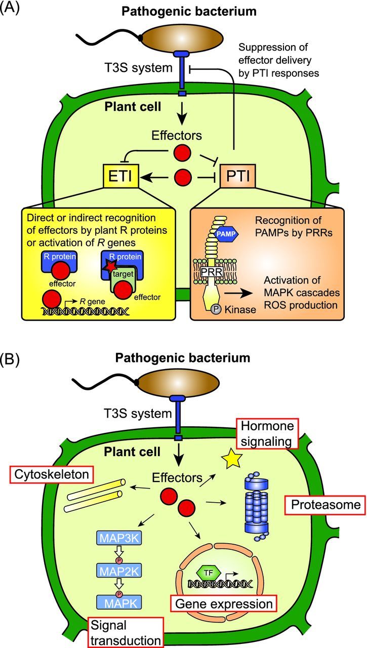
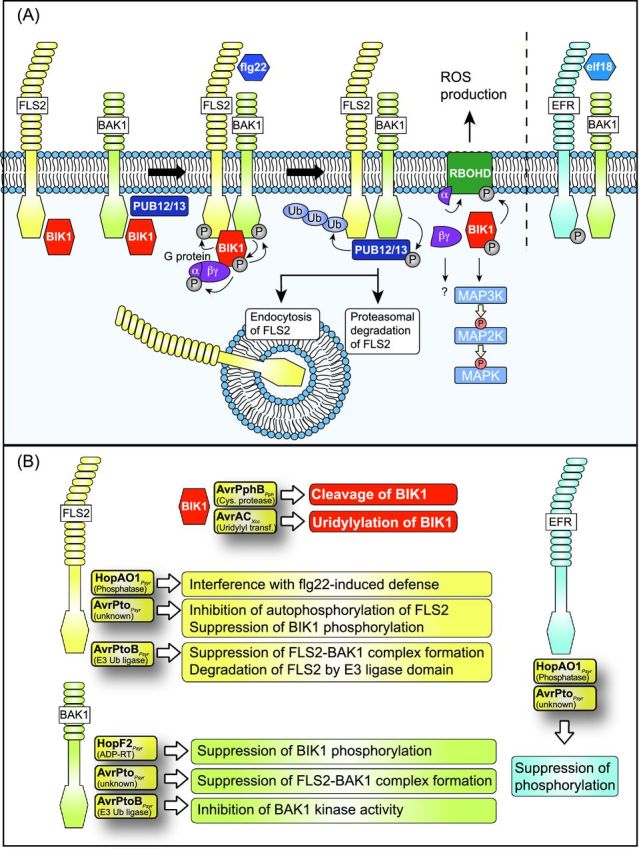
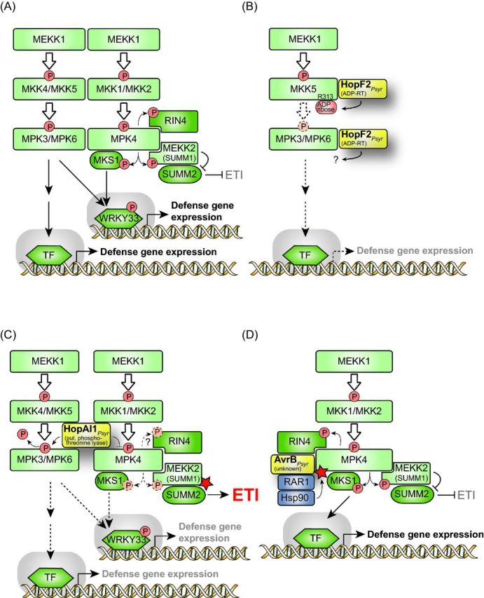
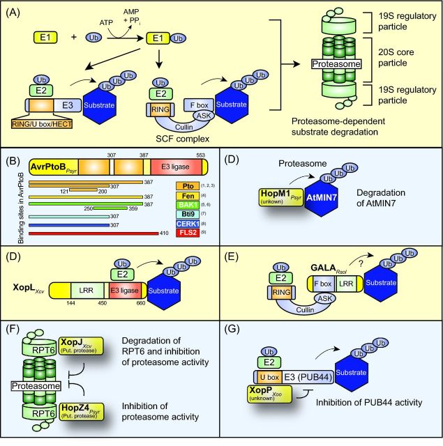
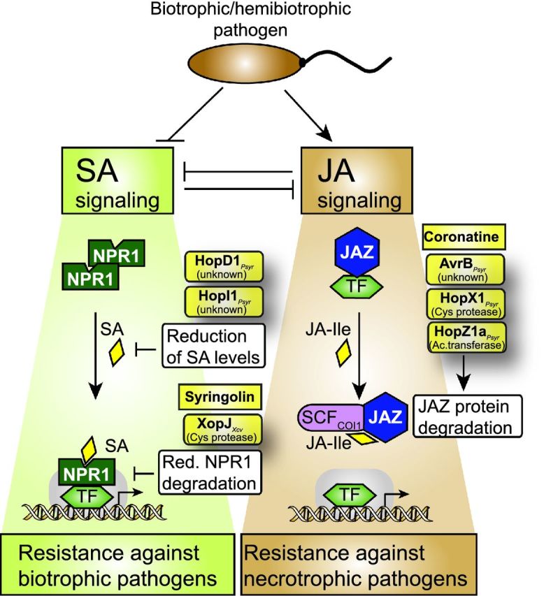
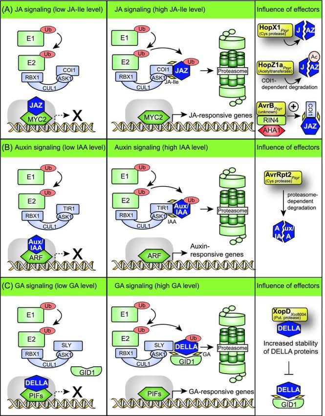
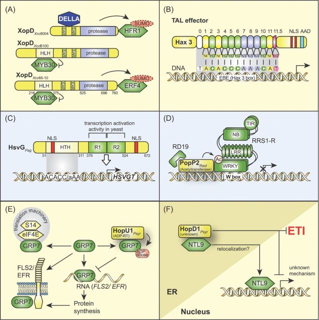
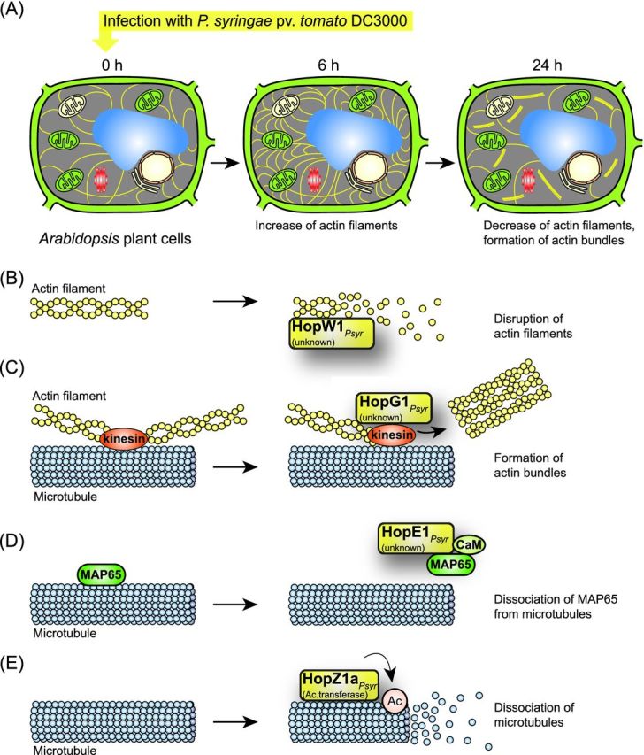
Pathogenicity of most Gram-negative plant-pathogenic bacteria depends on the type III secretion (T3S) system, which translocates bacterial effector proteins into plant cells. Type III effectors modulate plant cellular pathways to the benefit of the pathogen and promote bacterial multiplication. One major virulence function of type III effectors is the suppression of plant innate immunity, which is triggered upon recognition of pathogen-derived molecular patterns by plant receptor proteins. Type III effectors also interfere with additional plant cellular processes including proteasome-dependent protein degradation, phytohormone signaling, the formation of the cytoskeleton, vesicle transport and gene expression. This review summarizes our current knowledge on the molecular functions of type III effector proteins with known plant target molecules. Furthermore, plant defense strategies for the detection of effector protein activities or effector-triggered alterations in plant targets are discussed.

Keywords: type III effector; plant immunity; MAPK signaling; proteasome; cytoskeleton; phytohormones.


Figures

Figure 1.
Figure 1.
Interference of type III effector proteins with plant cellular pathways. (A) Type III effectors elicit and suppress plant defense reponses. Plant-pathogenic bacteria translocate effector proteins via the T3S system into plant cells. In resistant plants, individual effectors are directly or indirectly recognized by corresponding plant R proteins or activate plant R genes, and elicit defense responses, which are designated ETI. The indirect recognition of effector proteins by plant R proteins depends on effector-mediated modifications of plant target proteins, which are sensed by matching R proteins. Several type III effectors suppress ETI and/or PTI responses and thus promote bacterial virulence. PTI responses are triggered upon recognition of bacterial PAMPs by plant PRRs (see the text for details), and do not only interfere with pathogen survival but can also affect T3S-mediated delivery of effector proteins. (B) Overview on known plant targets of type III effectors. Translocated effector proteins interfere with the assembly of the cytoskeleton, MAPK cascades, gene expression, proteasome-dependent protein degradation or hormone signaling pathways (see the text for details).
Figure 2.
Figure 2.
Interference of type III effectors with FLS2- and EFR-dependent signaling pathways during PTI. (A) Schematic model of signaling pathways triggered by the flagellin receptor FLS2 and the EF-Tu receptor EFR. FLS2 and EFR consist of a cytoplasmic kinase, a transmembrane and an extracellular LRR domain and insert into the plant plasma membrane. Signaling by FLS2 and EFR involves the transmembrane kinase BAK1, which associates with FLS2 and EFR upon binding of their corresponding ligands. FLS2 and BAK1 are associated with the kinase BIK1, which is phosphorylated by BAK1 upon flg22 perception by FLS2 and subsequently phosphorylates FLS2 and BAK1. BIK1 also phosphorylates an associated heterotrimeric G protein, which stabilizes BIK1 and dissociates after flg22 perception. The Gα subunit interacts with the NADPH oxidase RBOHD, which is involved in ROS production. flg22 perception also leads to the dissociation of BIK1 from the FLS2/BAK1 complex. BIK1 subsequently phosphorylates RBOHD and activates MAPK signaling pathways. FLS2-associated BAK1 phosphorylates the E3 ubiquitin ligases PUB12 and PUB13, which interact with and ubiquitinate FLS2. The proteasomal degradation of FLS2 and the endocytosis of FLS2 after flg22 perception presumably prevent the constitutive activation of FLS2-mediated PTI responses. flg22, peptide of flagellin; elf18, peptide of elongation factor EF-Tu. (B) As indicated, several type III effectors target FLS2, BAK1, BIK1 and EFR. Known effector-triggered alterations in PRRs, BAK1 and BIK1 are listed in boxes (see the text for details). Contradictory data were published about the interaction of AvrPto with BAK1 (see the text for details).
Figure 3.
Figure 3.
Influence of type III effectors on MAPK signaling pathways. (A) Schematic overview on MAPK signaling pathways involved in plant defense responses. During plant defense responses, two MAPK signaling pathways are activated which involve (i) MPK3/MPK6 and MKK4/MKK5 and (ii) MPK4 and MKK1/MKK2, respectively. The MP2Ks of both pathways are activated by the MP3K MEKK1, however, MPK3 and MPK6 can also be activated independently of MEKK1 (Suarez-Rodriguez et al.2007). The activation of MAPKs directly or indirectly leads to the release of transcription factors (TFs), which trigger the expression of defense genes. Known substrates of MPK4 are MKS1, RIN4 and the MP3K MEKK2 (also designated SUMM1). Phosphorylation of MKS1 by MPK4 leads to the release of the MKS1-bound TF WRKY33, which subsequently activates gene expression. MPK4 also phosphorylates the MAP3K MEKK2 and presumably results in its inactivation. MPK4-mediated inactivation of MEKK2 leads to the suppression of ETI responses triggered by the CC-NB-LRR R protein SUMM2, which likely guards MEKK2 (see the text for details). (B) HopF2 from P. syringae interferes with MPK3/MPK6 signaling pathway. The mono-ADP-RT HopF2 ADP-ribosylates and thus inactivates MKK5 and suppresses the MPK3/MPK6-mediated signaling pathway (indicated by dashed arrows). HopF2 also interacts with MPK6, yet, the outcome of this interaction is unknown. (C) The putative phosphothreonine lyase HopAI1 from P. syringae inhibits the activity of MAPKs. HopAI1 dephosphorylates MPK3 and MPK6 and thus interferes with the MPK3/MPK6 signaling pathway. Furthermore, HopAI1 suppresses the kinase activity of MPK4 and thus the phosphorylation of the MPK4 substrates MKS1 and MEKK2. It has not yet been shown whether HopAI1 also interferes with the MPK4-mediated phosphorylation of RIN4 (indicated by a dashed arrow and a questionmark). The loss of MPK4 activity leads to the activation of MEKK2 (indicated by a red asterisk), which in turn triggers SUMM2-mediated ETI (see the text for details). (D) AvrB from P. syringae activates MPK4. AvrB interacts with MPK4 and leads to its phosphorylation and thus activation. The efficient interaction between AvrB and MPK4 depends on RAR1, which presumably acts as a linker between AvrB and Hsp90. Hsp90 promotes the activity of MPK4 as is indicated by a red asterisk (see the text for details).
Figure 4.
Figure 4.
Contribution of type III effectors to proteasome-dependent protein degradation. (A) Model of the proteasome-dependent protein degradation pathway. Ubiquitin (Ub) is activated by the ubiquitin-activating enzyme (E1) and transferred to the ubiquitin-conjugating enzyme (E2), which interacts with the ubiquitin ligase (E3). E3 ubiquitin ligases are divided into several classes according to the presence of a HECT, RING or U-box domain. RING domain-containing E3 ubiquitin ligases can be part of a multimeric protein complex such as the SCF complex, which consists of a RING-box protein, the molecular scaffold protein cullin, an Arabidopsis SKP1-like protein (ASK1) and an F-box protein, which binds the substrate of the E3 ligase. E3 enzymes mediate the transfer of ubiquitin molecules to the substrate, thus leading to the formation of poly-ubiquitin chains, which allow the targeting of proteins to the proteasome. The proteasome consists of two 19S regulatory and a 20S core particle and catalyzes the unfolding and degradation of polyubiquitinated proteins. (B) Domain structure of the effector protein AvrPtoB from P. syringae. The regions of AvrPtoB, which provide binding sites for the AvrPtoB interaction partners Pto, Bti9, Fen, FLS2 and BAK1, are indicated. References: (1) Dong et al. ; (2) Xiao et al. ; (3) Mathieu, Schwizer and Martin ; (4) Rosebrock et al. ; (5) Shan et al. ; (6) Cheng et al. ; (7) Zeng et al. ; (8) Gimenez-Ibanez et al. ; (9) Göhre et al.. Experimental evidence for the presence of two Pto-binding sites in AvrPtoB (indicated as orange boxes) was reported by Mathieu, Schwizer and Martin (2014). Numbers refer to amino acid positions in AvrPtoB. (C) HopM1 from P. syringae induces the degradation of its interaction partner AtMIN7. The HopM1-mediated degradation of AtMIN7 depends on the activity of the proteasome. (D) The effector protein XopL from X. campestris pv. vesicatoria triggers the ubiquitination of plant proteins. XopL contains an N-terminal LRR and a C-terminal E3 ubiquitin ligase domain. The plant targets of XopL are unknown. Numbers refer to amino acid positions in XopL. (E) GALA proteins from R. solancearum contain an F-box domain and were proposed to associate with components of the SCF complex. In agreement with this hypothesis, an interaction between GALA proteins and ASK proteins has been shown. A contribution of GALA proteins to the ubiquitination of substrates of the SCF complex remains to be demonstrated. (F) XopJ from Xanthomonas spp. and HopZ4 from P. syringae interact with the proteasome subunit RPT6 and suppress the activity of the proteasome. XopJ leads to the degradation of RPT6. (G) XopP from X. oryzae pv. oryzae interacts with the U box E3 ubiquitin ligase PUB44 from rice and inhibits its activity.
Figure 5.
Figure 5.
Interference of type III effectors with SA and JA signaling pathways. SA-dependent defense responses are required for plant resistance against biotrophic pathogens whereas JA-dependent defense is mounted against necrotrophic pathogens. SA and JA pathways thus act antagonistically and can suppress each other. Type III effectors from biotrophic or hemibiotrophic pathogens activate JA signaling pathways and suppress SA-mediated defense by the actions of translocated type III effectors. SA-dependent defense responses depend on NPR1 (non-expressor of PR genes), which is present in an oligomeric inactive state in the absence of SA. Upon binding of SA, monomeric NPR1 binds to and activates transcription factors (TF) and thus induces the expression of SA-dependent genes (Gimenez-Ibanez and Solano 2013). The effectors HopD1 and HopI1 from P. syringae lead to reduced SA levels whereas the bacterial toxin syringolin and the effector XopJ from X. campestris pv. vesicatoria interfere with the degradation of NPR1. The turnover of NPR1 is required for the expression of SA-responsive genes. Stabilization of NPR1, therefore, suppresses SA signaling (Robert-Seilaniantz, Grant and Jones 2011). JA signaling pathways involve JAZ proteins and the SCF complex. The bacterial toxin coronatine and the effector proteins AvrB, HopX1 and HopZ1 from P. syringae lead to the degradation of JAZ proteins and thus activate the expression of JA-responsive genes (see the text for details).
Figure 6.
Figure 6.
Modulation of JA, auxin and GA signaling pathways by type III effectors. (A) HopX1, HopZ1a and AvrB from P. syringae interfere with JA signaling pathways. Bioactive JA-Ile promotes the interaction between JAZ proteins and the F-box protein COI1, which is a component of the SCF complex. The subsequent degradation of JAZ proteins leads to the release of JAZ-interacting transcription factors (e.g. MYC2), which activate the expression of JA-responsive genes. The cysteine protease HopX1 directly or indirectly degrades several JAZ proteins independently of the JA receptor COI1 and thus activates the expression of JA-responsive genes. The acetyltransferase HopZ1a acetylates JAZ proteins and leads to their proteasome-dependent degradation. The effector protein AvrB from P. syrinage interacts with RIN4, which is a negative regulator of PTI and associates with the H+ ATPase AHA1. The interaction of AvrB with the RIN4-AHA1 complex promotes the interaction between JAZ proteins and COI1 and leads to the activation of JA-responsive genes. (B) Auxin signaling pathways are targeted by the cysteine protease AvrRpt2 from P. syringae. IAA promotes the interaction between Aux/IAA proteins and the F-box protein TIR1. This leads to the proteasome-dependent degradation of Aux/IAA proteins and to the release and activation of ARFs. ARFs subsequently activate the expression of auxin-responsive genes. The cysteine protease AvrRpt2 directly or indirectly induces the degradation of Aux/IAA proteins by the proteasome and thus activates the expression of auxin-responsive genes. (C) XopD from X. campestris pv. campestris strain 8004 interferes with the stability of DELLA proteins, which are negative regulators of GA-responsive genes. GA-dependent signaling is controlled by DELLA proteins, which inactivate PIF (phytochrome interacting factors) transcription factors. Binding of GA to its receptor GID1 leads to a conformational change in GID1, which subsequently binds to DELLA proteins. The formation of a GID1-DELLA complex promotes the interaction between DELLA proteins and the F-box protein SLY and thus the proteasome-dependent degradation of DELLA proteins. This leads to the release of PIF transcription factors, which activate the expression of GA-responsive genes. XopDXcc8004 presumably interferes with the binding of GID1 to DELLA proteins and delays the GA-induced degradation of the DELLA protein RGA. Notably, however, an influence of XopDXcc8004 on the transcription of GA-responsive genes has not yet been detected.
Figure 7.
Figure 7.
Interference of type III effector proteins with plant gene expression. (A) Domain organization of XopD proteins from Xanthomonas spp. XopD family members consist of a C-terminal cysteine protease domain and N-terminal EAR motifs. Additionally, XopD from X. campestris pv. vesicatoria strain 85-10 (XopDXcv85-10) and X. campestris pv. campestris strain B100 (XopDXccB100) contain N-terminal extensions and a central putative DNA-binding HLH domain. XopDXcc8004 was shown to interact with and stabilize DELLA proteins via the EAR motif-containing region. Furthermore, XopDXcc8004 interacts with and deSUMOylates the transcription factor HFR1. XopDXccB100 and XopDXcv85-10 bind to the transcription factor MYB30 via the HLH domain and suppress its transcriptional activity. XopDXcv deSUMOylates and thus destabilizes the transcription factor ERF4. Numbers refer to amino acid positions in XopDXcv85-10. (B) Domain organization and DNA-binding specificity of the TAL effector Hax (homolog of AvrBs3 in Xanthomonas) 3 from X. campestris pv. armoraciae. TAL effectors contain a C-terminal acidic activation domain (AAD), two NLSs and a central protein region with repeats. The RVDs of Hax3 and the matching bases in the EBE in the promoter regions of Hax3-induced genes are indicated. (C) Domain organization of HsvG from P. agglomerans (Pag). HsvG contains N- and C-terminal NLSs, an N-terminal HTH region and two repeats of 71 and 74 amino acids (R1 and R2), which confer transcription activation activity in yeast. The repeats determine the specificity of plant gene activation (indicated by a white arrow) but are dispensable for DNA binding of HsvG, which depends on the N-terminal region. Numbers refer to amino acid positions in HsvG. (D) Modification of RRS1-R by the effector protein PopP2 from R. solanacearum. The TIR-NB-LRR protein RRS1-R forms a dimer and binds via its WRKY domain to a DNA motif (W box) present in promoters of target genes of WRKY transcription factors. PopP2 interacts with and acetylates the WRKY domain of RRS1-R and thus interferes with its DNA binding. The additional PopP2 interaction partner RD19, which is a predicted protease, is presumably not acetylated by PopP2. RRS1-R also interacts with the R protein RPS4, which is required for the induction of ETI and is not shown in this figure (see the text for details). (E) The mono-ADP-RT HopU1 from P. syringae pv. tomato DC3000 ADP-ribosylates the RNA-binding protein GRP7, which interacts with components of the translational machinery including the cap-binding protein eIF4E and the ribosomal subunit S14. GRP7 also interacts with the PRRs FLS2 and EFR and with FLS2 and EFR transcripts, and was, therefore, assumed to promote PRR translation. ADP-ribosylation of GRP7 by HopU1 reduces the ability of GRP7 to bind to RNA and might suppress FLS2 and EFR protein synthesis. (F) HopD1 from P. syringae pv. tomato DC3000 interacts with the transcription factor NTL9 at the ER and leads to reduced expression of NTL9-induced genes during ETI. Furthermore, HopD1 suppresses ETI responses. The mechanisms underlying the HopD1-mediated inhibition of NTL9-dependent gene expression are unknown.
Figure 8.
Figure 8.
Influence of type III effectors on actin filaments and microtubules. (A) Infection of Arabidopsis cells with P. syringae pv. tomato DC3000 leads to alterations in the actin cytoskeleton. The infection with wild-type or T3S mutant strains leads to an increase in actin filaments 6 hours post infection. Twenty-four hours post infection, the wild-type strain induces the formation of actin bundles and leads to a reduced number of actin filaments. Actin filaments and bundles are indicated as yellow dashes. The plant cell wall is represented in green. The following cell organelles are shown: chloroplasts (green), mitochondria (beige), vacuole (blue), nucleus (beige), ER (light brown) and Golgi apparatus (red). (B) HopW1 leads to the disruption of actin filaments. The effector protein HopW1 binds to filamentous actin and leads to the disruption of actin filaments. (C) HopG1 induces the formation of actin bundles. HopG1 binds to a mitochondrial-localized kinesin motor protein, which associates with microtubules and presumably links microtubules to actin filaments. HopG1 induces the formation of actin bundles, presumably via its interaction with kinesin. (D) HopE1 leads to the dissociation of MAP65 from microtubules. HopE1 interacts with calmodulin (CaM) and the microtubule-associated protein MAP65 and leads to the dissociation of MAP65 from microtubules. No effect of HopE1 on the microtubule network was observed. (E) HopZ1a from P. syringae dissociates microtubules. The acetyltransferase HopZ1a binds to and acetylates tubulin and disrupts microtubules.
Figure 9.
Figure 9.
Activation of R protein-mediated defense responses by type III effectors. (A) Detection of effector protein-triggered modifications in plant target proteins by R proteins. Type III effectors interact with and modify plant target proteins to promote bacterial virulence. According to the guard model, plant R proteins in resistant plants detect effector-triggered modifications (indicated by yellow asterisks) in plant target molecules. Many R proteins consist of CC/TIR, NB and LRR domains. The activity of the NB domain is often regulated by intramolecular interactions with the LRR domain. The detection of effector-triggered modifications in plant targets leads to intramolecular rearrangements in the R proteins and thus to the activation of the NB domain (indicated by a red asterisk). According to an alternative model, plants have evolved decoy molecules, which resemble virulence targets of effectors but do not contribute to bacterial virulence. Effector-mediated modifications in plant decoys can also lead to the activation of R protein-mediated resistance. (B) Model of the activation of RRS1-R/RPS4-triggered plant defense responses. The TIR-NB-LRR proteins RPS4 and RRS1-R form a heterodimer in the plant nucleus, and RRS1-R negatively regulates RPS4. The WRKY domain of RRS1-R binds to DNA and presumably triggers the RRS1-R-dependent expression of plant genes. The effector proteins PopP2 from R. solanacearum and AvrRps4 from P. syringae interact with the WRKY domain of RRS1-R, which is acetylated by PopP2. Interactions with effectors and/or acetylation by PopP2 lead to molecular rearrangements in the RPS4/RRS1-R immune receptor complex and thus to the activation of RPS4 (indicated by a red asterisk). It has been suggested that the WRKY domain of RRS1-R acts as an integrated decoy, which allows the elicitation of AvrRps4- and PopP2-triggered ETI responses. The activated complex of RRS1-R and RPS4 was proposed to be a tetramer.
Figure 10.
Figure 10.
Model of Prf/Pto-triggered activation of plant defense responses. (A) AvrPtoB triggers Prf/Pto-dependent ETI responses. The CC-NB-LRR protein Prf contains an SD (Solanaceae domain) domain with weak homology to other solanaceous R proteins and an N-terminal domain (N), which interacts with the Pto kinase and mediates the Pto-independent dimerization of Prf (Gutierrez et al.2010). The Prf/Pto complex contains at least two molecules of each protein. Pto is autophosphorylated at S198 and required to maintain Prf in an inactive conformation. The sensor Pto molecule interacts with and phosphorylates the E3 ubiquitin ligase AvrPtoB from P. syringae and evades AvrPtoB-mediated degradation. Effector binding to the P+1 loop of the Pto sensor presumably triggers a conformational change in this loop, which activates the Pto helper protein. The Pto helper molecule transphosphorylates the sensor at amino acid residue T199. Transphosphorylation of the sensor Pto leads to the activation of Prf (indicated by a red asterisk) and thus to Prf/Pto-triggered ETI responses. (B) A kinase-inactive Pto helper molecule (PtoD164N) does not transphosphorylate the kinase-active Pto sensor and therefore does not activate Prf/Pto-dependent ETI responses.
Figure 11.
Figure 11.
Effector-triggered modifications of RIN4 and their contributions to PTI and ETI responses. (A) Domain organization of RIN4 and list of known plant interaction partners of RIN4. RIN4 contains N- and C-terminal NOI domains with conserved PxFGxW and Y/FTxxF amino acid motifs. The PxFGxW motif is the cleavage site of the effector protein AvrRpt2 from P. syringae. The Y/FTxxF motif contains a conserved threonine residue, which is phosphorylated by the effector proteins AvrB and AvrRpm1 from P. syringae. Additional important amino acids are indicated (see the text for details). Known plant interaction partners of RIN4 and their predicted functions are listed. References: 1, Liu et al. (2009); 2, Li et al. (2014b); 3, Day, Dahlbeck and Staskawicz (2006); 4, Cui et al. (2010); 5, Liu et al. (2011); 6, Mackey et al. (2002); 7, Axtell and Staskawicz (2003); 8, Luo et al. (2009). (B) Contribution of RIN4 to RPS2- and RPM1-triggered ETI responses. The ADP-RT HopF2 from P. syringae ADP-ribosylates RIN4 and suppresses the degradation of RIN4 by the cysteine protease AvrRpt2 from P. syringae. RIN4 is also degraded by AvrPto from P. syringae in the presence of Pto and Prf. The cleavage products of RIN4 are detected by the R protein RPS2, which triggers ETI. The effector proteins AvrRpm1 and AvrB from P. syringae lead to the phosphorylation of RIN4 and thus to the activation of the R protein RPM1 (indicated by a red asterisk). Effector-triggered phosphorylation of RIN4 presumably depends on the kinase RIPK, which interacts with RIN4 and AvrB and phosphorylates RIN4 at several amino acid residues including T166. The phosphorylation of T166 of RIN4 interferes with the interaction of RIN4 with the cyclophilin ROC1, which catalyzes the cis/trans isomerization of the proline residue at position 149 of RIN4. The cis/trans isomerization of P149 presumably leads to a specific conformational change in RIN4 and thus inhibits the activation of RPM1 and RPS2. Phosphorylation of RIN4 in the presence of AvrB likely induces conformational changes, which interfere with ROC1-mediated isomerization of RIN4 and lead to the activation of RPM1 (see the text for details). (C) Model of the role of RIN4 during PTI. RIN4 is a suppressor of PTI responses and interacts with and activates the H+ ATPases AHA1 and AHA2, which pump protons from the cytosol of guard cells into the apoplast. The activity of AHA1/2 leads to the establishment of a proton electrochemical gradient, which is used by channel and carrier proteins to mediate the uptake of ions into guard cells, thus leading to stomatal opening. Upon detection of flg22 during PTI, RIN4 is phosphorylated at amino acid S141, which leads to the derepression (= activation) of PTI. The phosphorylation of RIN4 at amino acid T166 by AvrB and AvrRpm1 from P. syringae presumably counteracts the effect of the S141 phosphorylation and restores the repression of PTI (see the text for details).
Figure 12.
Figure 12.
Detection of effector-triggered modifications in plant target molecules by the R proteins RPS5 and ZAR1. (A) RPS5 activates ETI upon cleavage of PBS1. The CC-NB-LRR protein RPS5 interacts via the CC domain with the kinase PBS1. Cleavage of PBS1 by the effector protein and cysteine protease AvrPphB leads to the activation of RPS5. Exchange of the AvrPphB cleavage site (indicated by a yellow triangle) against the recognition site of the cysteine protease AvrRpt2 (indicated by an orange triangle) leads to cleavage of PBS1 in the presence of AvrRpt2 and thus to the activation of RPS5. (B) The R protein ZAR1 detects modifications in ZED1 and PBL2. The CC-NB-LRR protein ZAR1 interacts via the CC domain with the pseudokinase ZED1. Acetylation of ZED1 by the effector protein HopZ1a leads to the activation of ZAR1 (indicated by a red asterisk) and thus to ETI responses. ZAR1 also interacts via the LRR domain with the ZED1-related pseudokinase RKS1. The ZED1-RKS1 complex detects modifications in PBL2, which is a target of the uridylyltransferase AvrAC from X. campestris pv. campestris (see the text for details). Uridylylation of PBL2 by AvrAC leads to the activation of ZAR1-dependent ETI responses.

Similar articles

Cited by

References

    1. Abramovitch RB, Janjusevic R, Stebbins CE, et al. Type III effector AvrPtoB requires intrinsic E3 ubiquitin ligase activity to suppress plant cell death and immunity. P Natl Acad Sci USA. 2006;103:2851–6. - PMC - PubMed
    1. Abramovitch RB, Kim YJ, Chen S, et al. Pseudomonas type III effector AvrPtoB induces plant disease susceptibility by inhibition of host programmed cell death. EMBO J. 2003;22:60–9. - PMC - PubMed
    1. Ade J, DeYoung BJ, Golstein C, et al. Indirect activation of a plant nucleotide binding site-leucine-rich repeat protein by a bacterial protease. P Natl Acad Sci USA. 2007;104:2531–6. - PMC - PubMed
    1. Afzal AJ, da Cunha L, Mackey D. Separable fragments and membrane tethering of Arabidopsis RIN4 regulate its suppression of PAMP-triggered immunity. Plant Cell. 2011;23:3798–811. - PMC - PubMed
    1. Afzal AJ, Kim JH, Mackey D. The role of NOI-domain containing proteins in plant immune signaling. BMC Genomics. 2013;14:327. - PMC - PubMed

Publication types

MeSH terms

Substances