. 2017 Apr 1;19(4):546-557.
doi: 10.1093/neuonc/now231.

Comprehensive molecular characterization of multifocal glioblastoma proves its monoclonal origin and reveals novel insights into clonal evolution and heterogeneity of glioblastomas

Affiliations

Comprehensive molecular characterization of multifocal glioblastoma proves its monoclonal origin and reveals novel insights into clonal evolution and heterogeneity of glioblastomas

Khalil Abou-El-Ardat et al. Neuro Oncol. .

Abstract

Background: The evolution of primary glioblastoma (GBM) is poorly understood. Multifocal GBM (ie, multiple synchronous lesions in one patient) could elucidate GBM development.

Methods: We present the first comprehensive study of 12 GBM foci from 6 patients using array-CGH, spectral karyotyping, gene expression arrays, and next-generation sequencing.

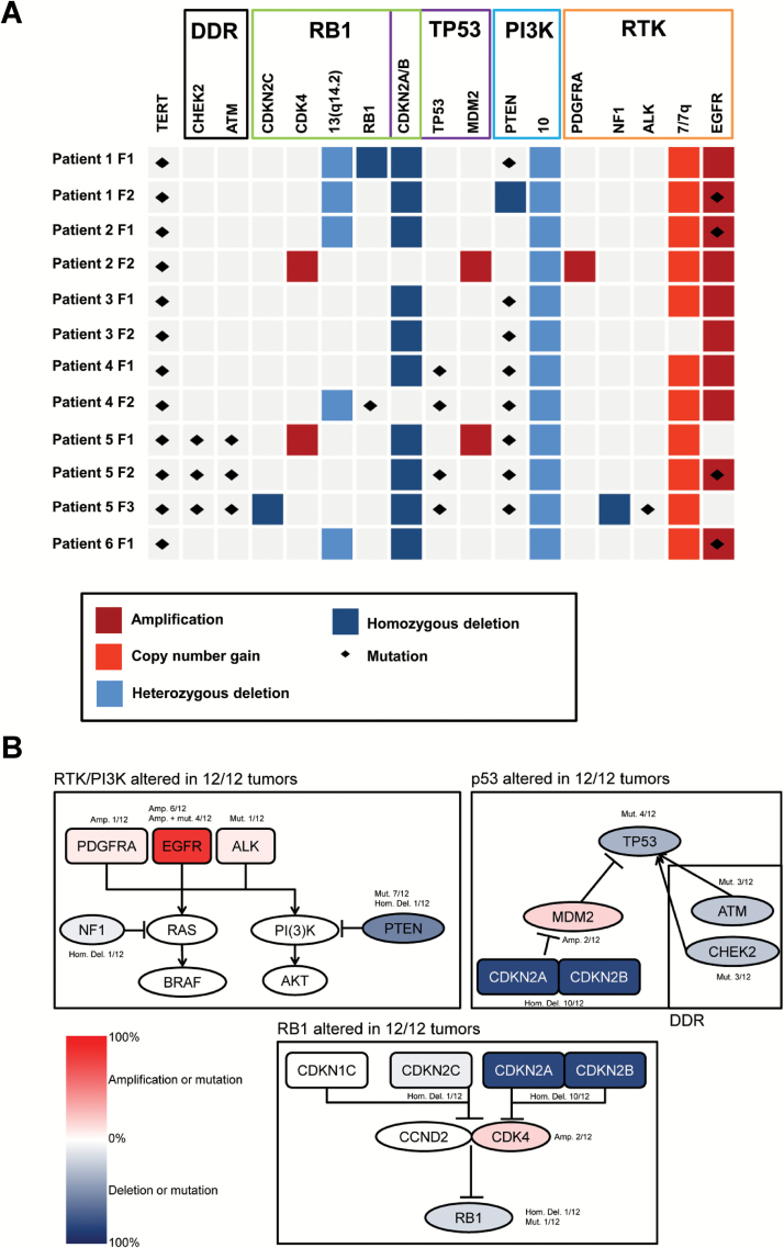
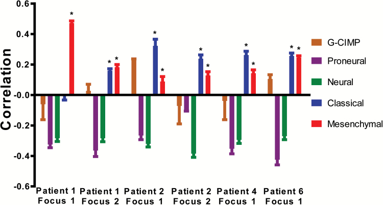
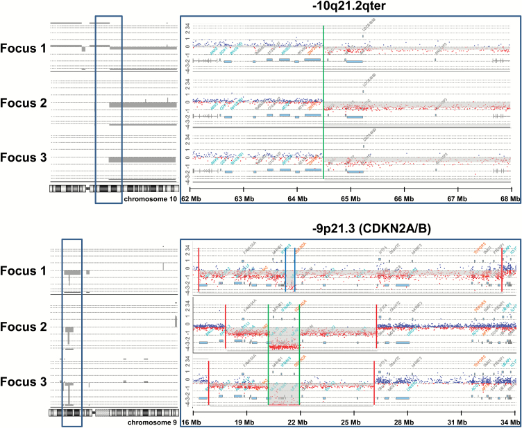
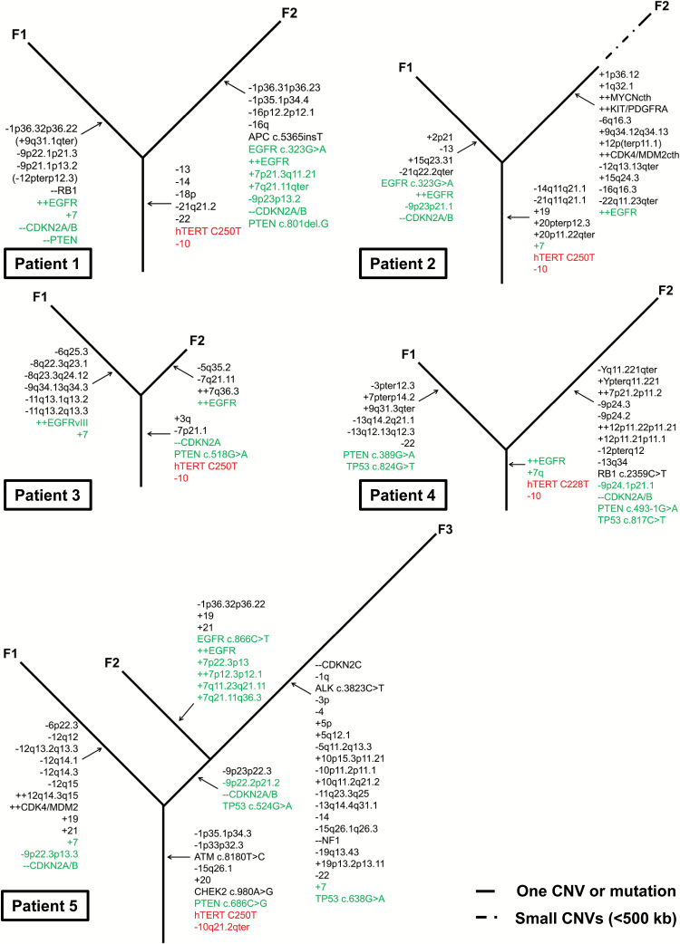
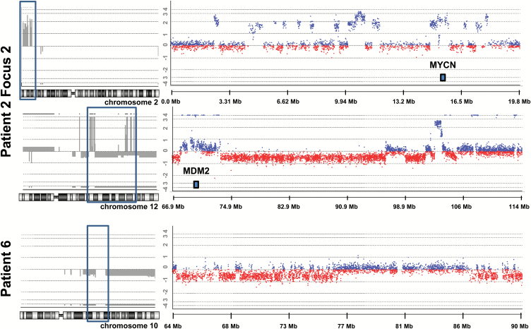
Results: Multifocal GBMs genetically resemble primary GBMs. Comparing foci from the same patient proved their monoclonal origin. All tumors harbored alterations in the 3 GBM core pathways: RTK/PI3K, p53, and RB regulatory pathways with aberrations of EGFR and CDKN2A/B in all (100%) patients. This unexpected high frequency reflects a distinct genetic signature of multifocal GBMs and might account for their highly malignant and invasive phenotype. Surprisingly, the types of mutations in these genes/pathways were different in tumor foci from the same patients. For example, we found distinct mutations/aberrations in PTEN, TP53, EGFR, and CDKN2A/B, which therefore must have occurred independently and late during tumor development. We also identified chromothripsis as a late event and in tumors with wild-type TP53. Only 2 events were found to be early in all patients: single copy loss of PTEN and TERT promoter point mutations.

Conclusions: Multifocal GBMs develop through parallel genetic evolution. The high frequency of alterations in 3 main pathways suggests that these are essential steps in GBM evolution; however, their late occurrence indicates that they are not founder events but rather subclonal drivers. This might account for the marked genetic heterogeneity seen in primary GBM and therefore has important implications for GBM therapy.

Keywords: monoclonal origin; multifocal glioblastoma; tumor evolution; tumor genetics; tumor heterogeneity.


Figures

Fig. 1
Fig. 1
Most common aberrations and affected pathways in 12 multifocal glioblastomas (GBMs) from 6 patients: (A) Most common copy number variations (CNVs) and mutations in our cohort per tumor focus (F) and patient (Y-axis) and pathway (X-axis). (B) Frequencies of copy number variations (CNVs) and mutations mapped to 3 core signaling pathways (framed in black). Abbreviations: Amp, amplification; DDR, DNA damage response; Hom. Del., homozygous deletion; Mut., mutation; RTK, receptor tyrosine kinase.
Fig. 2
Fig. 2
Association of gene expression profiles of 6 foci from 4 patients with the expression subtypes according to the Verhaak classification. Tumors were significantly associated with the Classical and Mesenchymal GBM subtypes (positive correlation) but not with the Proneural, G-CIMP, or Neural subtypes. Correlation scores (Y-axis) ranging from 1 (highly correlated) to −1 (highly anticorrelated) are obtained per sample for normalization with 3 different normal brain RNAs. Bars represent the median of correlation scores; error bars denote the interquartile range; * indicates significant correlations (P < .05).
Fig. 3
Fig. 3
Shared and different copy number variations (CNVs) between 3 tumor foci from patient 5 as detected by array-comparative genome hybridization (array-CGH). Above: Copy number loss of the long arm of chromosome 10 with identical breakpoints in all 3 foci from patient 5. The breakpoint area bordered by the blue frame is enlarged on the right and shows the identical breakpoint in all 3 foci at position [hg19]10q21.2(64,485,714) (indicated by green line). Below: Copy number loss in 9p (log-2-ratio approximately −1) and homozygous deletion of the area containing CDKN2A/B (log-2 ratio approximately −2) (indicated in grey on the left, enlargement of the region on the right). The breakpoints for the homozygous deletion of CDKN2A/B are identical between focus 1 and focus 2 (arr[hg19]9p21.3(21,531,275-22,086,857)x0 indicated by inner green lines) but dissimilar for focus 3 (arr[hg19]9p21.3(21,983,069-22,125,464)x0; blue lines). The region for loss on 9p (indicated by red lines) was different in all 3 foci, indicating 3 different independent and late events in tumor evolution.
Fig. 4
Fig. 4
Evolution in 5 patients with multifocal glioblastoma (GBM): A phylogenetic dendrogram showing the genetic evolution based on the shared and different alterations in the tumor foci from each patient. Loss of chromosome 10 and TERT promoter mutation were the only events that were shared between tumor foci in all patients (red). Frequent affected genes/regions considered important for mGBM development are marked in green. The dashed line in patient 2 focus 2 indicates the presence of numerous (14) small duplications and deletions (<500 kb). Abbreviations: -, deletion; -- , homozygous deletion; +, gain; ++, amplification; F, focus.
Fig. 5
Fig. 5
Chromothripsis in 2 tumors using array-comparative genome hybridization (array-CGH). A total of 34 breaks (17 copy number variations (CNVs) between 2 states (0 and 2) were observed on the short arm of chromosome 2 in focus 2 from patient 2 (topmost on the left framed in blue and enlarged on the right). The long arm of chromosome 12 in the same patient carried a total of 64 breaks (32 CNVs between 3 copy states: −1, 0, and 4; middle figure framed in blue on the left and enlarged on the right). Amplifications of MYCN (chromosome 2) and MDM2 (chromosome 12) are indicated in the figure. The tumor from patient 6 showed a large number of CNVs between 2 copy number states (0 and −1) involving loss on chromosome 10 (bottommost framed in blue on the left and enlarged on the right).

Comment in

Similar articles

Cited by

  • Multifocal glioblastoma-two case reports and literature review.
    Zhang ZX, Chen JX, Shi BZ, Li GH, Li Y, Xiang Y, Qin X, Yang L, Lv SQ. Zhang ZX, et al. Chin Neurosurg J. 2021 Jan 15;7(1):8. doi: 10.1186/s41016-020-00223-z. Chin Neurosurg J. 2021. PMID: 33446281 Free PMC article.
  • Integrated molecular and multiparametric MRI mapping of high-grade glioma identifies regional biologic signatures.
    Hu LS, D'Angelo F, Weiskittel TM, Caruso FP, Fortin Ensign SP, Blomquist MR, Flick MJ, Wang L, Sereduk CP, Meng-Lin K, De Leon G, Nespodzany A, Urcuyo JC, Gonzales AC, Curtin L, Lewis EM, Singleton KW, Dondlinger T, Anil A, Semmineh NB, Noviello T, Patel RA, Wang P, Wang J, Eschbacher JM, Hawkins-Daarud A, Jackson PR, Grunfeld IS, Elrod C, Mazza GL, McGee SC, Paulson L, Clark-Swanson K, Lassiter-Morris Y, Smith KA, Nakaji P, Bendok BR, Zimmerman RS, Krishna C, Patra DP, Patel NP, Lyons M, Neal M, Donev K, Mrugala MM, Porter AB, Beeman SC, Jensen TR, Schmainda KM, Zhou Y, Baxter LC, Plaisier CL, Li J, Li H, Lasorella A, Quarles CC, Swanson KR, Ceccarelli M, Iavarone A, Tran NL. Hu LS, et al. Nat Commun. 2023 Sep 28;14(1):6066. doi: 10.1038/s41467-023-41559-1. Nat Commun. 2023. PMID: 37770427 Free PMC article.
  • Natural Coevolution of Tumor and Immunoenvironment in Glioblastoma.
    Wu L, Wu W, Zhang J, Zhao Z, Li L, Zhu M, Wu M, Wu F, Zhou F, Du Y, Chai RC, Zhang W, Qiu X, Liu Q, Wang Z, Li J, Li K, Chen A, Jiang Y, Xiao X, Zou H, Srivastava R, Zhang T, Cai Y, Liang Y, Huang B, Zhang R, Lin F, Hu L, Wang X, Qian X, Lv S, Hu B, Zheng S, Hu Z, Shen H, You Y, Verhaak RGW, Jiang T, Wang Q. Wu L, et al. Cancer Discov. 2022 Dec 2;12(12):2820-2837. doi: 10.1158/2159-8290.CD-22-0196. Cancer Discov. 2022. PMID: 36122307 Free PMC article.
  • Survival and recurrence patterns of multifocal glioblastoma after radiation therapy.
    Syed M, Liermann J, Verma V, Bernhardt D, Bougatf N, Paul A, Rieken S, Debus J, Adeberg S. Syed M, et al. Cancer Manag Res. 2018 Oct 4;10:4229-4235. doi: 10.2147/CMAR.S165956. eCollection 2018. Cancer Manag Res. 2018. PMID: 30323678 Free PMC article.
  • Molecular and Circulating Biomarkers in Patients with Glioblastoma.
    Senhaji N, Squalli Houssaini A, Lamrabet S, Louati S, Bennis S. Senhaji N, et al. Int J Mol Sci. 2022 Jul 5;23(13):7474. doi: 10.3390/ijms23137474. Int J Mol Sci. 2022. PMID: 35806478 Free PMC article. Review.

References

    1. Ohgaki H, Kleihues P. The definition of primary and secondary glioblastoma. Clin Cancer Res. 2013;19(4):764–772. - PubMed
    1. Brennan CW, Verhaak RG, McKenna A, et al. ; TCGA Research Network. The somatic genomic landscape of glioblastoma. Cell. 2013;155(2):462–477. - PMC - PubMed
    1. Verhaak RG, Hoadley KA, Purdom E, et al. ; Cancer Genome Atlas Research Network. Integrated genomic analysis identifies clinically relevant subtypes of glioblastoma characterized by abnormalities in PDGFRA, IDH1, EGFR, and NF1. Cancer Cell. 2010;17(1):98–110. - PMC - PubMed
    1. Sturm D, Witt H, Hovestadt V, et al. Hotspot mutations in H3F3A and IDH1 define distinct epigenetic and biological subgroups of glioblastoma. Cancer Cell. 2012;22(4):425–437. - PubMed
    1. Patel AP, Tirosh I, Trombetta JJ, et al. Single-cell RNA-seq highlights intratumoral heterogeneity in primary glioblastoma. Science. 2014;344(6190):1396–1401. - PMC - PubMed