. 2017 Feb 15;17(1):35.
doi: 10.1186/s12866-017-0944-3.

Diffusible substances from lactic acid bacterial cultures exert strong inhibitory effects on Listeria monocytogenes and Salmonella enterica serovar enteritidis in a co-culture model

Affiliations

Diffusible substances from lactic acid bacterial cultures exert strong inhibitory effects on Listeria monocytogenes and Salmonella enterica serovar enteritidis in a co-culture model

Solomon H Mariam et al. BMC Microbiol. .

Abstract

Background: Food-borne infections cause huge economic and human life losses. Listeria monocytogenes and Salmonella enterica serovar Enteritidis are among the top ranking pathogens causing such losses. Control of such infections is hampered by persistent contamination of foods and food-processing environments, resistance of pathogens to sanitizing agents, existence of heterogeneous populations of pathogens (including culturable and viable but non-culturable cells) within the same food items, and inability to detect all such pathogens by culture-based methods. Modern methods such as flow cytometry allow analyses of cells at the single cell level within a short time and enable better and faster detection of such pathogens and distinctions between live and dead cells. Such methods should be complemented by control strategies including the use of beneficial bacteria that produce metabolites capable of inhibiting food-borne pathogens. In this study, broth cultures of lactic acid bacteria (LAB) isolated from fermented milk were tested for production of substances capable of inhibiting L. monocytogenes and S. Enteritidis in co-culture with LAB by assessment of colony-forming units (CFU) and live:dead cell populations by flow cytometry.

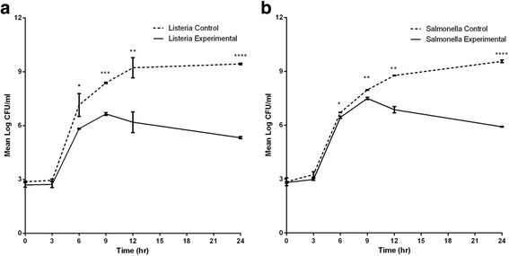
Results: The LAB isolates belonged to the species Lactococcus lactis, Enterococcus faecalis and Enterococcus faecium. Some LAB were effective in inhibition. Plating indicated up to 99% reduction in CFU from co-cultures compared to control cultures. Most of the bacteria in both cultures were in the viable but non-culturable state. The flow data showed that there were significantly higher dead cell numbers in co-cultures than in control cultures, indicating that such killing was caused by diffusible substances produced by the LAB cultures.

Conclusion: This study showed that metabolites from selected local LAB species can be used to significantly reduce pathogen load. However, conditions of use and application need to be further investigated and optimized for large-scale utilization.

Keywords: Co-culture; Inhibition; L. monocytogenes; Lactic acid bacteria; S. Enteritidis; Viable but nonculturable.


Figures

Fig. 1
Fig. 1
Hypervariable regions V1 to V5 (sequence 1 and sequence 2) of 16S rRNA gene amplified with universal primers for species level identification of LAB isolates used in this study
Fig. 2
Fig. 2
Gating strategy to identify and quantitate total and dead bacteria. Cultured L.monocytogenes or S. Enteritidis were either untreated (Panels b, c) or heat-killed (d, e), diluted in PBS, and counting beads and propidium iodide (Panels b through e) added prior to acquisition. Bacteria were defined as events present within gate P1, and were distinct from events in the PBS buffer + beads control (Panel a). Counting beads were utilized as an internal control to normalize acquisition volume between samples, and were identified in gate P2. Dead cells were defined in gate P3 as propidium iodide-fluorescent positive events among P1-gated cells
Fig. 3
Fig. 3
a 16S rRNA nucleotide sequence alignment of Lactococcus lactis isolates S2 and S6 with the sequence of Lactococcus lactis subsp. lactic ATCC 19435 (upper and middle panels) and with Lactococcus lactis subsp. lactis IL1403 (lower panel) (nucleotide differences are highlighted). b 16S rRNA nucleotide sequence alignment of Enterococcus faecium isolate S11 with the sequence of Enterococcus faecium type strains ATCC 700221, ATCC 19434, CECT 410 T and DSM 20477 (nucleotide differences are highlighted)
Fig. 4
Fig. 4
Log10CFU values from control cultures and co-cultures of L. monocytogenes (a) and S. Enteritidis (b) plated at 3 h intervals from Transwell control cultures and co-cultures. Hour 0 equals CFU values immediately after inoculation of cultures. The results at each time point are the means ± standard deviations for 3–4 replicate cultures. Asterisks indicate significantly higher CFU numbers of control cultures than those of co-cultures (experimental). *, P ≤ 0.05; **, P ≤ 0.01; ***, P ≤ 0.001; ****, P ≤ 0.0001
Fig. 5
Fig. 5
Representative results from flow cytometry analyses of dead cell staining of co-culture and control cultures. Upper panel: L.monocytogenes were either co-cultured with LAB (a, b) or cultured without LAB (control, c, d), and evaluated for dead cells (identified within P3) by PI staining as defined in Fig. 1. Dead cells (within P3) comprised 9.1% of P1- gated L.monocytogenes from co-cultures (b), whereas only 1.8% of P1-gated L.monocytogenes cells were identified as dead cells in control cultures (d). Lower panel: S. Enteritidis were either co-cultured with LAB (a, b) or cultured without LAB (control, c, d), and evaluated for dead cells (identified within P3) by PI staining as defined in Fig. 1. Dead cells (within P3) comprised 91% of P1-gated S. Enteritidis from co-cultures (b), whereas 60% of P1-gated S. Enteritidis cells were identified as dead cells in control cultures (d)

References

    1. Freitag NE, Port GC, Miner MD. Listeria monocytogenes—from saprophyte to intracellular pathogen. Nat Rev Microbiol. 2009;7:623–8. doi: 10.1038/nrmicro2171. - DOI - PMC - PubMed
    1. Pan Y, Breidt F, Kathariou S. Resistance of Listeria monocytogenes biofilms to sanitizing agents in a simulated food processing environment. Appl Environ Microbiol. 2006;72:7711–7. doi: 10.1128/AEM.01065-06. - DOI - PMC - PubMed
    1. Müller A, Rychli K, Muhterem-Uyar M, Zaiser A, Stessl B, Guinane CM, et al. Tn6188 - a novel transposon in Listeria monocytogenes responsible for tolerance to benzalkonium chloride. PLoS One. 2013;8:e76835. doi: 10.1371/journal.pone.0076835. - DOI - PMC - PubMed
    1. Ferreira V, Wiedmann M, Teixeira P, Stasiewicz MJ. Listeria monocytogenes persistence in food-associated environments: epidemiology, strain characteristics, and implications for public health. J Food Prot. 2014;77:150–70. doi: 10.4315/0362-028X.JFP-13-150. - DOI - PubMed
    1. Hitchins AD, Jinneman K. Detection and enumeration of Listeria monocytogenes in foods. Bacteriological analytical manual online. Washington: FDA; 2011.

MeSH terms

LinkOut - more resources