. 2017 Feb 15;12(1):34.
doi: 10.1186/s13023-017-0588-2.

Non-optic glioma in adults and children with neurofibromatosis 1

Affiliations

Non-optic glioma in adults and children with neurofibromatosis 1

Laura Sellmer et al. Orphanet J Rare Dis. .

Abstract

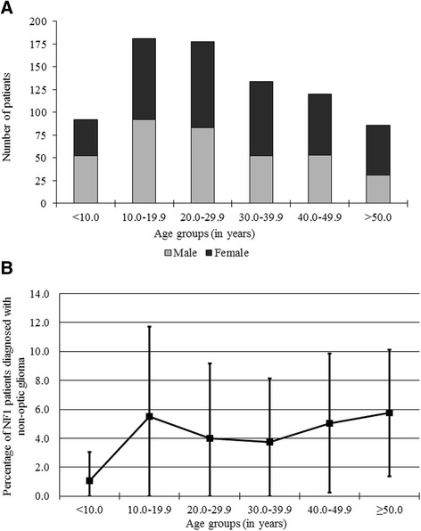
Background: Non-optic gliomas occur in 5% of children with NF1, but little is known about these tumours in adults. We aimed to investigate progression, spontaneous regression and the natural history of non-optic gliomas in adults and compare these findings to the results found in children.

Results: One thousand seven hundred twenty-two brain MRI scans of 562 unselected individuals with NF1 were collected at the NF outpatient department of the University Hospital Hamburg-Eppendorf between 2003 and 2015. The number of scans per patient ranged from one to 12; patients were followed for a median of 3.7 years. We identified 24 patients (4.3%) with non-optic gliomas. Median age at first scan with glioma was 21.2 years, much higher than in previous publications. Only seven of the 24 non-optic glioma patients were symptomatic. Five of 24 patients had multiple non-optic gliomas. Four individuals developed a new tumour, and 4 cases showed progression. The risk of new tumour development was 0.19% (95% confidence interval 0.06% to 0.52%) per patient year of follow-up for patients over 10 years. The rate of progressing non-optic gliomas per patient year of follow-up in the first 5 years after tumour diagnosis was 4.7% (95% confidence interval 1.5% to 12%).

Conclusions: Non-optic gliomas are twice as common in an unselected cohort of NF1 patients as previously reported. This is likely due to increased frequency of diagnosis of asymptomatic tumours when routine MRIs are performed and a higher prevalence in older individuals.

Keywords: Adults; Children; Cohort study; Glioma; Neurofibromatosis 1; Prospective.


Figures

Fig. 1
Fig. 1
Demographics and percentage of NF1 patients affected by non-optic glioma per age group. a Number of male and female NF1 patients per age group. Each person is counted once per age group, regardless of number of scans in that age group. Individuals may appear in more than one age group if scanned in more than one age range. b The overall prevalence of non-optic glioma appears stable in adulthood. Each person is counted once per age group, regardless of number of scans in that age group. Individuals may appear in more than one age group if scanned in more than one age range. Error bars are 1.96 standard deviations of a Poisson distribution
Fig. 2
Fig. 2
Newly-appearing glioma in the left cerebral peduncle in Patient 19. a There is no visible glioma on the patient’s first scan. b 4 years later, an enhancing glioma has appeared in the left cerebral peduncle measuring 0.5 cm3. c 5 years after the initial glioma-free scan, the glioma has increased to a volume of 0.8 cm3. d 7 years after the initial scan, the glioma measured 1.3 cm3. All images shown are FLAIR sequences. The patient remained asymptomatic during follow-up and also has an optic glioma (not visible in these images)
Fig. 3
Fig. 3
Progressing glioma in Patient 9. a First scan with glioma present in the left cerebral peduncle and left thalamus on FLAIR sequence. b Another scan performed 2 years after the previous one (again FLAIR sequence). The glioma has drastically increased in size and was treated with chemotherapy 1 month after this image was taken

Similar articles

Cited by

  • Expanding the clinical phenotype of individuals with a 3-bp in-frame deletion of the NF1 gene (c.2970_2972del): an update of genotype-phenotype correlation.
    Koczkowska M, Callens T, Gomes A, Sharp A, Chen Y, Hicks AD, Aylsworth AS, Azizi AA, Basel DG, Bellus G, Bird LM, Blazo MA, Burke LW, Cannon A, Collins F, DeFilippo C, Denayer E, Digilio MC, Dills SK, Dosa L, Greenwood RS, Griffis C, Gupta P, Hachen RK, Hernández-Chico C, Janssens S, Jones KJ, Jordan JT, Kannu P, Korf BR, Lewis AM, Listernick RH, Lonardo F, Mahoney MJ, Ojeda MM, McDonald MT, McDougall C, Mendelsohn N, Miller DT, Mori M, Oostenbrink R, Perreault S, Pierpont ME, Piscopo C, Pond DA, Randolph LM, Rauen KA, Rednam S, Rutledge SL, Saletti V, Schaefer GB, Schorry EK, Scott DA, Shugar A, Siqveland E, Starr LJ, Syed A, Trapane PL, Ullrich NJ, Wakefield EG, Walsh LE, Wangler MF, Zackai E, Claes KBM, Wimmer K, van Minkelen R, De Luca A, Martin Y, Legius E, Messiaen LM. Koczkowska M, et al. Genet Med. 2019 Apr;21(4):867-876. doi: 10.1038/s41436-018-0269-0. Epub 2018 Sep 7. Genet Med. 2019. PMID: 30190611 Free PMC article.
  • Increased prevalence of brain tumors classified as T2 hyperintensities in neurofibromatosis 1.
    Griffith JL, Morris SM, Mahdi J, Goyal MS, Hershey T, Gutmann DH. Griffith JL, et al. Neurol Clin Pract. 2018 Aug;8(4):283-291. doi: 10.1212/CPJ.0000000000000494. Neurol Clin Pract. 2018. PMID: 30140579 Free PMC article.
  • Brain tumors in Neurofibromatosis type 1.
    Costa AA, Gutmann DH. Costa AA, et al. Neurooncol Adv. 2019 Oct 26;1(1):vdz040. doi: 10.1093/noajnl/vdz040. eCollection 2019 May-Dec. Neurooncol Adv. 2019. PMID: 32642668 Free PMC article. Review.
  • A Systematic Review of Diagnostic Modalities and Strategies for the Assessment of Complications in Adult Patients with Neurofibromatosis Type 1.
    Rana S, Low CE, Karthikeyan M, Koh MJA, Ngeow J, Chiang J. Rana S, et al. Cancers (Basel). 2024 Mar 11;16(6):1119. doi: 10.3390/cancers16061119. Cancers (Basel). 2024. PMID: 38539455 Free PMC article. Review.
  • The molecular landscape of glioma in patients with Neurofibromatosis 1.
    D'Angelo F, Ceccarelli M, Tala, Garofano L, Zhang J, Frattini V, Caruso FP, Lewis G, Alfaro KD, Bauchet L, Berzero G, Cachia D, Cangiano M, Capelle L, de Groot J, DiMeco F, Ducray F, Farah W, Finocchiaro G, Goutagny S, Kamiya-Matsuoka C, Lavarino C, Loiseau H, Lorgis V, Marras CE, McCutcheon I, Nam DH, Ronchi S, Saletti V, Seizeur R, Slopis J, Suñol M, Vandenbos F, Varlet P, Vidaud D, Watts C, Tabar V, Reuss DE, Kim SK, Meyronet D, Mokhtari K, Salvador H, Bhat KP, Eoli M, Sanson M, Lasorella A, Iavarone A. D'Angelo F, et al. Nat Med. 2019 Jan;25(1):176-187. doi: 10.1038/s41591-018-0263-8. Epub 2018 Dec 10. Nat Med. 2019. PMID: 30531922 Free PMC article.

References

    1. Lammert M, Friedman JM, Kluwe L, Mautner V-F. Prevalence of neurofibromatosis 1 in German children at elementary school enrollment. Arch Dermatol. 2005;141:71–4. doi: 10.1001/archderm.141.1.71. - DOI - PubMed
    1. Neurofibromatosis 1 [http://www.ncbi.nlm.nih.gov/books/NBK1109/]. Access date 14 Feb 2017.
    1. Evans GD, O’Hara C, Wilding A, Ingham SL, Howard E, Dawson J, Moran A, Scott-Kitching V, Holt F, Huson SM. Mortality in neurofibromatosis 1: in North West England: an assessment of actuarial survival in a region of the UK since 1989. Eur J Hum Genet. 2011;19:1187–91. doi: 10.1038/ejhg.2011.113. - DOI - PMC - PubMed
    1. Rasmussen SA, Yang Q, Friedman JM. Mortality in neurofibromatosis 1: an analysis using U.S. death certificates. Am J Hum Genet. 2001;68:1110–8. doi: 10.1086/320121. - DOI - PMC - PubMed
    1. Gutmann DH, Rasmussen SA, Wolkenstein P, MacCollin M, Guha A, Inskip PD, North KN, Poyhonen M, Birch PH, Friedman JM. Gliomas presenting after age 10 in individuals with neurofibromatosis type 1 (NF1) Neurology. 2002;59:759–61. doi: 10.1212/WNL.59.5.759. - DOI - PubMed

Publication types