

Variability of radiation doses of cardiac diagnostic imaging tests: the RADIO-EVINCI study (RADIationdOse subproject of the EVINCI study)
- PMID: 28202051
- PMCID: PMC5311725
- DOI: 10.1186/s12872-017-0474-9
Variability of radiation doses of cardiac diagnostic imaging tests: the RADIO-EVINCI study (RADIationdOse subproject of the EVINCI study)
Abstract
Background: Patients with coronary artery disease can accumulate significant radiation dose through repeated exposures to coronary computed tomographic angiography, myocardial perfusion imaging with single photon emission computed tomography or positron emission tomography, and to invasive coronary angiography. Aim of the study was to audit radiation doses of coronary computed tomographic angiography, single photon emission computed tomography, positron emission tomography and invasive coronary angiography in patients enrolled in the prospective, randomized, multi-centre European study-EVINCI (Evaluation of Integrated Cardiac Imaging for the Detection and Characterization of Ischemic Heart Disease).
Methods: We reviewed 1070 tests (476 coronary computed tomographic angiographies, 85 positron emission tomographies, 310 single photon emission computed tomographies, 199 invasive coronary angiographies) performed in 476 patients (mean age 60 ± 9 years, 60% males) enrolled in 12 centers of the EVINCI. The effective doses were calculated in milli-Sievert (mSv) as median, interquartile range (IQR) and coefficient of variation of the mean.
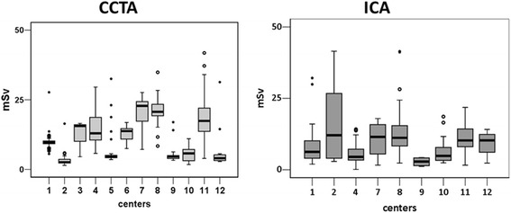
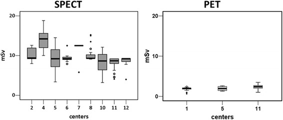
Results: Coronary computed tomographic angiography (476 exams in 12 centers) median effective dose was 9.6 mSv (IQR = 13.2 mSv); single photon emission computed tomography (310 exams in 9 centers) effective dose was 9.3 (IQR = 2.8); positron emission tomography (85 in 3 centers) effective dose 1.8 (IQR = 1.6) and invasive coronary angiography (199 in 9 centers) effective dose 7.4 (IQR = 7.3). Inter-institutional variability was highest for invasive coronary angiography (100%) and coronary computed tomographic angiography (54%) and lowest for single photon emission computed tomography (20%). Intra-institutional variability was highest for invasive coronary angiography (121%) and coronary computed tomographic angiography (115%) and lowest for single photon emission computed tomography (14%).
Conclusion: Coronary computed tomographic angiography and invasive coronary angiography doses vary substantially between and within centers. The variability in nuclear medicine procedures is substantially lower. The findings highlight the need to audit doses, to track cumulative exposures and to standardize doses for imaging techniques.
Trial registration: The study protocol is available at https://www.clinicaltrials.gov/ (ClinicalTrials.gov Identifier: NCT00979199 ). Information provided on September 16, 2009.
Keywords: CT; Effective dose; Medical imaging; Radiation dose exposure.
Figures
References
-
- Neglia D, Rovai D, Caselli C, Pietila M, Teresinska A, Aguadé-Bruix S, Pizzi MN, Todiere G, Gimelli A, Schroeder S, The EVINCI Study Investigators et al. Detection of significant coronary artery disease by noninvasive anatomical and functional imaging. Circ Cardiovasc Imaging. 2015;8:e002179. doi: 10.1161/CIRCIMAGING.114.002179. - DOI - PubMed
Publication types
MeSH terms
Associated data
LinkOut - more resources
Full Text Sources
Other Literature Sources
Medical
