. 2017 Feb 15;8(1):37.
doi: 10.1186/s13287-017-0491-8.

In vitro generation of Sertoli-like and haploid spermatid-like cells from human umbilical cord perivascular cells

Affiliations

In vitro generation of Sertoli-like and haploid spermatid-like cells from human umbilical cord perivascular cells

Ekaterina Shlush et al. Stem Cell Res Ther. .

Abstract

Background: First trimester (FTM) and term human umbilical cord-derived perivascular cells (HUCPVCs), which are rich sources of mesenchymal stem cells (MSCs), can give rise to Sertoli cell (SC)-like as well as haploid germ cell (GC)-like cells in vitro using culture conditions that recapitulate the testicular niche. Gamete-like cells have been produced ex vivo using pluripotent stem cells as well as MSCs. However, the production of functional gametes from human stem cells has yet to be achieved.

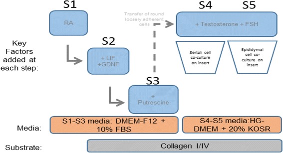
Methods: Three independent lines of FTM and term HUCPVCs were cultured using a novel 5-week step-wise in vitro differentiation protocol recapitulating key physiological signals involved in testicular development. SC- and GC-associated phenotypical properties were assessed by real-time polymerase chain reaction (RT-PCR), quantitative PCR immunocytochemistry, flow cytometry, and fluorescence in-situ hybridization (FISH). Functional spermatogonial stem cell-like properties were assessed using a xenotranplantation assay.

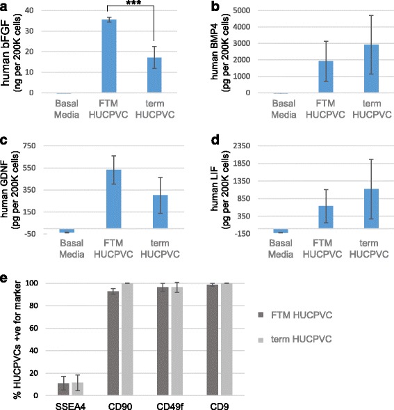
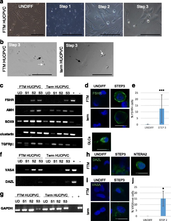
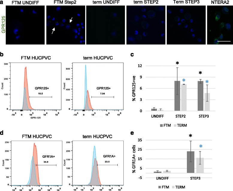
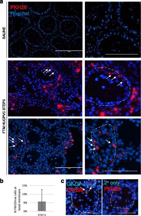
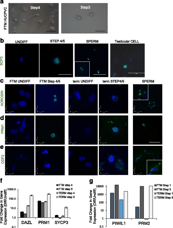
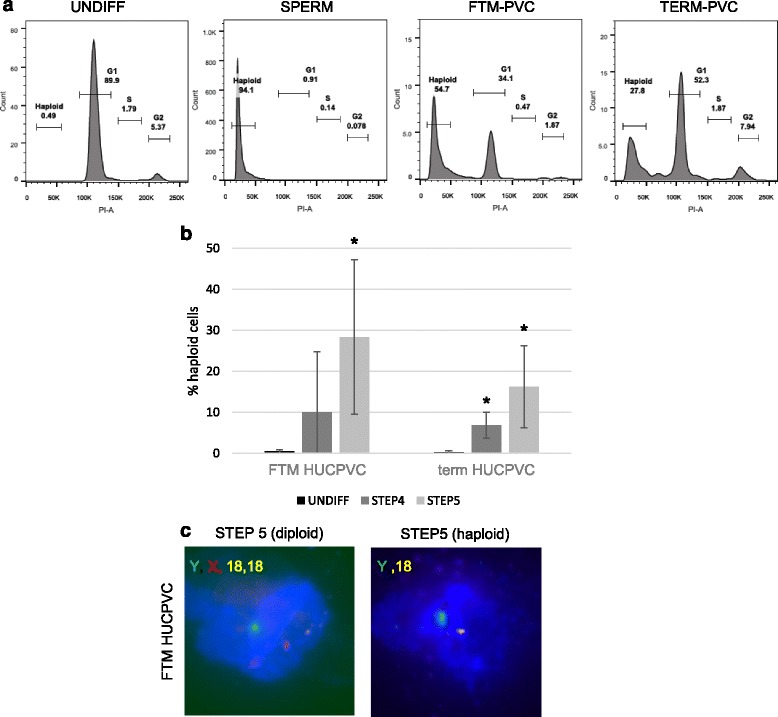
Results: Within 3 weeks of differentiation, two morphologically distinct cell types emerged including large adherent cells and semi-attached round cells. Both early GC-associated markers (VASA, DAZL, GPR125, GFR1α) and SC-associated markers (FSHR, SOX9, AMH) were upregulated, and 5.7 ± 1.2% of these cells engrafted near the inner basal membrane in a xenograft assay. After 5 weeks in culture, 10-30% of the cells were haploid, had adopted a spermatid-like morphology, and expressed PRM1, Acrosin, and ODF2. Undifferentiated HUCPVCs secreted key factors known to regulate spermatogenesis (LIF, GDNF, BMP4, bFGF) and 10-20% of HUCPVCs co-expressed SSEA4, CD9, CD90, and CD49f. We hypothesize that the paracrine properties and cellular heterogeneity of HUCPVCs may explain their dual capacity to differentiate to both SC- and GC-like cells.

Conclusions: HUCPVCs recapitulate elements of the testicular niche including their ability to differentiate into cells with Sertoli-like and haploid spermatid-like properties in vitro. Our study supports the importance of generating a niche-like environment under ex vivo conditions aiming at creating mature GC, and highlights the plasticity of HUCPVCs. This could have future applications for the treatment of some cases of male infertility.

Keywords: Ex-vivo spermatogenesis; Germ cell; Human mesenchymal stem cell; Human umbilical cord; Niche; Paracrine; Perivascular cell; Sertoli cell; Spermatogonial stem cell; Xenograft.


Figures

Fig. 1
Fig. 1
Schematic of the optimized step-wise 5-week differentiation protocol that we developed for HUCPVC differentiation towards the Sertoli and germ cell lineages. DMEM Dulbecco’s modified Eagle’s medium, FBS fetal bovine serum, FSH follicle-stimulating hormone, GDNF glial cell-derived neurotrophic factor, HG high glucose, KOSR knockout serum replacement, LIF leukemia inhibitory factor, RA retinoic acid
Fig. 2
Fig. 2
First trimester (FTM) and term human umbilical cord perivascular cells (HUCPVCs) express and secrete testis niche-associated growth factors. ELISA analysis of the human growth factors a basic fibroblast growth factor (bFGF), b bone morphogenetic protein 4 (BMP4), c glial cell-derived neurotrophic factor (GDNF), and d leukemia inhibitory factor (LIF) in FTM HUCPVC- and term HUCPVC-conditioned media. Basal media (DMEM-F12) was used as a control. The figure shows the average of two independent lines of FTM HUCPVCs and term HUCPVCs each analyzed in duplicate. Error bars indicate standard deviations. *** p = 0.005. e Percentage of cells expressing testicular progenitor-associated markers including stage-specific embryonic antigen 4 (SSEA4), CD9, CD90, and CD49f as determined by flow cytometry (n = 3 independent FTM and term lines each)
Fig. 3
Fig. 3
First trimester (FTM) and term human umbilical cord perivascular cells (HUCPVCs) differentiate towards both Sertoli- and germ-like cells within the same culture environment. a Undifferentiated FTM HUCPVCs and FTM HUCPVCs at the end of Step 1, Step 2, and Step 3. b FTM HUCPVCs (left) and term HUCPVCs (right) at Step 3 of the differentiation protocol showing adherent cells (black arrow) and small round loosely attached cells (white arrow). c RT-PCR for Sertoli-associated gene expression including follicle stimulating hormone receptor (FSHR), anti-müllerian hormone (AMH), SOX9, clusterin (CLU), and transforming growth factor receptor beta (TGFRβ1) in FTM HUCPVCs and term HUCPVCs at the undifferentiated stage (UD), Step 1 (S1), Step 2 (S2), and Step 3 (S3); granulosa cells used as positive control (+). – indicates no cDNA controls. d Undifferentiated (UNDIFF) FTM HUCPVCs (negative control) (top, left) and term HUCPVCs (bottom, left) (negative control) immunostained for FSHR (green, undetected) and counterstained with Hoechst to show live nuclei. Granulosa cells (GLCs) were used as a positive control. e Flow cytometric quantification of the percentage of FTM HUCPVCs that upregulated FSHR at the end of Step 2 when compared to undifferentiated controls. f RT-PCR for early germ cell-associated gene expression of VASA and DAZL in the same samples as panel c. g RT-PCR for GAPDH results shown in panels c and f. h Undifferentiated FTM HUCPVCs (left) immunostained for DAZL (green, undetected) and counterstained with Hoechst to show live nuclei. i Undifferentiated FTM HUCPVCs (top, left) and term HUCPVCs (bottom, left) immunostained for VASA (green, undetected) and counterstained with Hoechst. j Flow cytometric quantification of the percentage of FTM HUCPVCs that upregulated VASA at the end of Step 2 when compared to undifferentiated controls
Fig. 4
Fig. 4
Human spermatogonial stem cell-associated markers are upregulated in first trimester (FTM) and term human umbilical cord perivascular cells (HUCPVCs) in the early steps of differentiation. a Representative micrograph of undifferentiated (UNDIFF) FTM HUCPVCs (left) and at Step 2 (second panel), undifferentiated term HUCPVCs (third panel) and at Step 2 and Step 3 (fourth and fifth panel, respectively) immunostained for GPR125 (green, undetected in undifferentiated PVCs) and counterstained with Hoechst to show live nuclei. GPR125 (green) was detected in FTM HUCPVCs and term HUCPVCs at the end of Steps 2 and 3. Human testicular cells were used as a positive control. Scale bar = 50 μm. b Representative flow cytometry analysis plots for GPR125 in FTM HUCPVCs (left) and term HUCPVCs (right), comparing the undifferentiated stage to Step 3. c Quantification of the percentage of FTM HUCPVCs and term HUCPVCs that upregulated GPR125 at Steps 2 and 3, in comparison to undifferentiated controls (p < 0.05 for all comparisons to undifferentiated). n = 3 independent lines of each FTM and term HUCPVC. d Representative flow cytometry plots for GFR1α expression analysis (also known as GDNFR) in FTM HUCPVCs (left) and term HUCPVCs (right), comparing the undifferentiated stage to Step 3. e Quantification of the percentage of FTM HUCPVCs and term HUCPVCs that upregulated GFR1α at Step 3 in comparison to undifferentiated controls (p < 0.05 for all comparisons to undifferentiated). n = 3 independent lines of each FTM and term HUCPVC
Fig. 5
Fig. 5
A portion of Step 3-induced first trimester (FTM) human umbilical cord perivascular cells (HUCPVCs) display functional properties of SSCs in a modified xenograft assay. a Representative micrographs of xenograft assay tissue sections including saline control (top row, with low magnification on left and high magnification on right). Scale bars = 200 μm and 50 μm, respectively. Arrows indicate PKH26-positive cells near the basal membrane of the seminiferous tubule, the contour of which is delineated by the dotted line. b Quantification of the percentage of total PKH26-positive cells that localize within seminiferous tubules and near the basal membrane as opposed to the interstitial regions. c Representative micrograph of DAZL (green) immunohistochemistry on frozen sections containing PKH26-labeled human cells (red) (left panel) and the corresponding no primary antibody negative control (right panel). Scale bar = 100 μm. Arrows indicate PKH26-positive/DAZL+ cells near the basal membrane of the seminiferous tubule, the contour of which is delineated by the dotted line
Fig. 6
Fig. 6
Second stage spermatocyte- and spermatid-associated markers are upregulated in first trimester (FTM) human umbilical cord perivascular cells (HUCPVCs) and term HUCPVCs at Step 4 and Step 5. a Bright field representative micrographs of FTM HUCPVCs at Step 4 and Step 5 of the differentiation protocol. b Representative micrograph of undifferentiated (UNDIFF) HUCPVCs (negative control) immunostained for SYCP3 (green, undetected) and counterstained with Hoechst to show live nuclei. SYCP3 (green) was detected in and Step 4 and Step 5 (showing term at Step 5). Spermatozoa (SPERM) were used as a positive control. Scale bars = 100 μm. ce Representative micrographs of undifferentiated (negative control) and Step 5 FTM HUCPVCs and undifferentiated (negative control) and Step 5 term HUCPVCS immunostained for the spermatid-associated proteins Acrosin, Protamine 1 (PRM1) and ODF2 (green) and counterstained for Hoechst to visualize nuclear integrity. Sperm was used as a positive control. All images are at the same magnification, with scale bars shown, with the exception of insets for sperm markers which were magnified approximately 6 times. f qPCR analysis of differentiated (Diff) FTM and term HUCPVCs at Step 4 and Step 5 showing fold-change of late spermatogenesis marker (DAZL, PRM1, SYCP3) gene expression when compared to undifferentiated HUCPVCs. g qPCR analysis of differentiated HUCPVCs isolated at Step 1 and Step 5, showing fold-change in gene expression compared to undifferentiated cells for early and late spermatogenesis markers PIWIL1 and PRM2, respectively
Fig. 7
Fig. 7
First trimester (FTM) and term human umbilical cord perivascular cells (HUCPVCs) generate haploid cells. a Representative flow cytometry plots for cell cycle analysis of diploid undifferentiated (UNDIFF) HUCPVCs (showing FTM HUCPVCs, left panel), haploid spermatozoa (SPERM, second panel) and FTM HUCPVCs and term HUCPVCs at the end of Step 5 (right panels, respectively). b Quantification of the percentage of haploid cells in FTM HUCPVCs and term HUCPVCs, comparing undifferentiated HUCPVCs with HUCPVCs at the end of Step 4 and Step 5. Each condition was analyzed in three to four independent experiments. *p < 0.05. c Representative micrographs of FTM HUCPVCs processed for fluorescence in-situ hybridization (FISH) at Step 5, showing an example of a diploid cell observed at Step 5 displaying chromosome X (red), Y (green) and 2 chromosome 18 (yellow) and an example of a haploid cells at Step 5 displaying chromosome Y (green) and one chromosome 18 (yellow)

Similar articles

Cited by

References

    1. Sato T, Katagiri K, Gohbara A, Inoue K, Ogonuki N, Ogura A, Kubota Y, Ogawa T. In vitro production of functional sperm in cultured neonatal mouse testes. Nature. 2011;471(7339):504–7. doi: 10.1038/nature09850. - DOI - PubMed
    1. Dobrinski I, Avarbock MR, Brinster RL. Transplantation of germ cells from rabbits and dogs into mouse testes. Biol Reprod. 1999;61(5):1331–9. doi: 10.1095/biolreprod61.5.1331. - DOI - PubMed
    1. Hermann BP, Sukhwani M, Winkler F, Pascarella JN, Peters KA, Sheng Y, Valli H, Rodriguez M, Ezzelarab M, Dargo G, et al. Spermatogonial stem cell transplantation into rhesus testes regenerates spermatogenesis producing functional sperm. Cell Stem Cell. 2012;11(5):715–26. doi: 10.1016/j.stem.2012.07.017. - DOI - PMC - PubMed
    1. Easley CA, Phillips BT, McGuire MM, Barringer JM, Valli H, Hermann BP, Simerly CR, Rajkovic A, Miki T, Orwig KE, et al. Direct differentiation of human pluripotent stem cells into haploid spermatogenic cells. Cell Rep. 2012;2(3):440–6. doi: 10.1016/j.celrep.2012.07.015. - DOI - PMC - PubMed
    1. Chuang CY, Lin KI, Hsiao M, Stone L, Chen HF, Huang YH, Lin SP, Ho HN, Kuo HC. Meiotic competent human germ cell-like cells derived from human embryonic stem cells induced by BMP4/WNT3A signaling and OCT4/EpCAM (epithelial cell adhesion molecule) selection. J Biol Chem. 2012;287(18):14389–401. doi: 10.1074/jbc.M111.338434. - DOI - PMC - PubMed

MeSH terms