Case Reports
. 2017 Feb 15;17(1):17.
doi: 10.1186/s12893-017-0214-1.

Isolated IgG4-related sclerosing cholangitis misdiagnosed as malignancy in an area with endemic cholangiocarcinoma: a case report

Affiliations
Case Reports

Isolated IgG4-related sclerosing cholangitis misdiagnosed as malignancy in an area with endemic cholangiocarcinoma: a case report

Narongsak Rungsakulkij et al. BMC Surg. .

Abstract

Background: The most common cause of perihilar obstruction is cholangiocarcinoma, especially in Thailand. Benign perihilar stricture represents less than 20% of cases. IgG4-related disease and IgG4-related sclerosing cholangitis, however, have been receiving increased recognition. Isolated IgG4-related sclerosing cholangitis is less common. The preoperative diagnosis of IgG4-related sclerosing cholangitis without pancreatic involvement is very difficult because the clinical presentation and preoperative evaluation are extremely difficult to distinguish from perihilar cholangiocarcinoma.

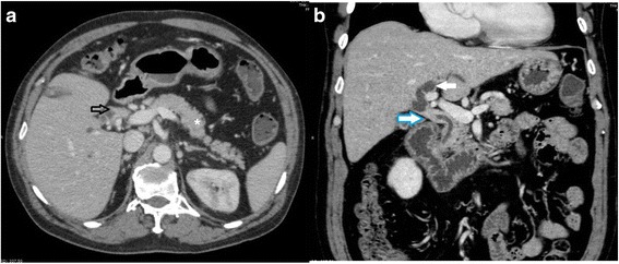
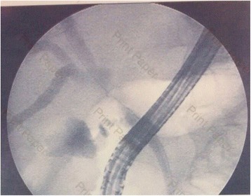
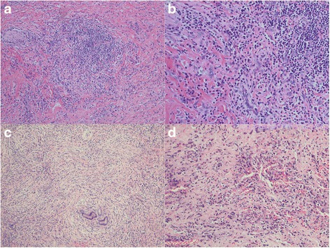
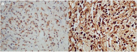
Case presentation: We report the case of a 56-year-old man who presented with obstructive jaundice with preoperative imaging showing proximal common bile duct obstruction. He underwent right lobe liver hepatectomy with extrahepatic bile duct resection and regional lymph node dissection due to high suspicion of malignancy. The pathological report showed severe acute and chronic inflammation of the bile duct with morphology and immunohistochemistry suggestive of IgG4-related sclerosing cholangitis.

Conclusions: IgG4-related sclerosing cholangitis with perihilar obstruction should be considered even in areas where cholangiocarcinoma is endemic.

Keywords: Benign biliary stricture; Cholangiocarcinoma; IgG4, IgG4-related sclerosing cholangitis; Sclerosing cholangitis.


Figures

Fig. 1
Fig. 1
Abdominal CT showing focal short segment sheet-like thickening of proximal common bile duct with enhancement. a: axial view showing sheet-like thickening of common hepatic duct wall (arrow); note the normal pancreas (star). b: coronal view showing focal short segment, sheet-like, symmetric thickening of common hepatic duct causing nearly complete obstruction of common hepatic duct just below bifurcation (white arrow with blue edge) of right and left intrahepatic bile ducts and dilatation of left intrahepatic duct (white arrow)
Fig. 2
Fig. 2
ERCP show stricture of common hepatic duct just below hepatic confluence
Fig. 3
Fig. 3
Intraoperative findings and gross specimen. a: tumor at hepatic hilum (arrow). b: left main intrahepatic duct (arrow). c: gross specimen after complete resection showing mass at proximal common bile duct just below hepatic confluence (arrow). d: diffuse bile duct thickening with marked focal thickening at common hepatic duct (arrow)
Fig. 4
Fig. 4
Left hepatic duct along with common bile duct showing fibrosis and plasma cells. a, H&E x100; b. H&E x200; c, H&E x100 storiform fibrosis with lymphocytes and plasma cells; d, H&E x200 obliterative phlebitis H&E, Hematoxylin and eosin stain
Fig. 5
Fig. 5
Left hepatic duct along with common bile duct showing positive plasma cells. a. IHC x400 of IgG-positive plasma cells. b. IHC x400 of IgG4-positive plasma cells (80 cells/HPF) IHC, immunohistochemistry

References

    1. Sripa B, Bethony JM, Sithithaworn P, Kaewkes S, Mairian E, Loukas A, et al. Opisthorchiasis and Opisthorchis-associated cholangiocarcinoma in Thailand and Laos. Acta Trop. 2011;120:S158–68. doi: 10.1016/j.actatropica.2010.07.006. - DOI - PMC - PubMed
    1. Are C, Gonen M, D’Angelica M, DeMatteo RP, Fong Y, Blumgart LH, Jarnagin WR. Differential diagnosis of proximal biliary obstruction. Surgery. 2006;140:756–63. doi: 10.1016/j.surg.2006.03.028. - DOI - PubMed
    1. Gerhards MF, Vos P, van Gulik TM, Rauws AJ, Bosma A, Gouma DJ. Incidence of benign lesions in patients resected for suspicious hilar obstruction. Br J Surg. 2001;88:48–51. doi: 10.1046/j.1365-2168.2001.01607.x. - DOI - PubMed
    1. Koea J, Holden A, Chau K, McCall J. Differential diagnosis of stenosis lesions at the hepatic hilus. World J Surg. 2004;28:466–70. doi: 10.1007/s00268-004-7034-z. - DOI - PubMed
    1. Corvera CU, Blumgart LH, Darvishian F, Klimstra DS, DeMatteo R, Fong Y, et al. Clinical and pathologic features of proximal biliary strictures masquerading as hilar cholangiocarcinoma. J Am Coll Surg. 2005;201:862–9. doi: 10.1016/j.jamcollsurg.2005.07.011. - DOI - PubMed

Publication types

Substances