. 2017 Feb 15;14(1):31.
doi: 10.1186/s12985-017-0707-7.

The DNA damage response promotes polyomavirus JC infection by nucleus to cytoplasm NF- kappaB activation

Affiliations

The DNA damage response promotes polyomavirus JC infection by nucleus to cytoplasm NF- kappaB activation

Martyn K White et al. Virol J. .

Abstract

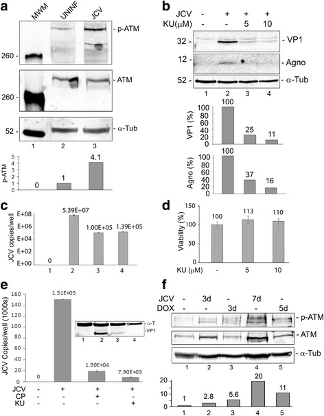
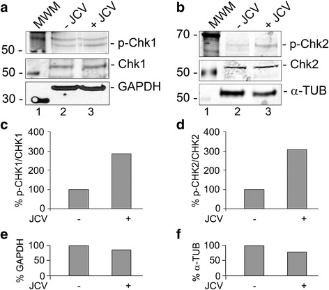
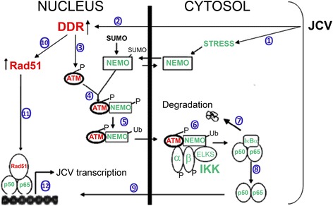
Background: Infection of glial cells by human neurotropic polyomavirus JC (JCV), the causative agent of the CNS demyelinating disease progressive multifocal leukoencephalopathy (PML), rapidly inflicts damage to cellular DNA. This activates DNA damage response (DDR) signaling including induction of expression of DNA repair factor Rad51. We previously reported that Rad51 co-operates with the transcription factor NF-κB p65 to activate JCV early transcription. Thus Rad51 induction by JCV infection may provide positive feedback for viral activation early in JCV infection. DDR is also known to stimulate NF-κB activity, a phenomenon known as nucleus to cytoplasm or "inside-out" NF-κB signaling, which is initiated by Ataxia telangiectasia mutated (ATM) protein, a serine/threonine kinase recruited and activated by DNA double-strand breaks. Downstream of ATM, there occurs a series of post-translational modifications of NF-κB essential modulator (NEMO), the γ regulatory subunit of inhibitor of NF-κB (IκB) kinase (IKK), resulting in NF-κB activation.

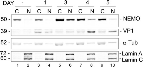
Methods: We analyzed the effects of downstream pathways in the DDR by phosphospecific Western blots and analysis of the subcellular distribution of NEMO by cell fractionation and immunocytochemistry. The role of DDR in JCV infection was analyzed using a small molecule inhibitor of ATM (KU-55933). NEMO sumoylation was investigated by Western and association of ATM and NEMO by immunoprecipitation/Western blots.

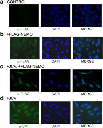
Results: We show that JCV infection caused phosphorylation and activation of ATM while KU-55933 inhibited JCV replication. JCV infection caused a redistribution of NEMO from cytoplasm to nucleus. Co-expression of JCV large T-antigen and FLAG-tagged NEMO showed the occurrence of sumoylation of NEMO, while co-expression of ATM and FLAG-NEMO demonstrated physical association between ATM and NEMO.

Conclusions: We propose a model where JCV infection induces both overexpression of Rad51 protein and activation of the nucleus to cytoplasm NF-κB signaling pathway, which then act together to enhance JCV gene expression.

Keywords: DNA damage response; Nuclear factor kappa-B; Polyomavirus JC; Progressive multifocal leukoencephalopathy.


Figures

Fig. 1
Fig. 1
Role of ATM phosphorylation in JCV infection. a Effect of JCV infection on ATM phosphorylation. SVGA cells were uninfected or infected, as indicated, with the Mad-1 strain of JCV at moi = 1 for 5 days and harvested. Fifty micrograms of total cell extract were loaded onto a 6% polyacrylamide SDS gel, electrophoresed and analyzed by Western blot for phospho-ATM and total ATM. Alpha-tubulin (α-Tub) was the loading control. The intensity of the p-ATM band in each lane was quantified using the ImageQuant software (Molecular Dynamics, GE Healthcare Bio-Sciences, Pittsburgh PA) and these are shown in the lower part of the panel. b Effect of ATM inhibition on JCV infection. SVGA were infected with the Mad-1 of JCV and treated with or without KU-55933(2-morpholin-4-yl-6-thianthren-1-yl-pyran-4-one; 5 μM or 10 μM), which is ATP competitive inhibitor of ATM, as indicated. Cells were harvested and expression of VP1 and agnoprotein measured by Western blot. The loading control was α-Tubulin. The intensity of the VP1 and Agno bands in each lane was quantified using the ImageQuant software and these are shown in the lower part of the panel. c Culture supernatants from the cells in Panel b were assayed for virus using qPCR. Each viral load was measured in triplicate and presented as a histogram with the standard deviation shown as an error bar. d The viability of SVGA cells was assayed at different concentrations of KU-55933 by MTT assay as described in the Methods section. Each viability was measured in triplicate and presented as a histogram with the standard deviation shown as an error bar. e Effect of different ATM inhibitors on JCV infection. SVGA were infected with the Mad-1 of JCV and treated with or without KU-55933 or CP466722 as indicated. Cells and supernatants were harvested and viral copy number in the culture medium assayed by QPCR. Expression of VP1 was measured by Western blot with α-Tubulin as the loading control (shown inset). f Effect of JCV infection or doxorubicin treatment on ATM phosphorylation. SVGA cells were uninfected or infected, as indicated, with the Mad-1 strain of JCV at moi = 1 or treated with 2.5 μM doxorubicin for the times indicated and harvested. Western blots were performed for phospho-ATM and total ATM. Alpha-tubulin was the loading control. The intensity of the p-ATM band in each lane was quantified using the ImageQuant software and these are shown in the lower part of the panel.
Fig. 2
Fig. 2
Phosphorylation of CHK1 and CHK2 in JCV infection. SVGA cells were infected with Mad-1 JCV at an moi of 1 and cultured for 5 days. Total cell lysates were harvested and analyzed by Western blots for phospho-Chk1(Ser 317), Panel a and phospho-Chk2(Thr 68), Panel b. The Western membranes were reprobed for total Chk1 (a) and Chk2 (b) and then for a loading control, which was either glyceraldehyde-3-phosphate dehydrogenase (GAPDH, a) or α-tubulin (α-TUB, b). The intensity of each band was quantified using the IMAGEQUANT software (Molecular Dynamics, GE Healthcare Bio-Sciences, Pittsburgh PA) and the ratio of phospho-Chk1 to Chk1 (c) and phospho-Chk2 to Chk2 (d) calculated and shown as histograms expressed as a percentage of the signal in uninfected cells. Similarly, the loading controls were quantified (e and f)
Fig. 3
Fig. 3
Role of NEMO cytoplasm to nucleus translocation in JCV infection: Analysis of the effect of JCV infection on the subcellular distribution of NEMO by cell fractionation. SVGA cells were transfected with expression plasmid for FLAG-NEMO and infected with wild-type Mad-1 JCV 24 h later. Cells were harvested at 1, 3, 4 and 5 d after infection and fractionated into cytoplasmic and nuclear fractions. Western blot for α-FLAG was used to monitor the subcellular distribution of NEMO. Western blots for α-tubulin and lamin A/C were used to monitor cell fraction purity and for VP1 to monitor infection
Fig. 4
Fig. 4
Role of NEMO cytoplasm to nucleus translocation in JCV infection: Analysis of the effect of JCV infection on the subcellular distribution of NEMO by immunocytochemistry. SVGA cells were uninfected and untransfected (a) or uninfected and transfected with expression plasmid for FLAG-tagged NEMO (b). SVGA cells were also infected with wild-type Mad-1 JCV and transfected twice with expression plasmid for FLAG-NEMO on days 2 and 5 postinfection (c). Control cells were infected with JCV but not transfected (d). Immunocytochemistry for FLAG and VP1 was performed on day 7 postinfection as described in the Methods section
Fig. 5
Fig. 5
Role of NEMO sumoylation in JCV infection and NEMO association with ATM. a Effect of JCV T-antigen on Sumoylation of NEMO. TC620 cells were transfected with expression plasmids for FLAG-tagged NEMO and/or T-Ag. Western blots were performed with antibody to T-Ag and anti-FLAG antibody to detect NEMO and sumoylated NEMO (arrowhead) as indicated. The quantified intensity of the sumoylated NEMO band is indicated in the histogram directly below the Western. The intensity of the sumoylated band as a percentage of total NEMO with and without T-Ag is presented in the lower right-hand part of the panel. b Physical association between ATM and NEMO. TC620 cells were transfected with expression plasmids for ATM and FLAG-NEMO. Whole cell extract (Input, lane 1) was immunoprecipitated with rabbit anti-ATM (lane 3). Nonimmune rabbit serum (NRS) was the negative control (lane 2). Western blot was with α-FLAG antibody. c In a reciprocal immunoprecipitation, whole cell extract from Panel b (Input, lane 1) was immunoprecipitated with mouse anti-FLAG (lane 3). Nonimmune mouse serum (NMS) was the negative control (lane 2). Western blot was with α-ATM antibody
Fig. 6
Fig. 6
Schematic representation of the nucleus to cytoplasm NF-κB activation pathway in JCV infection. Infection of glial cells by JCV results in stress leading to NEMO sumoylation in the nucleus ① and activation of the DDR ②, which phosphorylates and activates ATM ③. Activated ATM binds NEMO and leads to phosphorylation of NEMO on Ser85 ④, which is involved in NEMO ubiquitination and export from the nucleus ⑤ to the cytoplasm. ATM and NEMO activate IKK ⑥, which phosphorylates IκB leading to IκB degradation ⑦ and release of NF-κB ⑧ to migrate to the nucleus ⑨. Meanwhile, Rad51 is induced by the DDR ⑩ and associates with nuclear NF-κB ⑪ to activate JCV transcription ⑫

Similar articles

Cited by

References

    1. Padgett BL, Walker DL, ZuRhein GM, Eckroade RJ, Dessel BH. Cultivation of papova-like virus from human brain with progressive multifocal leukoencephalopathy. Lancet. 1971;i:1257–60. doi: 10.1016/S0140-6736(71)91777-6. - DOI - PubMed
    1. White MK, Khalili K. Pathogenesis of progressive multifocal leukoencephalopathy – revisited. J Infect Dis. 2011;203:578–86. doi: 10.1093/infdis/jiq097. - DOI - PMC - PubMed
    1. Wollebo HS, White MK, Gordon J, Berger JR, Khalili K. Persistence and pathogenesis of the neurotropic polyomavirus JC. Ann Neurol. 2015;77:560–70. doi: 10.1002/ana.24371. - DOI - PMC - PubMed
    1. Kedar S, Berger JR. The changing landscape of progressive multifocal leukoencephalopathy. Curr Infect Dis Rep. 2011;13:380–6. doi: 10.1007/s11908-011-0196-6. - DOI - PubMed
    1. Berger JR. Progressive multifocal leukoencephalopathy and newer biological agents. Progressive multifocal leukoencephalopathy and newer biological agents. Drug Saf. 2010;33:969–83. doi: 10.2165/11537510-000000000-00000. - DOI - PubMed

Publication types

MeSH terms

LinkOut - more resources