

Exendin-4 in combination with adipose-derived stem cells promotes angiogenesis and improves diabetic wound healing
- PMID: 28202074
- PMCID: PMC5311833
- DOI: 10.1186/s12967-017-1145-4
Exendin-4 in combination with adipose-derived stem cells promotes angiogenesis and improves diabetic wound healing
Abstract
Background: Diminished wound healing is a major complication of diabetes mellitus and can lead to foot ulcers. However, there are limited therapeutic methods to treat this condition. Exendin-4 (Ex-4), a glucagon-like peptide-1 receptor agonist, is known to have many beneficial effects on diabetes. In addition, mesenchymal stem cells are known to have wound healing effects. We investigated the effects of Ex-4 in combination with human adipose tissue-derived stem cells (ADSCs) on diabetic wound healing in a diabetic animal model.
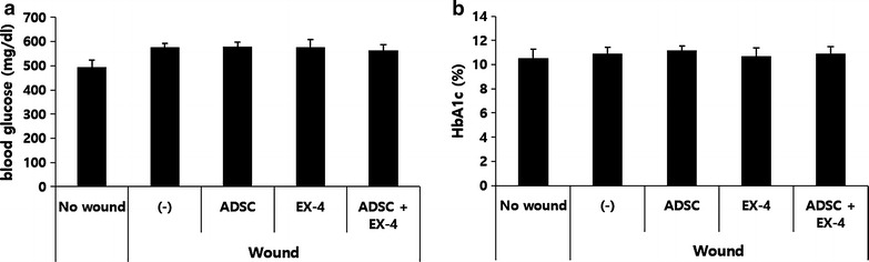
Methods: Diabetic db/db (blood glucose levels, >500 mg/dl) or C57BL/6 mice were subjected to wounding on the skin of the back. One day after wounding, each wound received ADSCs (2.5 × 10⁵ cells) injected intradermally around the wound and/or Ex-4 (50 μl of 100 nM Ex-4) topically applied on the wound with a fine brush daily. Wound size was monitored and wound histology was examined. Human endothelial cells and keratinocyte cells were used to assess angiogenesis and vascular endothelial growth factor expression in vitro.
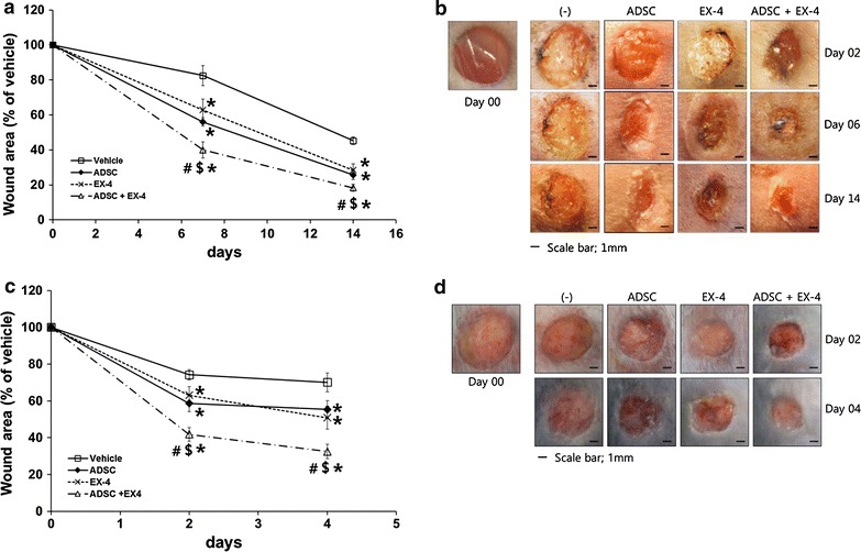
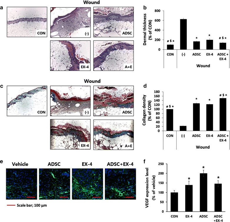
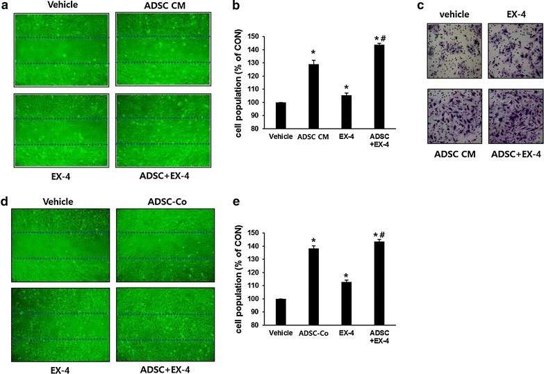
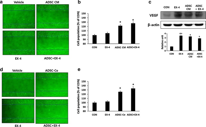
Results: Topical administration of Ex-4 or injection of ADSCs resulted in a rapid reduction of wound size in both diabetic and normoglycemic animals compared with vehicle treatment. Histological analysis also showed rapid skin reconstruction in Ex-4-treated or ADSC-injected wounds. A combination of Ex-4 and ADSCs showed a significantly better therapeutic effect over either treatment alone. In vitro angiogenesis assays showed that both Ex-4 and ADSC-conditioned media (CM) treatment improved migration, invasion and proliferation of human endothelial cells. ADSC-CM also increased migration and proliferation of human keratinocytes. In addition, both Ex-4 and ADSC-CM increased the expression of vascular endothelial growth factor. Co-culture with ADSCs increased migration and proliferation of these cells similar to that found after ADSC-CM treatment.
Conclusions: We suggest that Ex-4 itself is effective for the treatment of diabetic skin wounds, and a combination of topical treatment of Ex-4 and injection of ADSCs has a better therapeutic effect. Thus, a combination of Ex-4 and ADSCs might be an effective therapeutic option for the treatment of diabetic wounds, such as foot ulcers.
Keywords: Adipose-derived stem cells; Angiogenesis; Diabetic wound; Exendin-4; GLP-1.
Figures
Similar articles
-
VAP-PLGA microspheres (VAP-PLGA) promote adipose-derived stem cells (ADSCs)-induced wound healing in chronic skin ulcers in mice via PI3K/Akt/HIF-1α pathway.Bioengineered. 2021 Dec;12(2):10264-10284. doi: 10.1080/21655979.2021.1990193. Bioengineered. 2021. PMID: 34720043 Free PMC article.
-
Adipose-derived stem cells seeded in Pluronic F-127 hydrogel promotes diabetic wound healing.J Surg Res. 2017 Sep;217:63-74. doi: 10.1016/j.jss.2017.04.032. Epub 2017 May 5. J Surg Res. 2017. PMID: 28595815
-
Galectin-1 from conditioned medium of three-dimensional culture of adipose-derived stem cells accelerates migration and proliferation of human keratinocytes and fibroblasts.Wound Repair Regen. 2018 Dec;26 Suppl 1:S9-S18. doi: 10.1111/wrr.12579. Epub 2017 Nov 8. Wound Repair Regen. 2018. PMID: 28857355
-
Adipose-derived stem cells: Effectiveness and advances in delivery in diabetic wound healing.Biomed Pharmacother. 2018 Nov;107:625-633. doi: 10.1016/j.biopha.2018.08.013. Epub 2018 Aug 14. Biomed Pharmacother. 2018. PMID: 30118878 Review.
-
The Basic Mechanism of Hair Growth Stimulation by Adipose-derived Stem Cells and Their Secretory Factors.Curr Stem Cell Res Ther. 2017;12(7):535-543. doi: 10.2174/1574888X12666170829161058. Curr Stem Cell Res Ther. 2017. PMID: 28875863 Review.
Cited by
-
Adipose-derived stem cells in diabetic foot care: Bridging clinical trials and practical application.World J Diabetes. 2024 Jun 15;15(6):1162-1177. doi: 10.4239/wjd.v15.i6.1162. World J Diabetes. 2024. PMID: 38983804 Free PMC article. Review.
-
Human adipose-derived stem cells promote seawater-immersed wound healing via proangiogenic effects.Aging (Albany NY). 2021 Mar 26;13(13):17118-17136. doi: 10.18632/aging.202773. Epub 2021 Mar 26. Aging (Albany NY). 2021. PMID: 33819183 Free PMC article.
-
Stem Cells and Angiogenesis: Implications and Limitations in Enhancing Chronic Diabetic Foot Ulcer Healing.Cells. 2022 Jul 25;11(15):2287. doi: 10.3390/cells11152287. Cells. 2022. PMID: 35892584 Free PMC article. Review.
-
Secretome of Adipose Tissue-Derived Stem Cells (ASCs) as a Novel Trend in Chronic Non-Healing Wounds: An Overview of Experimental In Vitro and In Vivo Studies and Methodological Variables.Int J Mol Sci. 2019 Jul 30;20(15):3721. doi: 10.3390/ijms20153721. Int J Mol Sci. 2019. PMID: 31366040 Free PMC article. Review.
-
Advances and perspective on animal models and hydrogel biomaterials for diabetic wound healing.Biomater Transl. 2022 Sep 28;3(3):188-200. doi: 10.12336/biomatertransl.2022.03.003. eCollection 2022. Biomater Transl. 2022. PMID: 36654776 Free PMC article. Review.
References
-
- Ricco JB, Thanh Phong L, Schneider F, Illuminati G, Belmonte R, Valagier A, Regnault DL. The diabetic foot: a review. J Cardiovasc Surg. 2013;54:755–762. - PubMed
MeSH terms
Substances
LinkOut - more resources
Full Text Sources
Other Literature Sources
Medical
Miscellaneous
