. 2017 Feb 15;15(1):34.
doi: 10.1186/s12967-017-1143-6.

Bone Morphogenetic Proteins and myostatin pathways: key mediator of human sarcopenia

Affiliations

Bone Morphogenetic Proteins and myostatin pathways: key mediator of human sarcopenia

Manuel Scimeca et al. J Transl Med. .

Abstract

Background: Sarcopenia, osteoporosis and osteoarthritis are the most frequent musculoskeletal disorders affecting older people. The main aim of this study was to test the hypothesis that the balance between BMPs and myostatin pathways regulates the age-related muscle degeneration in OP and OA patients. To this end, we investigated the relationship among the expression of BMP-2/4-7, myostatin and phosphorylated Smads1-5-8 and the muscle quality, evaluated in term of fibers atrophy and satellite cells activity.

Methods: In this retrospective study, we collected 123 biopsies of vastus lateralis: 48 biopsies from patients who underwent hip arthroplasty for subcapital fractures of the femur (OP), 55 biopsies from patients who underwent hip arthroplasty for osteoarthritis (OA) and 20 biopsies from patients who underwent hip arthroplasty for high-energy hip fractures (CTRL). Muscle biopsies were fixed in 4% paraformaldehyde and paraffin embedded. Serial sections were used for morphometrical and immunohistochemical analysis (BMP/2/4-7, myostatin, Smads1-5-8, Pax7 and myogenin). In addition, 1 mm3 of muscle tissue of each patient was embedded in epon for ultrastructural study.

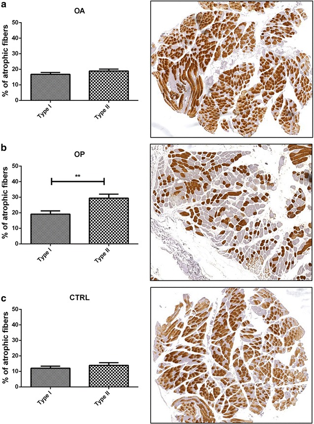
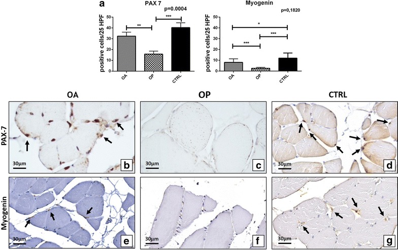
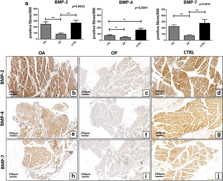
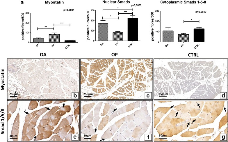
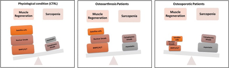
Results: Morphometric data indicated an increase of the number of atrophic fibers in OP patients compare to OA. In line with these data, we found an high regenerative potential in muscle tissues of OA patients due to the significant amount of both Pax7 and myogenin positive satellite cells detected in OA group. In addition, our data showed the decrease of BMP2/4 and -7 expression in OP patients compared to both OA group and CTRL. Conversely, OP patients were characterized by high levels of myostatin expression. A different expression profile was also found for phosphorylated Smad1-5-8 between OP and OA patients. In particular, OP patients showed a low number of positive phosphorylated Smad1-5-8 nuclei.

Conclusion: The identification of molecular pathways involved in the pathogenesis of sarcopenia open new prospective for the development of drugs able to prevent/treat the muscle impairment that occur in elderly. Results here reported, highlighting the role of BMPs and myostatin pathways in physio-pathogenesis of human sarcopenia, allow us to propose human recombinant BMP-2/7 and anti-myostatin antibodies as a possible therapeutic option for the sarcopenia.


Figures

Fig. 1
Fig. 1
Mophometric analysis. Graphs show the percentage of muscle atrophic fibers in OA (a), OP (b) and CTRL (c) patients. Immunohistochemical analysis display type II fibers in OA (a), OP (b) and CTRL (c) group
Fig. 2
Fig. 2
Satellite cells study. a Graph display the number of Pax7 and myogenin satellite cells in OA, OP and CTRL patients. b Image shows numerous Pax7 positive cells in a muscle biopsy of OA patient (arrows) (40×). c Muscle tissue of OP patient characterize by no/rare Pax7 positive satellite cells (40×). d Image displays numerous Pax7 positive cells in a muscle biopsy of a CTRL patient (arrows) (40×). e Arrows mark myogenin positive cells in a muscle biopsy of OA patient (40×). f Muscle tissue of OP patient characterize by no/rare myogenin positive satellite cells (40×). g Image displays numerous myogenin positive satellite cells in a muscle biopsy of a CTRL patient (arrows) (40×)
Fig. 3
Fig. 3
Ultrastructural analysis of muscle fiber and satellite cell niches. a Image shows muscle fibers of OA patient (2500×). b Well conserved sarcomere structure of a muscle fibers of OA patient (20,000). c Image displays satellite cells (arrows) strongly associated among them and fused to form a syncytium (5000×). d Image shows atrophic fibers in OP patient (1000×). e Misaligned sarcomere in muscle tissue of OP patient (20,000×). f Satellite cells with obvious mark of degeneration (square) (5000×)
Fig. 4
Fig. 4
Immunohistochemical analysis of BMP2/4-7 expression. a Graph display the number of BMP2/4-7 muscle fibers in OA, OP and CTRL patients. b Image shows a muscle biopsy of OA patient with numerous BMP-2 positive fibers (4×). c Rare BMP-2 positive fibers in a muscle tissue of OP patient (4×). d Numerous BMP-2 positive fibers in a muscle biopsy of CTRL patient (4×). e Immunohistochemical reaction shows several BMP-4 positive fiber in a muscle biopsy of OA patient (4×). f Image shows a muscle biopsy of OP patient with rare/no BMP-4 positive fibers (4×). g Muscle of a CTRL patient characterize by high expression of BMP4 (4×). h Several BMP-7 positive fibers in a muscle tissue of OA patient (4×). i Immunohistochemical reaction shows no/rare BMP-7 positive fiber in a muscle biopsy of OP patient (4×). j, d Numerous BMP-7 positive fibers in a muscle biopsy of CTRL patient (4×)
Fig. 5
Fig. 5
Analysis of the expression of myostatin and phosphorylated Smad1-5-8. a Graph display the expression of myostatin and phosphorylated Smad1-5-8 in muscle biopsies of OA, OP and CTRL patients. b Rare myostatin positive fibers in a muscle tissue of OA patient (4×). c Muscle tissue of OP patient characterizes by numerous myostatin positive fibers (4×). d Muscle of a CTRL patient characterize by no expression of myostatin (4×). e Image shows muscle tissue of OA patient with numerous phosphorylated Smad1-5-8 positive nuclei (arrows) (40×). f Muscle of OP patient with phosphorylated Smad1-5-8 negative nuclei (arrows) (40×). g Image displays muscle biopsy of CTRL patient with numerous phosphorylated Smad1-5-8 positive nuclei (arrows)
Fig. 6
Fig. 6
Cellular and molecular characteristics of Sarcopenia in CTRL, OA and OP patients

Similar articles

Cited by

References

    1. Rosenberg IH. Summary comments. Am J Clin Nutr. 1989;50:1231–1233.
    1. Rosenberg IH. Sarcopenia: origins and clinical relevance. J Nutr. 1997;127(5 Suppl):990S–991S. - PubMed
    1. Cruz-Jentoft AJ, Baeyens JP, Bauer JM, Boirie Y, Cederholm T, Landi F, et al. Sarcopenia: European consensus on definition and diagnosis: report of the European Working Group on Sarcopenia in Older People. Age Ageing. 2010;39(4):412–423. doi: 10.1093/ageing/afq034. - DOI - PMC - PubMed
    1. Piscitelli P, Tarantino U, Chitano G, Argentiero A, Neglia C, Agnello N, Saturnino L, Feola M, Celi M, Raho C, Distante A, Brandi ML. Updated incidence rates of fragility fractures in Italy: extension study 2002–2008. Clin Cases Miner Bone Metab. 2011;8(3):54–61. - PMC - PubMed
    1. Piscitelli P, Brandi ML, Tarantino U, Baggiani A, Distante A, Muratore M, Grattagliano V, Migliore A, Granata M, Guglielmi G, Gimigliano R, Iolascon G. Incidence and socioeconomic burden of hip fractures in Italy: extension study 2003–2005. Reumatismo. 2010;62(2):113–118. - PubMed

Publication types

LinkOut - more resources