. 2017 Feb 15;14(1):37.
doi: 10.1186/s12974-017-0813-x.

Aprepitant limits in vivo neuroinflammatory responses in a rhesus model of Lyme neuroborreliosis

Affiliations

Aprepitant limits in vivo neuroinflammatory responses in a rhesus model of Lyme neuroborreliosis

Alejandra N Martinez et al. J Neuroinflammation. .

Abstract

Background: Substance P (SP) is produced at high levels in the central nervous system (CNS), and its target receptor, neurokinin 1 receptor (NK-1R), is expressed by glia and leukocytes. This tachykinin functions to exacerbate inflammatory responses at peripheral sites. Moreover, SP/NK-1R interactions have recently been associated with severe neuroinflammation and neuronal damage. We have previously demonstrated that NK-1R antagonists can limit neuroinflammatory damage in a mouse model of bacterial meningitis. Furthermore, we have since shown that these agents can attenuate Borrelia burgdorferi-induced neuronal and glial inflammatory mediator production in non-human primate brain explants and isolated neuronal cells.

Methods: In the present study, we have assessed the role played by endogenous SP/NK-1R interactions in damaging CNS inflammation in an established rhesus macaque model that faithfully reproduces the key clinical features of Lyme neuroborreliosis, using the specific NK-1R antagonist, aprepitant. We have utilized multiplex ELISA to quantify immune mediator levels in cerebrospinal fluid, and RT-PCR and immunoblot analyses to quantify cytokine and NK-1R expression, respectively, in brain cortex, dorsal root ganglia, and spinal cord tissues. In addition, we have assessed astrocyte number/activation status in brain cortical tissue by immunofluorescence staining and confocal microscopy.

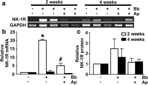
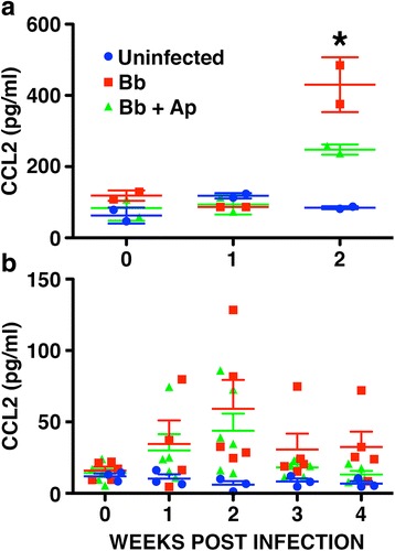
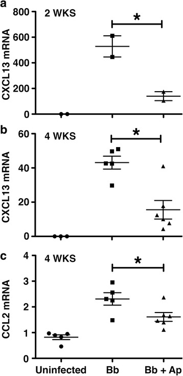
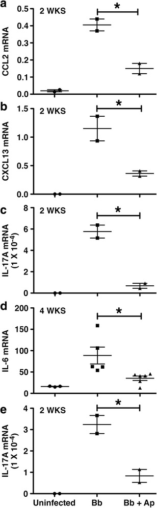
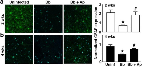
Results: We demonstrate that aprepitant treatment attenuates B. burgdorferi-induced elevations in CCL2, CXCL13, IL-17A, and IL-6 gene expression in dorsal root ganglia, spinal cord, and/or cerebrospinal fluid of rhesus macaques at 2 to 4 weeks following intrathecal infection. In addition, we demonstrate that this selective NK-1R antagonist also prevents increases in total cortical brain NK-1R expression and decreases in the expression of the astrocyte marker, glial fibrillary acidic protein, associated with B. burgdorferi infection.

Conclusions: The ability of a centrally acting NK-1R inhibitor to attenuate B. burgdorferi-associated neuroinflammatory responses and sequelae raises the intriguing possibility that such FDA-approved agents could be repurposed for use as an adjunctive therapy for the treatment of bacterial CNS infections.

Keywords: Borrelia burgdorferi; Lyme neuroborreliosis; NK-1R antagonist; Neuroinflammation; Substance P.


Figures

Fig. 1
Fig. 1
In vivo NHP infection with B. burgdorferi increases NK-1R expression in the CNS, and such increases are prevented by treatment with the NK-1R antagonist aprepitant. Rhesus macaques were uninfected (n = 2 animals) or infected intrathecally with B. burgdorferi (Bb, 1 × 108 bacteria; n = 8), and infected animals were either untreated (n = 4) or treated with aprepitant (n = 4) for 2 or 4 weeks prior to euthanasia. Expression of mRNA encoding NK-1R in frontal cortical tissue samples was determined by RT-PCR (a) and relative expression normalized to GAPDH levels was determined by densitometric analysis (b). NK-1R protein expression was determined in tissue samples by immunoblot analysis and normalized to β-actin expression (c). Data is expressed as the mean ± SD. Asterisk and pound symbols indicate statistically significant difference from uninfected animals and untreated infected animals, respectively (p < 0.05)
Fig. 2
Fig. 2
Aprepitant treatment prevents B. burgdorferi-induced increases in CCL2 protein levels in the CSF of NHPs. Rhesus macaques were uninfected (n = 5 animals) or infected intrathecally with B. burgdorferi (Bb, 1 × 108 bacteria; n = 15) and were untreated (n = 7) or treated with aprepitant (Ap, n = 8) for 2 (a) or 4 (b) weeks. Expression of CCL2 in CSF was determined by multiplex analysis. Data is expressed as the mean ± SD and asterisks indicate statistically significant differences between the untreated and treated groups (p < 0.05)
Fig. 3
Fig. 3
Aprepitant treatment prevents B. burgdorferi-induced increases in CCL2 and CXCL13 mRNA expression in the dorsal root ganglia of NHPs. Rhesus macaques were uninfected (n = 5 animals) or infected intrathecally with B. burgdorferi (Bb, 1 × 108 bacteria; n = 15), and were untreated (n = 7) or treated with aprepitant (Ap, n = 8) for 2 (a) or 4 (b, c) weeks. The level of expression of mRNA encoding CXCL13 (a, b) and CCL2 (c) in the dorsal root ganglia was determined by qPCR. Data is expressed as the mean ± SD and asterisks indicate statistically significant differences between the untreated and treated groups (p < 0.05)
Fig. 4
Fig. 4
Aprepitant treatment prevents B. burgdorferi-induced increases in CCL2, CXCL13, IL-17A, and IL-6 mRNA expression in the spinal cord of NHPs. Rhesus macaques were uninfected (n = 5 animals) or infected intrathecally with B. burgdorferi (Bb, 1 × 108 bacteria; n = 15) and were untreated (n = 7) or treated with aprepitant (Ap, n = 8) for 2 (ac, e) or 4 (d) weeks. The level of expression of mRNA encoding CCL2 (a), CXCL13 (b), IL-17A (c, e), and IL-6 (d) in the spinal cord cervical (ad) and thoracic (e) regions was determined by qPCR. Data is expressed as the mean ± SD and asterisks indicate statistically significant differences between the untreated and treated infected animal groups (p < 0.05)
Fig. 5
Fig. 5
Aprepitant treatment attenuates B. burgdorferi infection-induced reductions in astrocyte activity/numbers. Rhesus macaques were uninfected orx infected intrathecally with B. burgdorferi (Bb, 1 × 108 bacteria) and were untreated or treated with aprepitant (Ap) for 2 or 4 weeks prior to euthanasia. GFAP expression in frontal cortical tissue samples at 2 (a) and 4 (b) weeks following infection was determined by immunofluorescence microscopy. Relative GFAP expression in two fields of three sections from an animal in each group is shown and data is expressed as the mean ± SD. Asterisk and pound symbols indicate statistically significant difference from uninfected animals and untreated infected animals, respectively (p < 0.05)

Similar articles

Cited by

References

    1. Martinez AN, Philipp MT. Substance P and antagonists of the neurokinin-1 receptor in neuroinflammation associated with infectious and neurodegenerative diseases of the central nervous system. J Neurol Neuromedicine. 2016;1:29–36. - PMC - PubMed
    1. Johnson MB, Young A, Marriott I. The therapeutic potential of targeting substance P/NK-1R interactions in inflammatory CNS disorders. Front Cell Neurosci. 2016;In Press. - PMC - PubMed
    1. Bost KL. Tachykinin-modulated anti-viral responses. Front Biosci. 2004;9:1994–8.4. doi: 10.2741/1376. - DOI - PubMed
    1. Robinson P, Garza A, Moore J, Eckols TK, Parti S, Balaji V, Vallejo J, Tweardy DJ. SP is required for the pathogenesis of EMCV infection in mice. Int J Clin Exp Med. 2009;2:76–86. - PMC - PubMed
    1. Kennedy PG, Rodgers J, Jennings FW, Murray M, Leeman SE, Burke JM. A SP antagonist, RP-67,580, ameliorates a mouse meningoencephalitic response to Trypanosoma brucei brucei. Proc Natl Acad Sci U S A. 1997;94:4167–70. doi: 10.1073/pnas.94.8.4167. - DOI - PMC - PubMed

MeSH terms