. 2017 Mar 15;6(3):381-389.
doi: 10.1242/bio.024661.

A key centriole assembly interaction interface between human PLK4 and STIL appears to not be conserved in flies

Affiliations

A key centriole assembly interaction interface between human PLK4 and STIL appears to not be conserved in flies

Matthew A Cottee et al. Biol Open. .

Abstract

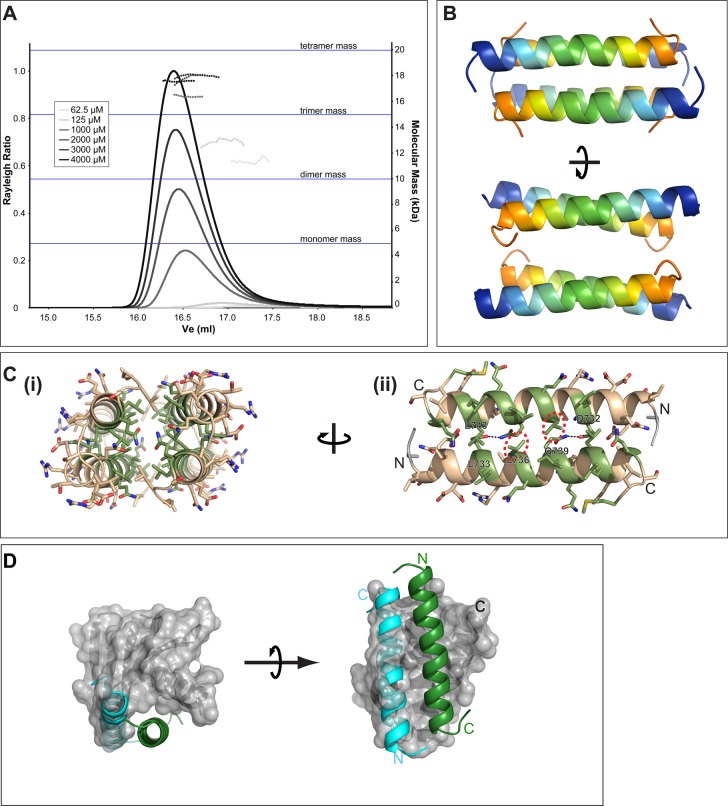
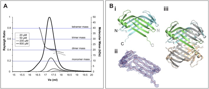
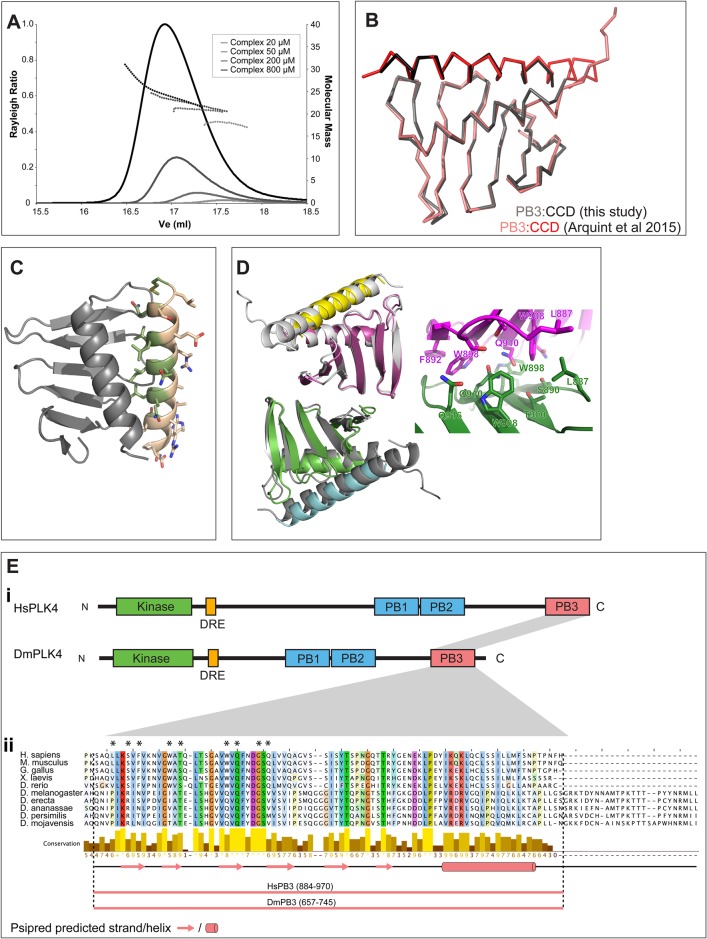
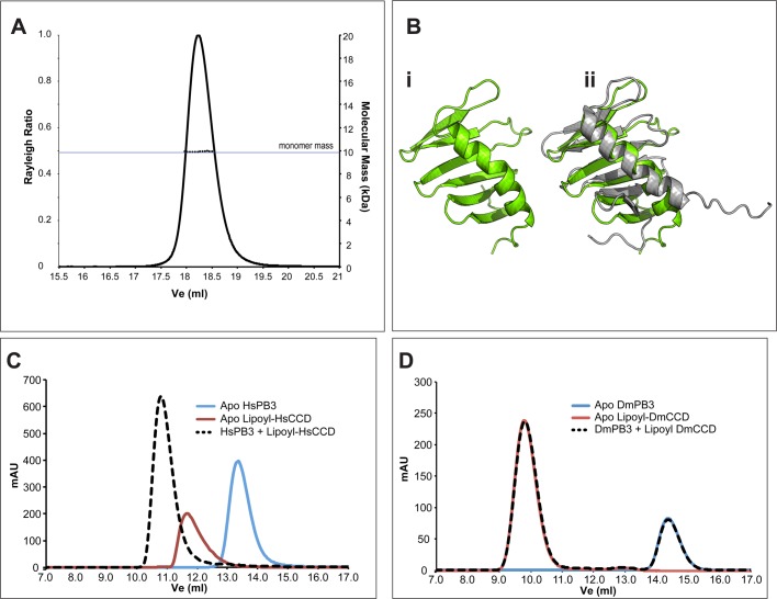
A small number of proteins form a conserved pathway of centriole duplication. In humans and flies, the binding of PLK4/Sak to STIL/Ana2 initiates daughter centriole assembly. In humans, this interaction is mediated by an interaction between the Polo-Box-3 (PB3) domain of PLK4 and the coiled-coil domain of STIL (HsCCD). We showed previously that the Drosophila Ana2 coiled-coil domain (DmCCD) is essential for centriole assembly, but it forms a tight parallel tetramer in vitro that likely precludes an interaction with PB3. Here, we show that the isolated HsCCD and HsPB3 domains form a mixture of homo-multimers in vitro, but these readily dissociate when mixed to form the previously described 1:1 HsCCD:HsPB3 complex. In contrast, although Drosophila PB3 (DmPB3) adopts a canonical polo-box fold, it does not detectably interact with DmCCD in vitro Thus, surprisingly, a key centriole assembly interaction interface appears to differ between humans and flies.

Keywords: Cartwheel; Centriole duplication; Centrosome.


Conflict of interest statement

Competing interests

The authors declare no competing or financial interests.

Figures

Fig. 1.
Fig. 1.
Sequence analysis of CCD domains of STIL/Ana2 family proteins. (A) Schematic illustration of the domain topologies of Homo sapiens STIL and Drosophila melanogaster Ana2. The sequences of the CCD domains are shown; regions predicted to be helical (Jones, 1999) are highlighted in orange. (B) Multiple sequence alignments of CCD regions from (i) Drosophila, (ii) vertebrates, and (iii) Caenorhabditis, coloured according to the ClustalX scheme. Sequences within each phylum/genus align unambiguously, but alignments between these groups are poor and are ambiguous. (C) When STIL/Ana2 family proteins are included in multiple sequence alignments, the CCD regions often align, but the exact register of these alignments is often different. i, ii, iii show alignments of the HsSTIL and DmAna2 CCD domain sequences extracted from different multiple sequence alignments. Asterisks represent identical residues and dots indicate similar residues. Residues known to be involved in either tetramerisation (Ana2) or in PB3 binding (STIL) are coloured yellow or green, respectively. The structures of the Ana2 CCD tetramer (PDB ID: 5AL6) and the STIL CCD in complex with PLK4-PB3 (PDB ID: 4YYP) were analysed by the PISA server (Krissinel and Henrick, 2007). In each alignment, CCD residues involved in the relevant interfaces are coloured according to the legend. Each alignment is unique, but in each case a similar number of residues appear conserved. No single alignment appears more feasible than any other, so it is not possible to unambiguously align these sequences.
Fig. 2.
Fig. 2.
The STIL CCD forms unstable oligomers in solution and crystallises as an antiparallel dimer of dimers. (A) SEC-MALS analysis of the STIL CCD (aa 717-758). This construct differs slightly from that used in our previous study (Cottee et al., 2015) (see Materials and Methods). Different injected protein concentrations are indicated by different shades of grey, as indicated. Solid lines represent the relative Rayleigh ratio and dashed lines show the measured masses across each peak. For reference, horizontal blue lines indicate the masses of a monomer, dimer, trimer and tetramer. The STIL CCD can be seen to self-associate in solution. The average mass of these assemblies increases with concentration and varies between dimeric to nearly tetrameric. 100 µl of each sample was injected over an S200 10/300 column. (B) The crystal structure of the STIL CCD (aa 726-750) at 0.91 Å reveals a symmetric, anti-parallel coiled-coil dimer of dimers generated by crystallographic symmetry. Each helix is shown as a cartoon coloured blue→orange, N→C. (C) (i) End-on view of the CCD anti-parallel dimer of dimers, shown as a tan cartoon and stick representation; residues that form the CCD:PB3 interface are coloured in green. (ii) Expanded view of the most closely associated dimer. Highlighted by a dashed red circle is residue L736, which is involved in both the dimerisation and PB3 interfaces. Mutation of this residue affects both STIL self-oligomerisation and PB3 binding (David et al., 2016). (D) Superposition of the dimer of HsCCD onto the previously published HsPB3:HsCCD structure (4YYP). The first HsCCD helix is modelled as a green cartoon in the HsPB3 binding site. The second copy of the STIL-CCD helix is shown as a blue cartoon and clashes with several PB3 loops (grey surface), indicating that HsCCD self-association and binding to PB3 are likely mutually exclusive.
Fig. 3.
Fig. 3.
apoHsPB3 forms a strand-swapped dimer of dimers. (A) SEC-MALS analysis of apoHsPB3 (aa884-970). Apo HsPB3 eluted as a single peak. Solid lines represent the relative Rayleigh ratio and dashed lines show the measured masses across each peak. 100 µl of apo HsPB3 at 200 µM was injected over a Superdex 200 10/300 column. (B) (i) Apo HsPB3 crystallised as a strand-swapped dimer. 60 chains were present in the asymmetric unit (ASU) of the crystal, forming 30 virtually identical strand-swapped dimers, exhibiting very strong non-crystallographic symmetry. Chains Y (green) and Z (blue) are shown in cartoon representation. (ii) The tertiary structure of the strand swapped dimers is unambiguous. Electron density map (blue mesh) carved around apo HsPB3 chain Y, at 1.3σ, showing contiguous density throughout the backbone of the chain. apo HsPB3 chain Y is shown in red ribbon representation. (iii) The HsPB3 strand-swapped dimer (green, blue and tan cartoons) forms a tetramer that is very similar to that seen in the mouse PB3 crystal (MmPB3, grey cartoon).
Fig. 4.
Fig. 4.
HsPB3 and HsCCD form a complex. (A) SEC-MALS analysis of HsPB3 mixed with HsCCD at various concentrations. Solid lines represent the relative Rayleigh ratio and dashed lines show the measured masses across each peak. 100 µl of each sample was injected over an S200 10/300 column. (B) Ribbon overlay of the PB3:CCD complex (grey:black, this study) with that previously reported (Arquint et al., 2015) (pink:red). The complexes overlay with a root-mean-square deviation (RMSD) of 0.535±0.053 Å over 85±4 Cα atoms. (C) The complex of HsPB3 (grey) with the STIL CCD (tan) in cartoon representation (this study). Three such copies were evident in the crystal ASU. CCD residues interfacing with PB3 are coloured green. (D) Overlay of a dimer of heterodimers from the HsPB3:STIL-CCD crystal (coloured cartoon) with an equivalent assembly observed in the earlier structure 4YYP (grey cartoon). Inset is a zoom on the dimer interface highlighting the highly hydrophobic nature of the interaction. (E) (i) Schematic illustration showing the domain topologies of the PLK4 orthologues from humans and D. melanogaster. (ii) Multiple sequence alignment of the PLK4 PB3 domain sequences from five vertebrates and five Drosophila species. The sequences align well and are predicted (Jones, 1999) to share similar secondary structures as annotated below the alignment. Shown below this are the domain boundaries of the HsPB3 and DmPB3 constructs used in this study. These boundaries were chosen to be topologically equivalent to other PB3 constructs used in previous studies (Arquint et al., 2015; Leung et al., 2002). Residues involved in the HsPB3:HsPB3 interaction interface shown in D are highlighted with asterisks.
Fig. 5.
Fig. 5.
Drosophila PB3 and CCD do not detectably interact. (A) SEC-MALS analysis of DmPB3. Solid lines represent the relative Rayleigh ratio and dashed lines show the measured masses across each peak. 100 µl of DmPB3 at 500 µM was injected over an S200 10/300 column. (B) (i) The crystal structure of DmPB3 at 1.53 Å shown in green in cartoon representation. The domain exhibits a canonical Polo domain fold, forming a sequential six-stranded beta sheet with an alpha helix packed against one side. (ii) As in i, but with the NMR structure of HsPB3 (Arquint et al., 2015) (PDB ID: 2N19) superimposed as a grey cartoon (RMSD=1.3 Å over 58 Cα atoms). (C) Overlaid chromatograms of analytical gel filtration (AGF) experiments on the apo HsPB3 (blue trace) and apo lipoyl-HsCCD (red trace) domains. When the two proteins were mixed, the apo peaks were no longer evident, and a larger peak (dashed black trace) was evident indicating the formation of a complex. All proteins were injected at 500 µM in each experiment. (D) Overlaid chromatograms showing the equivalent experiment to C but carried out with apo DmPB3 (blue trace) and apo lipoyl-DmCCD (red trace). When these proteins were mixed (dashed black trace), the behaviour of the apo-proteins was not detectably altered.

Similar articles

Cited by

References

    1. Afonine P. V., Grosse-Kunstleve R. W., Echols N., Headd J. J., Moriarty N. W., Mustyakimov M., Terwilliger T. C., Urzhumtsev A., Zwart P. H. and Adams P. D. (2012). Towards automated crystallographic structure refinement with phenix.refine. Acta Crystallogr. D Biol. Crystallogr. 68, 352-367. 10.1107/S0907444912001308 - DOI - PMC - PubMed
    1. Arquint C., Gabryjonczyk A.-M., Imseng S., Böhm R., Sauer E., Hiller S., Nigg E. A. and Maier T. (2015). STIL binding to Polo-box 3 of PLK4 regulates centriole duplication. eLife 4, e07888 10.7554/elife.07888 - DOI - PMC - PubMed
    1. Bettencourt-Dias M., Hildebrandt F., Pellman D., Woods G. and Godinho S. A. (2011). Centrosomes and cilia in human disease. Trends Genet. 27, 307-315. 10.1016/j.tig.2011.05.004 - DOI - PMC - PubMed
    1. Chen V. B., Arendall W. B., Headd J. J., Keedy D. A., Immormino R. M., Kapral G. J., Murray L. W., Richardson J. S. and Richardson D. C. (2010). MolProbity: all-atom structure validation for macromolecular crystallography. Acta Crystallogr. D Biol. Crystallogr. 66, 12-21. 10.1107/S0907444909042073 - DOI - PMC - PubMed
    1. Conduit P. T., Wainman A. and Raff J. W. (2015). Centrosome function and assembly in animal cells. Nat. Rev. Mol. Cell Biol. 16, 611-624. 10.1038/nrm4062 - DOI - PubMed

LinkOut - more resources