. 2017 Feb 15;6(2):165-175.
doi: 10.1242/bio.020040.

The Crumbs_C isoform of Drosophila shows tissue- and stage-specific expression and prevents light-dependent retinal degeneration

Affiliations

The Crumbs_C isoform of Drosophila shows tissue- and stage-specific expression and prevents light-dependent retinal degeneration

Stephanie Spannl et al. Biol Open. .

Abstract

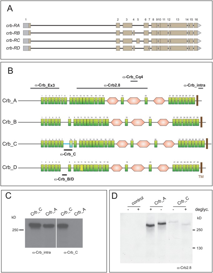
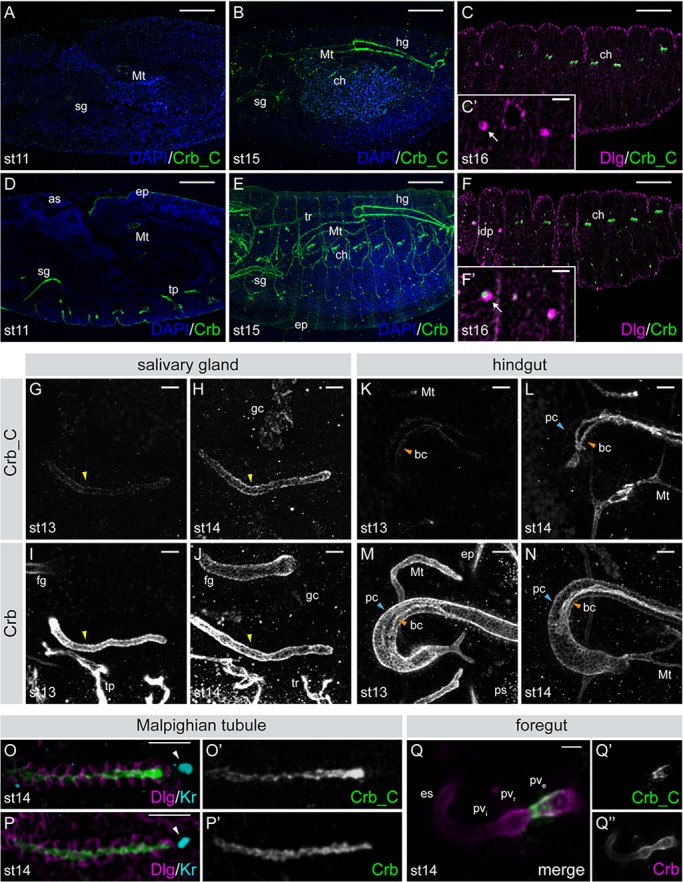
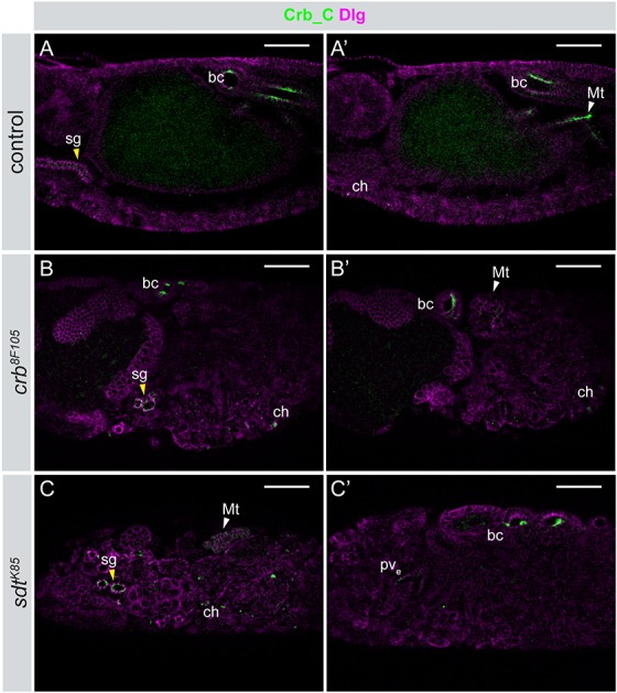
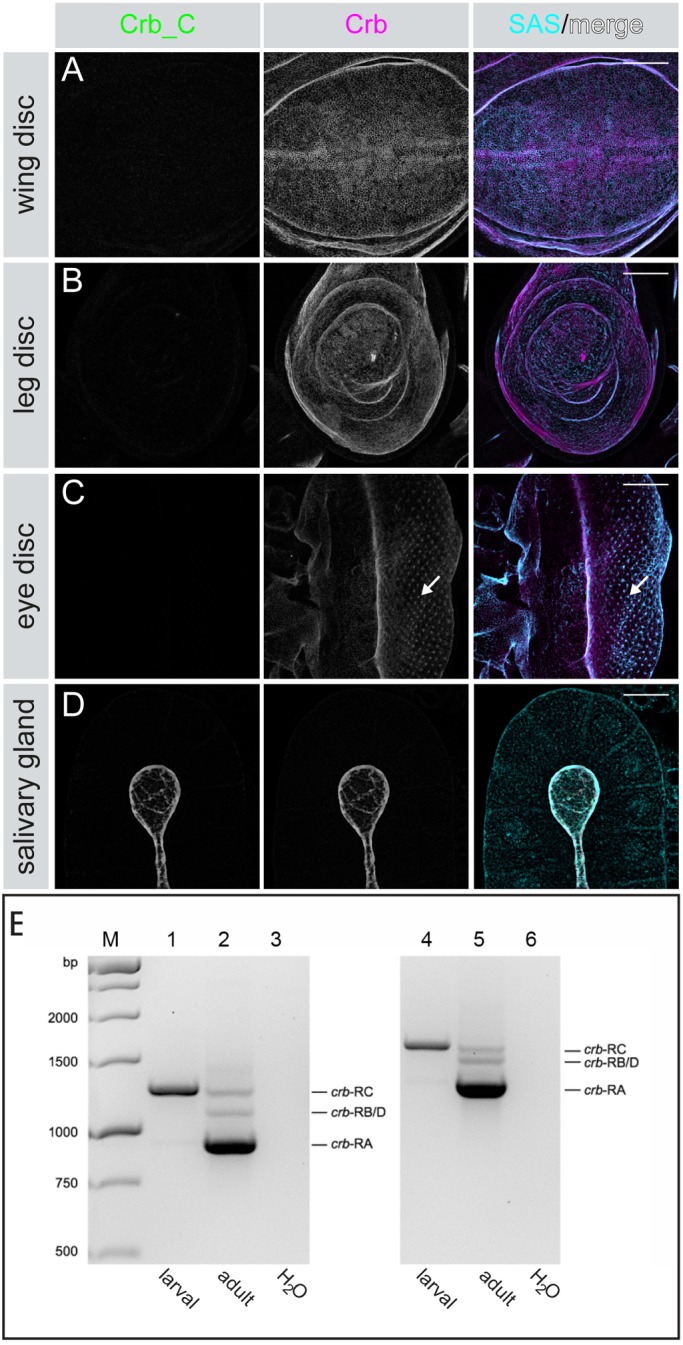
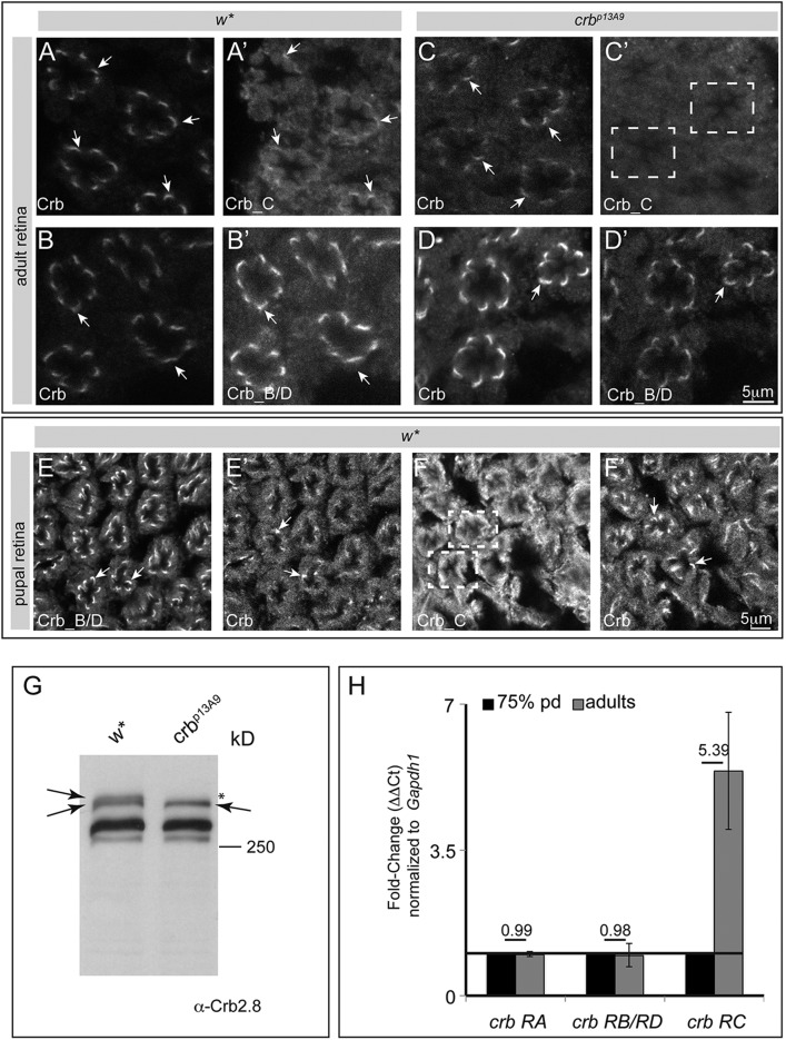
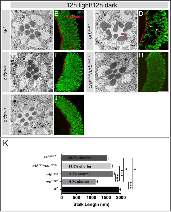
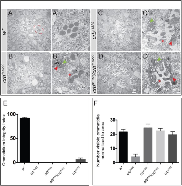
Drosophila Crumbs (Crb) is a key regulator of epithelial polarity and fulfils a plethora of other functions, such as growth regulation, morphogenesis of photoreceptor cells and prevention of retinal degeneration. This raises the question how a single gene regulates such diverse functions, which in mammals are controlled by three different paralogs. Here, we show that in Drosophila different Crb protein isoforms are differentially expressed as a result of alternative splicing. All isoforms are transmembrane proteins that differ by just one EGF-like repeat in their extracellular portion. Unlike Crb_A, which is expressed in most embryonic epithelia from early stages onward, Crb_C is expressed later and only in a subset of embryonic epithelia. Flies specifically lacking Crb_C are homozygous viable and fertile. Strikingly, these flies undergo light-dependent photoreceptor degeneration despite the fact that the other isoforms are expressed and properly localised at the stalk membrane. This allele now provides an ideal possibility to further unravel the molecular mechanisms by which Drosophila crb protects photoreceptor cells from the detrimental consequences of light-induced cell stress.

Keywords: Alternative splicing; EGF-like repeat; Epithelial polarity; Mutually exclusive exon.


Conflict of interest statement

Competing interests

The authors declare no competing or financial interests.

Figures

Fig. 1.
Fig. 1.
The crb locus encodes four different protein isoforms. (A) Four crb mRNA variants are the result of alternative splicing. Exons 1-3, 6 and 8-16 are shared. Exon 4 is specific for crb-RB and crb-RD, exon 5 is only found in crb-RC and exon 7 only in crb-RD. Grey boxes indicate 5′ and 3′ UTRs. (Adapted from Flybase.) (B) The four predicted Crb isoforms. Green rectangle: EGF-like repeat (numbering based on Crb_A), pink hexagon: repeat with similarity to the globular domain of laminin A, TM: transmembrane domain. The four isoforms have the same N-terminus, including EGF-like repeat #1-6, and share the C-terminus from EGF-like repeat #8 onwards, except for EGF-like repeat #11, which carries a 12 amino acid insertion in Crb_D. Isoforms B and D lack EGF-like repeat #7, isoform C carries an additional EGF-like repeat (#7a). The horizontal bars indicate the regions used as epitopes for immunization. Antibodies indicated on top detect all isoforms. α-Crb_C is specific for isoform C, α-Crb_B/D is specific for isoforms B and D. The blue line in isoform C indicates the isoform-specific insertion (see also Fig. S1). (The current version of SMART used does not suggest an EGF-like repeat between #8 and #9, nor an additional laminin A-like repeat N-terminal to EGF-like repeat #1 as shown in some previous publications.) (C) Western blot to demonstrate the specificity of α-Crb_C antibody. Crb_A and Crb_C were overexpressed in Drosophila S2R+ cells and cell extracts were probed with either α-Crb_intra, which detects both isoforms, or α-Crb_C. Upon strong overexpression, α-Crb_C detects two bands, which presumably represents the non-glycosylated precursor and the mature glycoprotein, as upon deglycosylation only a single band is detected (shown in D). (D) Crb_A and Crb_C were overexpressed in Drosophila S2R+ cells, cell extracts were deglycosylated and probed with α-Crb2.8. Crb_C runs slightly slower than Crb_A, independent of whether it is glycosylated or not.
Fig. 2.
Fig. 2.
Expression of Crb isoforms during embryogenesis. (A-F) Lateral view of wild-type embryos at stage 11 (A,D), stage 15 (B,E), and stage 16 (C,F) stained with α-Crb_C or α-Crb (green) and α-Discs large (Dlg; magenta); nuclei (DAPI, blue). Scale bars: 50 µm. (C′,F′) Higher magnification of the precursors of the imaginal disc (idp; arrow). Scale bars:10 µm. (G-N) Dorsal views of stage 13 and 14 embryos showing the salivary gland (sg; G-J, yellow arrowheads) and hindgut (hg; K-N). Crb_C expression increases during sg development. In the hg, expression of Crb_C is first detected in the boundary cells at stage 13 (orange arrowheads in K-N). Crb_C expression level gradually increases during embryogenesis (L). The principal cells (pc; blue arrowheads) express very low levels of Crb_C (L). (O,P) Dorsal view of the Malpighian tubules (Mt) at stage 14 stained with α-Crb_C (O,O′) or α-Crb (P,P′). Dlg (magenta) marks the lateral membranes, Krüppel (Kr, cyan) the nucleus of the distal tip cell (white arrowheads). Crb_C is predominantly expressed at the distal tip. (Q) Ventral view of the foregut stained with α-Crb_C (green, Q′) and α-Crb (magenta, Q″). α-Crb_C only stains the external portion of the proventriculus (pve), while α-Crb stains all parts. as, amnioserosa; bc, boundary cells; ch, chordotonal organs; ep, epidermis; es, esophagus; fg, foregut; gc, garland cells; hg, hindgut; idp, imaginal disc precursors; Mt, Malpighian tubule; pc, principal cells; ps, posterior spiracle; pve, external portion of the proventriculus; pvi, internal portion of the proventriculus; pvr, recurrent portion of the proventriculus; sg, salivary gland; tp, tracheal pits; tr, tracheal tree. Anterior to the left. Scale bars: 10 µm.
Fig. 3.
Fig. 3.
Apical localisation of Crb_C is independent of the Crb/Sdt interaction in Drosophila embryos. Different optical sections of control (crbGX24/+) (A/A′), homozygous crb8F105 (B/B′) and sdtK85 (C/C′) embryos stained with α-Crb_C (green) and α-Dlg (magenta). Crb_C localizes normally in the boundary cells (bc), the chordotonal organs (ch) and the salivary glands (sg; yellow arrowhead in A,B, and C) in both mutants, whereas it is delocalised in the Malpighian tubules (Mt; white arrowhead in A′,B′ and C). pve, external portion of the proventriculus. Anterior is to the left. Scale bars: 50 μm.
Fig. 4.
Fig. 4.
Postembryonic expression of Crb isoforms. (A-D) Expression of Crb isoforms in tissues of wild-type third instar larvae stained with α-Crb_C (green), α-Crb_Ex3 (magenta), and α-Stranded at Second (SAS, cyan) as apical marker. Crb localises to the apical membrane. In the eye disc, Crb is predominantly expressed behind the morphogenetic furrow, and is strongly enriched at the apical membranes of differentiating photoreceptor cells (arrow in C). Crb_C expression is not detected in the examined imaginal discs (A-C), but is observed in the larval salivary gland (D). Scale bars: 50 µm. (E) Gel electrophoresis of RT-PCR experiments to identify transcribed crb mRNAs. Larval, RNA from larval large intestine; adult, RNA from adult flies; H2O, negative control (no DNA). For primers used, see Table S2. M: 1 kb DNA Ladder. 1, 2, 3: primer pair 1, expected sizes crb-RA 919 bp, crb-RB/D 1048 bp, crb-RC 1240 bp. 4, 5, 6: primer pair 2, expected sizes crb-RA 1285 bp, crb-RB/D 1414 bp, crb-RC 1606 bp.
Fig. 5.
Fig. 5.
Expression of Crb_C in pupal and adult wild-type and mutant flies. (A-F′) Confocal images of representative optical sections from retinal whole mounts of adult (1 day old female flies; A-D′) and pupae (72 h APF; E-F′) from w* and crb p13A9 animals, stained for Crb. (A-D) α-Crb_Cq4 detects all isoforms, α-Crb_B/D is specific for Crb_B/D and α-Crb_C is specific for Crb_C. Arrows indicate stalk membrane of PRCs. Dashed white boxes in C′ and F indicate ommatidial clusters with no obvious α-Crb_C immunoreactivity at stalk membranes. Scale bar: 5 μm. (G) Western blot of protein extracts isolated from control (w*) and crbp13A9 homozygous mutant adult heads (2 days old female flies), probed with α-Crb2.8, detecting all isoforms. The upper arrow points to a slower migrating protein present in head extracts of wild-type flies, which is missing in the mutant (*). The lower arrow points to a protein that is detected in w* and crbp13A9, which co-migrates with overexpressed Crb_A (data not shown). The identity of the other bands cannot unambiguously be determined. (H) Graph depicts fold-change (on y-axis, quantified as Δ ΔCt) after normalisation with housekeeping gene Gapdh1, for different crb transcripts (on x-axis) from heads between 72 h APF (pupal stages) and newly eclosed adult. Whilst there is negligible change in crb-RA (fold-change=0.99) and crb-RB/D transcripts (fold-change=0.98), crb-RC transcript levels increase by 5.39-fold between the last day of pupal development and at eclosion (72 h APF and adulthood). Error bars depict s.e.m.
Fig. 6.
Fig. 6.
crb_C mutant photoreceptor cells exhibit normal morphology. (A-J) Representative TEM images of retinal cross sections (A,C,E,G,I) and confocal images of longitudinal retinal sections (B,D,F,H,J) stained for Chaoptin (green) and Phalloidin (red) of adult flies, prepared 2 days after eclosion (light/dark cycle). crb11A22 show mosaic eyes, all other eyes were from flies of the indicated allele/allelic combination. Fused rhabdomeres (red arrow) and incompletely elongated rhabdomeres (white arrows) are only seen in crb11A22 mosaic retinas. Scale bars: 1.7 µm (A,C,E,G,I), 50 µm (B,D,F,H,J). (K) Mean stalk length (nm)±s.e.m. of PRCs of different genotypes. Statistically significant changes between genotypes (highlighted by a black line) are indicated with ***P<0.05E-10 and *P<0.05E-4 following ANOVA and post hoc Bonferroni test. The average reduction in stalk length is 20.7% for crbp17F5, 14.5% for crbp13A9/crb11A22, 9.9% for crbp13A9, and 41% for crb11A22 with respect to the genetic control (w*).
Fig. 7.
Fig. 7.
crb_C mutant photoreceptor cells undergo light-dependent degeneration. (A-D′) Representative TEM images of retinal cross sections of adult flies of the indicated genotypes after 7 days continuous light exposure. A-D are overview images, A′-D′ are higher magnifications of one ommatidium. The stereotypic trapezoid arrangement of the seven rhabdomeres (w*) is lost in all crb alleles. Signs of degeneration include loss of rhabdomeric integrity (red asterisk), accumulation of electron dense debris (red arrowheads) and extensive vacuolization (green arrowheads). The dashed red circle in A outlines one ommatidium. Scale bars: 4 µm (A,B,C,D), 1 µm (A′,B′,C′,D′). (E) Bar graph depicting the mean Ommatidium Integrity Index (OII)±s.e.m. Ommatidial integrity index is the percent ratio of ommatidia (with no obvious signs of intracellular debris and vacuolization) to the total number of ommatidia normalized to area for the genotypes indicated on the x-axis. The significantly reduced OII in all crb alleles highlights retinal degeneration in these genotypes. (F) Bar graph depicting the mean number±s.e.m. of remnant ommatidia, normalized to area, in each of the genotypes indicated on the x -axis. Although there is degeneration in all crb mutant alleles, the phenotype is most severe in the loss-of-function allele crb11A22 (large clone mosaics), evident from the reduced number of identifiable ommatidia per unit area (compare with sections shown in the TEM panel above). Sample size consists of three biological replicates, from which at least 100 ommatidia were evaluated (with the exception of crb11A22, in which most of the ommatidia completely degenerate and only 25 ommatidia could be identified for quantification).

Similar articles

Cited by

References

    1. Alves C. H., Pellissier L. P., Vos R. M., Garcia Garrido M., Sothilingam V., Seide C., Beck S. C., Klooster J., Furukawa T., Flannery J. G. et al. (2014). Targeted ablation of Crb2 in photoreceptor cells induces retinitis pigmentosa. Hum. Mol. Genet. 23, 3384-3401. 10.1093/hmg/ddu048 - DOI - PubMed
    1. Appella E., Weber I. T. and Blasi F. (1988). Structure and function of epidermal growth factor-like regions in proteins. FEBS Lett. 231, 1-4. 10.1016/0014-5793(88)80690-2 - DOI - PubMed
    1. Bachmann A., Schneider M., Grawe F., Theilenberg E. and Knust E. (2001). Drosophila Stardust is a partner of Crumbs in the control of epithelial cell polarity. Nature 414, 638-643. 10.1038/414638a - DOI - PubMed
    1. Bazellieres E., Assemat E., Arsanto J. P., Le Bivic A. and Massey-Harroche D. (2009). Crumbs proteins in epithelial morphogenesis. Front. Biosci. 14, 2149-2169. 10.2741/3368 - DOI - PubMed
    1. Bennett E. P., Mandel U., Clausen H., Gerken T. A., Fritz T. A. and Tabak L. A. (2012). Control of mucin-type O-glycosylation: a classification of the polypeptide GalNAc-transferase gene family. Glycobiology 22, 736-756. 10.1093/glycob/cwr182 - DOI - PMC - PubMed

LinkOut - more resources