. 2017 Feb 15;6(2):290-295.
doi: 10.1242/bio.022376.

Oocyte shuttle, a recombinant protein transporting donor DNA into the Xenopus oocyte in situ

Affiliations

Oocyte shuttle, a recombinant protein transporting donor DNA into the Xenopus oocyte in situ

Duri Rungger et al. Biol Open. .

Abstract

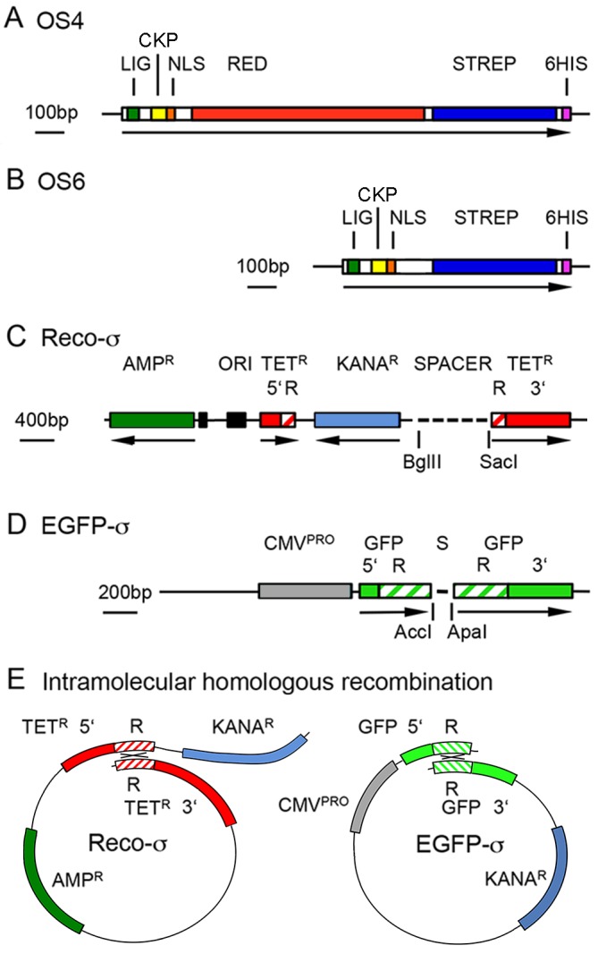
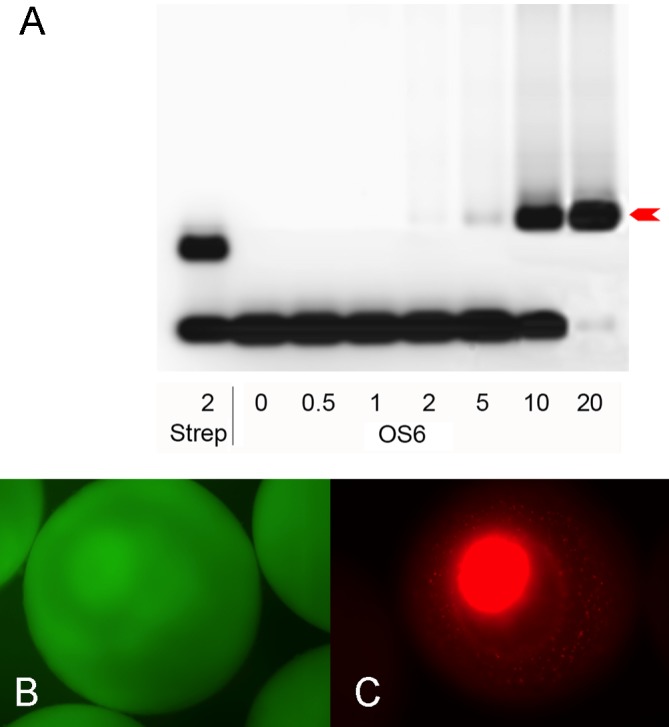
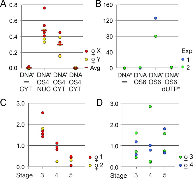
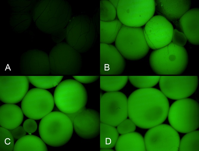
The newly developed oocyte shuttle protein contains a streptavidin moiety that tightly binds biotinylated DNA. Injected intravenously into adult Xenopus females, the protein-DNA complex is rapidly transported through the bloodstream and, within the ovary, the vitellogenin ligand present in the protein binds to the receptors at the surface of the oocytes. The bound complex is internalized and translocates into the oocyte nucleus thanks to an SV40 nuclear localization signal, enhanced by an adjacent casein kinase phosphorylation site. Functioning of the shuttle protein is documented by transporting DNA molecules that, upon intramolecular homologous recombination within the oocyte nucleus, express easily traceable markers such as green fluorescence or tetracycline resistance.

Keywords: Gene delivery; Homologous recombination; Oogenesis; Vitellogenin pathway.


Conflict of interest statement

Competing interests

The authors declare no competing or financial interests.

Figures

Fig. 1.
Fig. 1.
Recombinant plasmids. (A,B) Constructs encoding the shuttle proteins OS4 (A) and OS6 (B). LIG, vitellogenin ligand; CKP, casein kinase phosphorylation site; NLS, SV40 nuclear localization signal; RED, red fluorescent protein (in OS4 only); STREP, streptavidin core; 6HIS, His tag. The different segments were either cut from corresponding plasmids, synthetic oligonucleotides, or synthesized by PCR, adding appropriate linkers. For sequences see Fig. S1 (OS4) and Fig. S2 (OS6). (C) Donor plasmid Reco-σ with unique 5′ segment (5′), internal repeat (R) and unique 3′ segment (3′). AmpR and KanaR, genes coding for ampicillin and kanamycin resistance respectively. (D) Plasmid pEGFP-σ contains two 320 bp repeats of the coding segment of pEGFP-C1 between the BclI and GsuI sites, separated by a linker (S) with AccI and ApaI restriction sites. 5′, R, 3′, as in C. (E) Intramolecular homologous recombination within linearized Reco-σ and EGFP-σ DNAs. Crossing-over (×) takes place between the homologous repeats (R).
Fig. 2.
Fig. 2.
DNA binding and nuclear translocation. (A) Gel retardation by commercial streptavidin and OS6 protein (arrow) of an oligonucleotide that was biotinylated at the 5′ end of one strand and carried the fluorescent dye Cy5 on the 5′ end of the other. The numbers on the abscissa correspond to nominal excess of streptavidin or OS6 molecules if the extracts were 100% pure. From this titration the amount needed to bind a given number of donor DNA molecules was calculated. (B) After injection of pEGFP-C1 into albino oocytes, the green fluorescence expressed from this plasmid remains in the cytoplasm. (C) The red fluorescent shuttle protein, produced from a plasmid expressing OS4 under the control of a CMV promoter, migrates to the oocyte nucleus.
Fig. 3.
Fig. 3.
Transport of biotinylated DNA. (A) Transport from cytoplasm to nucleus in oocytes collected from two females (X,Y). Linearized Reco-σ, biotinylated (DNA*) or untreated (DNA), was injected without (–) or with OS4 shuttle protein into the cytoplasm (CYT) or nucleus (NUC). The numbers of TetR colonies obtained with DNA recovered from these oocytes were normalized (ratio) to the number of colonies obtained in the same series by direct nuclear injection of linearized Reco-σ. Red bars (Avg) depict mean values in each experimental point. (B) Transport from blood to oocyte nucleus in situ. Two independent competition experiments in which biotinylated Reco-σ alone (DNA*), non-biotinylated Reco-σ (DNA), or biotinylated Reco-σ mixed with OS6 protein, or a mixture of OS6 protein pre-incubated with excess 14biotindUTP was injected, each mix into two females. Values indicated are TetR colonies per plate. (C,D) Biotinylated Reco-σ (DNA*) linked to OS6 protein was injected into four females. After two days, DNA was extracted from batches of stage 3, 4 or 5 oocytes and used to transform bacteria that were first grown on ampicillin and then replica-plated on tetracycline plates. The uptake of donor DNA was highest in stage 3 oocytes of females 1 and 2 (C), whereas in females 3 and 4 (D), the oocytes of all stages took up variable amounts of DNA with no clear preference for a given stage. The values are given as the ratio of TetR colonies per batch over the average number of TetR colonies in all batches of the same female.
Fig. 4.
Fig. 4.
Expression of transported EGFP-σ in ovary. Fluorescent micrographs. (A) Control ovary recovered from a female injected two weeks earlier with biotinylated EGFP-σ without shuttle protein into the bloodstream. (B) Ovary and (C,D) isolated oocytes from three different females injected with biotinylated EGFP-σ bound to OS6 protein. Exposition and image treatment of the four shots were equal.

Similar articles

References

    1. Amaya E. and Kroll K. L. (1999). A method for generating transgenic frog embryos. Methods Mol. Biol. 97, 393-414. 10.1385/1-59259-270-8:393 - DOI - PubMed
    1. Bertrand P., Akhmedov A. T. and Lopez B. S. (1995). Characterization of Reca mediated homologous pairing on nitrocellulose membrane. Biochimie 77, 840-847. 10.1016/0300-9084(95)90001-2 - DOI - PubMed
    1. Bibikova M., Carroll D., Segal D. J., Trautman J. K., Smith J., Kim Y.-G. and Chandrasegaran S. (2001). Stimulation of homologous recombination through targeted cleavage by chimeric nucleases. Mol. Cell. Biol. 21, 289-297. 10.1128/mcb.21.1.289-297.2001 - DOI - PMC - PubMed
    1. Blitz I. L., Biesinger J., Xie X. and Cho K. W. Y. (2013). Biallelic genome modification in F0 Xenopus tropicalis embryos using the CRISPR/Cas system Genesis 51, 827-834. 10.1002/dvg.22719 - DOI - PMC - PubMed
    1. Carroll D. (1996). Homologous genetic recombination in Xenopus: mechanism and implications for gene manipulation. Prog. Nucleic Acid Res. Mol. Biol. 54, 101-125. 10.1016/S0079-6603(08)60361-X - DOI - PubMed

LinkOut - more resources