. 2017 Jul;28(7):2119-2132.
doi: 10.1681/ASN.2016111166. Epub 2017 Feb 15.

Deficiency of the Angiotensinase Aminopeptidase A Increases Susceptibility to Glomerular Injury

Affiliations

Deficiency of the Angiotensinase Aminopeptidase A Increases Susceptibility to Glomerular Injury

Juan Carlos Q Velez et al. J Am Soc Nephrol. 2017 Jul.

Abstract

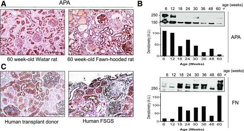
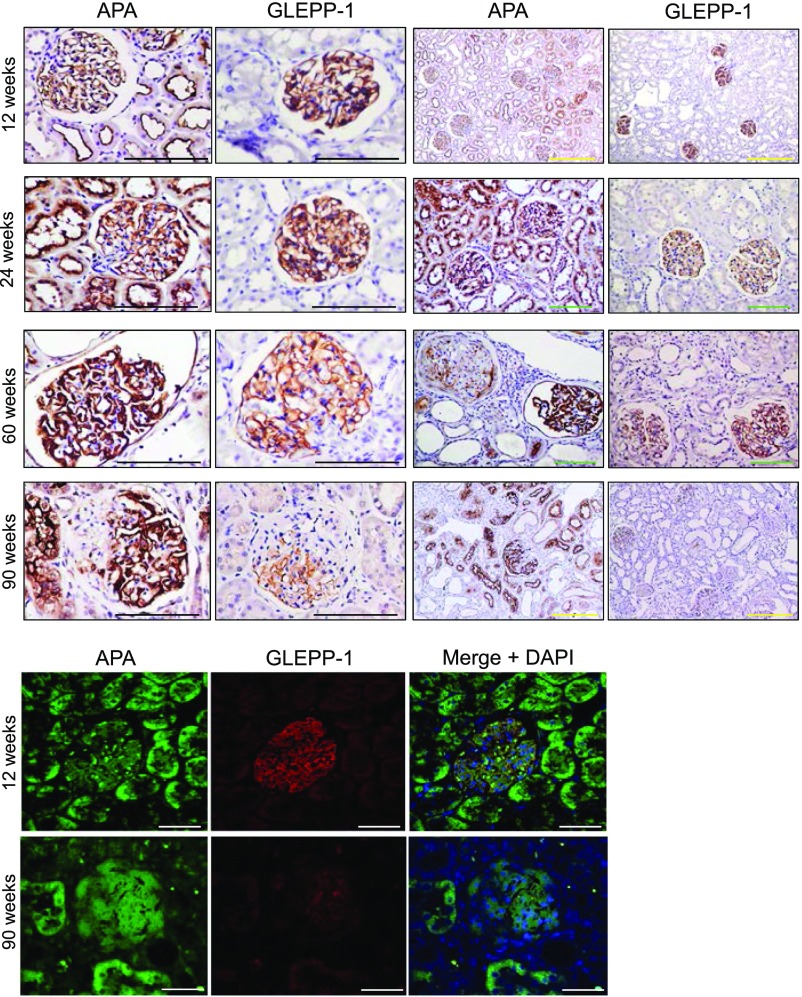
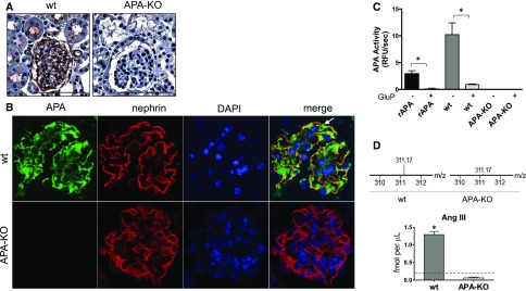
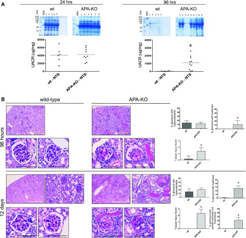
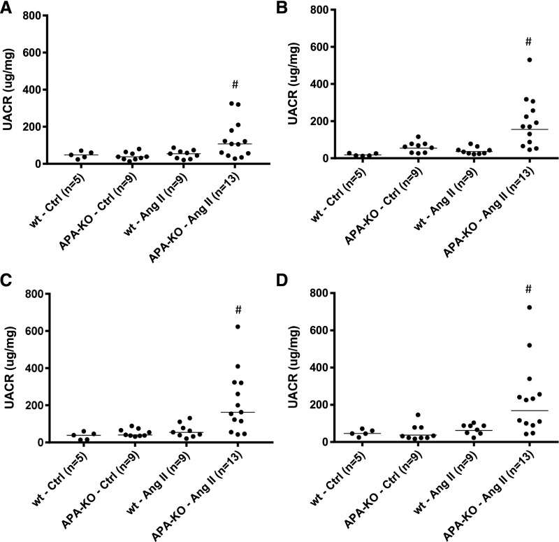
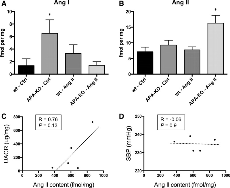
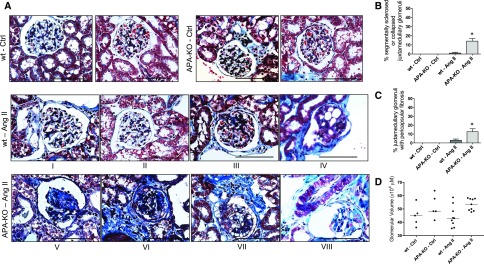
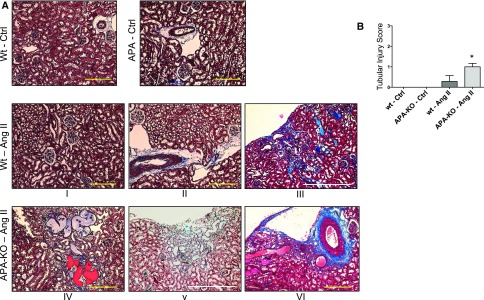
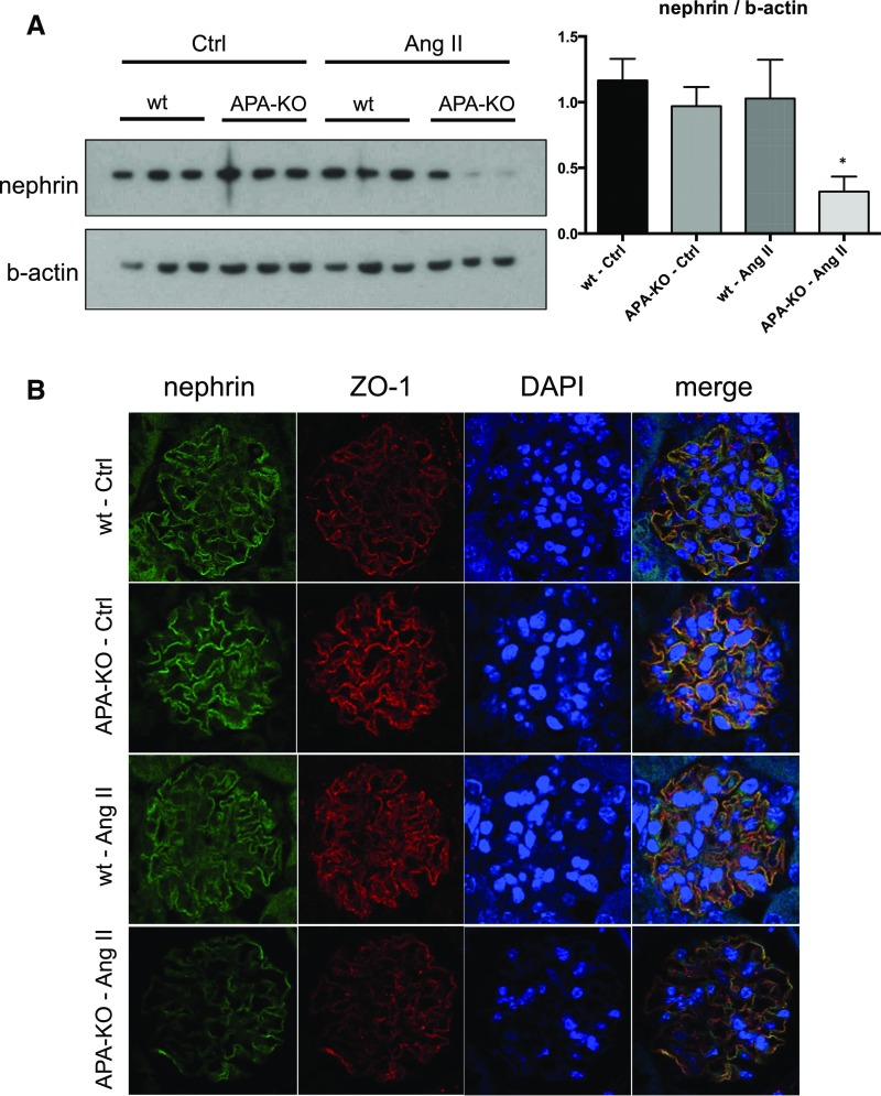
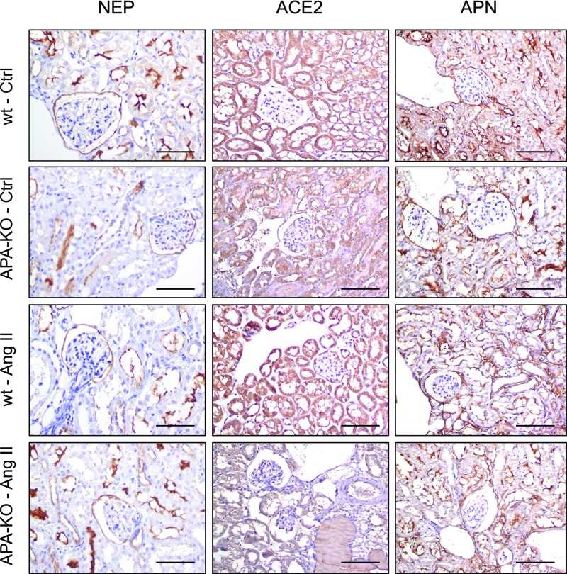
Aminopeptidase A (APA) is expressed in glomerular podocytes and tubular epithelia and metabolizes angiotensin II (AngII), a peptide known to promote glomerulosclerosis. In this study, we tested whether APA expression changes in response to progressive nephron loss or whether APA exerts a protective role against glomerular damage and during AngII-mediated hypertensive kidney injury. At advanced stages of FSGS, fawn-hooded hypertensive rat kidneys exhibited distinctly increased APA staining in areas of intact glomerular capillary loops. Moreover, BALB/c APA-knockout (KO) mice injected with a nephrotoxic serum showed persistent glomerular hyalinosis and albuminuria 96 hours after injection, whereas wild-type controls achieved virtually full recovery. We then tested the effect of 4-week infusion of AngII (400 ng/kg per minute) in APA-KO and wild-type mice. Although we observed no significant difference in achieved systolic BP, AngII-treated APA-KO mice developed a significant rise in albuminuria not observed in AngII-treated wild-type mice along with increased segmental and global sclerosis and/or collapse of juxtamedullary glomeruli, microcystic tubular dilation, and tubulointerstitial fibrosis. In parallel, AngII treatment significantly increased the kidney AngII content and attenuated the expression of podocyte nephrin in APA-KO mice but not in wild-type controls. These data show that deficiency of APA increases susceptibility to glomerular injury in BALB/c mice. The augmented AngII-mediated kidney injury observed in association with increased intrarenal AngII accumulation in the absence of APA suggests a protective metabolizing role of APA in AngII-mediated glomerular diseases.

Keywords: albuminuria; angiotensin II; glomerulosclerosis; intrarenal; murine; podocyte.


Figures

Figure 1.
Figure 1.
Pattern of glomerular APA expression changes during progressive glomerulosclerosis. Examination of glomerular APA expression in 60-week-old FHH rat kidneys by (A) immunohistochemistry. In comparison with normal age-matched Wistar rat kidneys that revealed uniform glomerular APA staining, FHH rat glomeruli showed a heterogeneous pattern of expression, with a mixture of globally sclerotic glomeruli showing virtually no APA expression, segmentally sclerotic glomeruli with enhanced APA staining in the nonsclerotic segments, and fully preserved glomeruli with overall increased APA staining. Examination of APA expression in glomerular extracts (pooled from three rats per lane) by (B) Western blotting showed progressive loss of total APA over time along with a parallel progressive increase in fibronectin (FN). The upper band for APA represents a mature transmembrane domain, whereas the lower band corresponds to an immature endoplasmic reticulum fraction. Examination of APA expression in (C) human kidneys of a transplant healthy donor and an individual with FSGS also revealed a similar distinct patter of APA expression. Scale bars, 200 µm.
Figure 2.
Figure 2.
Podocyte APA and GLEPP-1 expression evolve during early and advanced stages of FSGS. Glomerular APA expression examined by immunohistochemistry in kidney sections from FHH rats in specimens obtained at 12, 24, 60, and 90 weeks of age. Sections revealed contrasting degrees of glomerular APA expression at advanced stages. Although APA staining increased in intact glomeruli as well as intact areas of segmentally sclerosed glomeruli, it was absent in globally sclerotic glomeruli. Staining for GLEPP-1 did not show a similar expression pattern. Representative immunofluorescence images show APA staining along with faint GLEPP-1 staining in a segmentally sclerotic glomerulus at advanced stages of disease. Scale bars, 200 μm in black and green; 500 μm in yellow; 50 μm in white.
Figure 3.
Figure 3.
Lack of expression and activity of APA were verified in APA-KO mice. Examination of APA expression by (A) immunohistochemistry in kidney sections of wild-type (wt) and APA-KO mice showed glomerular and apical tubular distribution of APA in wt mice and negative staining in APA-KO mice. Scale bars, 200 µm. Examination by (B) immunofluorescence revealed colocalization of APA with nephrin, a podocyte marker, in wt mice and lack of glomerular APA expression in APA-KO mice. The white arrow indicates the body of the podocyte only showing APA staining (green), whereas colocalization of APA and nephrin corresponds to the podocyte slit diaphragms (yellow). DAPI, 4′,6-diamidino-2-phenylindole. Fluorometry-based APA enzymatic activity (C) was measured in whole-kidney homogenates harvested from wt or APA-KO mice. Bars represent mean values, and error bars show SEMs. APA activity was measured with or without the presence of the APA inhibitor glutamate phosphonate (GluP; 10 μm). Recombinant aminopeptidase A (rAPA) was used as a positive control. (D) Conversion of AngII into AngIII was verified by LC-MS/MS, showing detection of AngIII (represented by the +3 parent ion 311.17 m/z) in glomerular suspensions of wt mice and almost complete absence of AngIII in those of APA-KO mice. The horizontal dashed line in the bar graph denotes the lower limit of detection. *P<0.001.
Figure 4.
Figure 4.
Urine albumin excretion and glomerular injury persist in APA-KO mice injected with NTS. (A) Persistence of albuminuria was observed in APA-KO mice 96 hours after injection of NTS but not in wild-type (wt) mice as shown by albumin gel electrophoresis and UACR by ELISA obtained 24 and 96 hours after NTS injections. (B) Histologic examination shows exacerbated renal parenchymal injury in APA-KO mice injected with NTS. Representative light microscopy images of kidney sections stained with periodic acid–Schiff. Tissue was harvested from wt or APA-KO mice 96 hours or 12 days after treatment with NTS injection. At 96 hours, mesangial hypercellularity was observed in both groups. However, only APA-KO mouse glomeruli showed hyalinosis, tuft adhesion to the Bowman’s capsule, and microaneurysms. Tubular atrophy with protein lakes was also seen in APA-KO mice. At 12 days, in addition to the aforementioned changes, parietal cell hyperplasia was noted in APA-KO mice. Scale bars, 200 μm in black and green; 500 μm in yellow; 2 mm in white. *P<0.001; #P<0.001.
Figure 5.
Figure 5.
Global deficiency of APA leads to increased albuminuria during chronic AngII infusion. Values of urinary albumin excretion collected during a 4-week infusion of AngII (400 ng/kg per minute) in APA-KO mice. Urinary albumin excretion rates expressed in UACR in wild-type (wt) or APA-KO mice treated with chronic subcutaneous infusion of either vehicle control (Ctrl) or AngII (400 ng/kg per minute). Data presented in scatter dot plots of values obtained at (A) 1, (B) 2, (C) 3, and (D) 4 weeks showed greater magnitude of UACR in AngII-treated APA-KO mice. #P<0.01 versus all three groups.
Figure 6.
Figure 6.
Kidney content of Ang peptides after chronic AngII infusion is altered in APA-KO mice. Quantification of tissue content of (A) AngI and (B) AngII performed by LC-MS/MS in whole-kidney homogenates from wild-type (wt) or APA-KO mice treated with chronic infusion of either vehicle control (Ctrl) or AngII (400 ng/kg per minute) for 4 weeks (n=5 per group). *P<0.05. Correlation between kidney AngII content and (C) UACR and (D) SBP) is shown.
Figure 7.
Figure 7.
Histologic examination shows exacerbated glomerular injury in APA-KO mice after chronic AngII infusion. (A) Representative light microscopy images of kidney sections stained with Masson trichrome. Tissue was harvested from wild-type (wt) or APA-KO mice treated with chronic subcutaneous infusion of either vehicle control (Ctrl) or AngII (400 ng/kg per minute) for 4 weeks. (B and C) Bar graphs show comparison of the percentages of juxtamedullary glomeruli with segmental (III, IV, and VII) or global (V and VI) sclerosis, collapse (V and VIII), or microaneurysms (IV and VIII). (D) Evidence of glomerular enlargement was compared between groups. *P<0.01. Scale bars, 200 μm.
Figure 8.
Figure 8.
Histologic examination shows exacerbated tubulointerstitial injury in APA-KO mice after chronic AngII infusion. (A) Representative light microscopy images of kidney sections stained with Masson trichrome. Tissue was harvested from wild-type (wt) or APA-KO mice treated with chronic subcutaneous infusion of either vehicle (Ctrl) or AngII (400 ng/kg per minute) for 4 weeks. (B) Bar graph shows comparison of tubulointerstitial injury scores that accounted for patchy subcapsular fibrosis (III and V) and microcystic tubular dilation (III–VI). *P<0.01. Scale bars, 500 μm in yellow; 2 mm in white.
Figure 9.
Figure 9.
Podocyte protein expression changes observed in APA-KO mice after chronic AngII infusion. Representative images depicting the effect of chronic infusion of AngII on (A) nephrin and β-actin as examined by Western blotting in whole-kidney homogenates and (B) nephrin and ZO-1 expression as examined by immunofluorescence in 5 µm kidney sections. Graph shown next to the Western blot reflects quantification of band densities from five individual mouse kidney homogenates from the wild-type (wt) control (Ctrl) group and six of each of the other three treatment groups. *P<0.01.
Figure 10.
Figure 10.
RAS peptidase expression does not change in APA-KO mice after chronic AngII infusion. Representative images depicting the effect of chronic infusion of AngII on expression of NEP, ACE2, and APN as examined by immunofluorescence. Glomerular parietal epithelial and tubular epithelial cell distribution was noted for all three enzymes, with faint podocyte localization. No difference in degree of staining was observed across treatment groups. Ctrl, control; wt, wild type. Scale bars, 200 μm.

Similar articles

Cited by

References

    1. Bauer J, Berthold H, Schaefer F, Ehmke H, Parekh N: Quantification of conversion and degradation of circulating angiotensin in rats. Am J Physiol 277: R412–R418, 1999 - PubMed
    1. Velez JC, Bland AM, Arthur JM, Raymond JR, Janech MG: Characterization of renin-angiotensin system enzyme activities in cultured mouse podocytes. Am J Physiol Renal Physiol 293: F398–F407, 2007 - PubMed
    1. Velez JC: Prolyl carboxypeptidase: A forgotten kidney angiotensinase. Focus on “Identification of prolyl carboxypeptidase as an alternative enzyme for processing of renal angiotensin II using mass spectrometry.” Am J Physiol Cell Physiol 304: C939–C940, 2013 - PMC - PubMed
    1. Yang HY, Erdös EG, Chiang TS: New enzymatic route for the inactivation of angiotensin. Nature 218: 1224–1226, 1968 - PubMed
    1. Allred AJ, Diz DI, Ferrario CM, Chappell MC: Pathways for angiotensin-(1---7) metabolism in pulmonary and renal tissues. Am J Physiol Renal Physiol 279: F841–F850, 2000 - PubMed

Substances