Review
. 2017 Jan 15;31(2):101-126.
doi: 10.1101/gad.291518.116.

PARPs and ADP-ribosylation: recent advances linking molecular functions to biological outcomes

Affiliations
Review

PARPs and ADP-ribosylation: recent advances linking molecular functions to biological outcomes

Rebecca Gupte et al. Genes Dev. .

Abstract

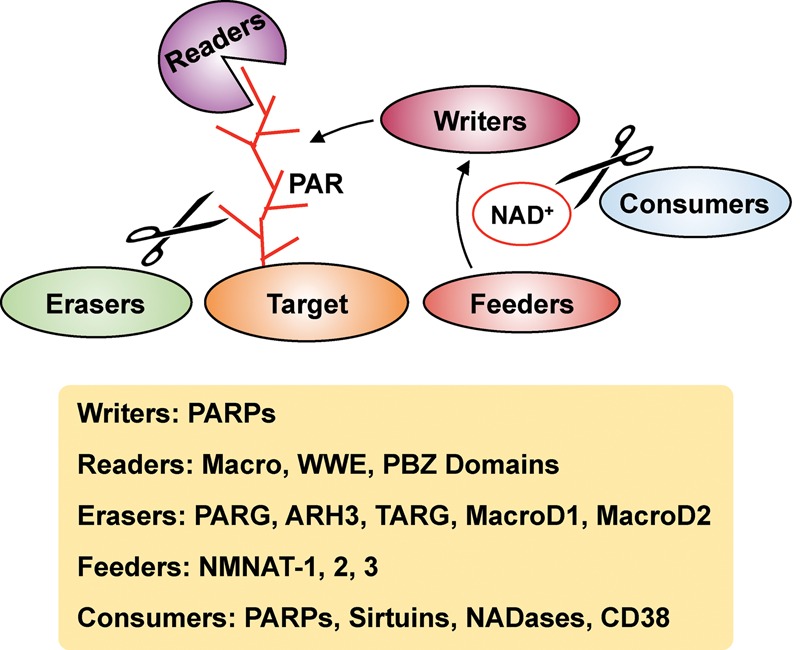
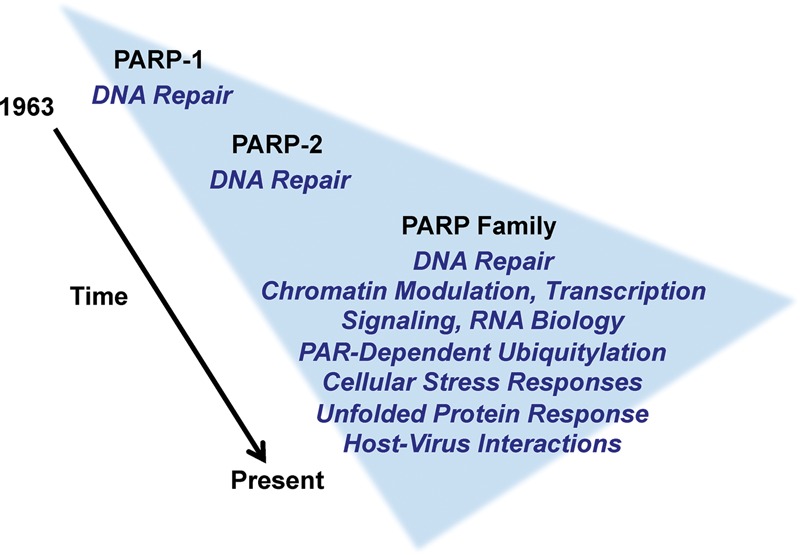
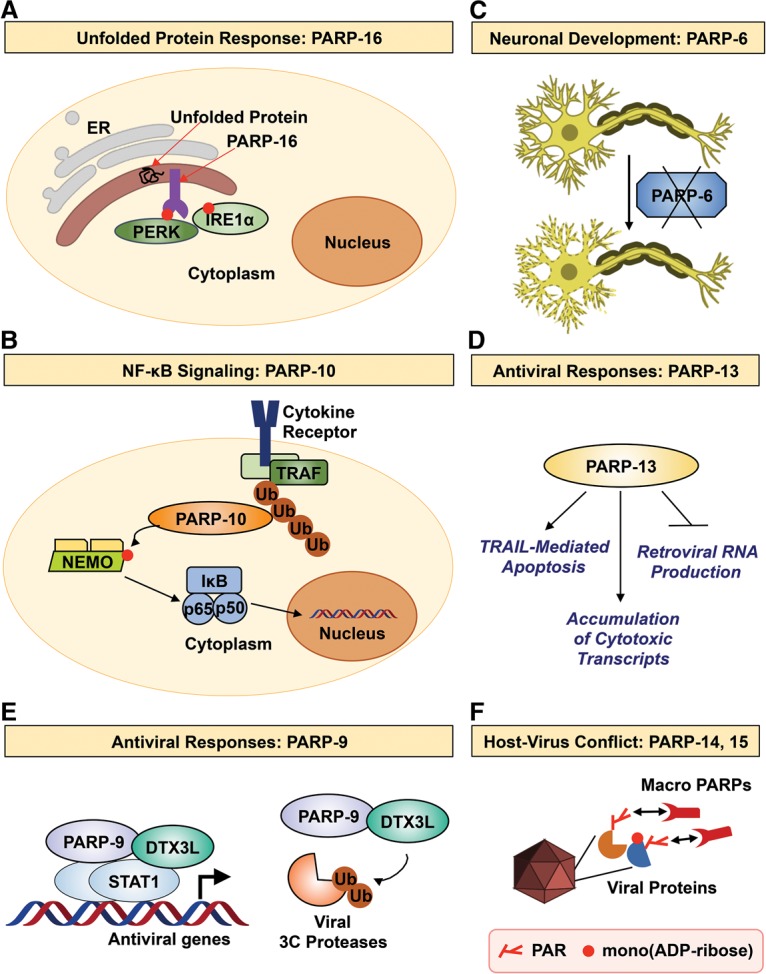
The discovery of poly(ADP-ribose) >50 years ago opened a new field, leading the way for the discovery of the poly(ADP-ribose) polymerase (PARP) family of enzymes and the ADP-ribosylation reactions that they catalyze. Although the field was initially focused primarily on the biochemistry and molecular biology of PARP-1 in DNA damage detection and repair, the mechanistic and functional understanding of the role of PARPs in different biological processes has grown considerably of late. This has been accompanied by a shift of focus from enzymology to a search for substrates as well as the first attempts to determine the functional consequences of site-specific ADP-ribosylation on those substrates. Supporting these advances is a host of methodological approaches from chemical biology, proteomics, genomics, cell biology, and genetics that have propelled new discoveries in the field. New findings on the diverse roles of PARPs in chromatin regulation, transcription, RNA biology, and DNA repair have been complemented by recent advances that link ADP-ribosylation to stress responses, metabolism, viral infections, and cancer. These studies have begun to reveal the promising ways in which PARPs may be targeted therapeutically for the treatment of disease. In this review, we discuss these topics and relate them to the future directions of the field.

Keywords: DNA repair; RNA biology; gene regulation; mono(ADP-ribose) (MAR); poly(ADP-ribose) (PAR); poly(ADP-ribose) polymerase (PARP).


Figures

Figure 1.
Figure 1.
A variety of effectors mediate intracellular ADP-ribosylation dynamics. PARPs act as “writers” that add ADP-ribose moieties to target proteins. The NAD+ required for these PARP-mediated ADP-ribosylation reactions is supplied by nicotinamide mononucleotide adenylyl transferases (NMNATs; “feeders”). The ADP-ribose units on target protein can be recognized by “readers” containing macro, WWE, or PAR-binding zinc finger (PBZ) domains. The removal of ADP-ribose chains is catalyzed by “erasers,” which include PAR glycohydrolase (PARG), ADP-ribosyl hydrolase 3 (ARH3), TARG, and MacroD1/D2. NAD+ levels can be modulated by NAD+ “consumers,” such as PARPs, sirtuins, NADases, and CD38, which hydrolyze NAD+.
Figure 2.
Figure 2.
Structures of ARBDs. (A) The ARBDs shown include a macrodomain from Archaeoglobus fulgidus Af1521 (Protein Data Bank [PDB] 2BFQ), a WWE domain from human RNF146 (PDB 3V3L), and a PBZ motif from human CHFR (PDB 2XOY). The ARBDs are shown in blue, and the ADP-ribose ligands are highlighted in red. (B) Schematic showing the structures of example proteins containing ARBDs: human PARG (macrodomain), human RNF146 (WWE domain), and human CHFR (PBZ motif). The different domains in the proteins are indicated.
Figure 3.
Figure 3.
A time line of discoveries for PARPs and ADP-ribosylation. The schematic illustrates our expanding understanding of the functions of PARP family members in different biological processes. Details are discussed in the section “Recurring Themes in PARP Biology: DNA Repair, Transcription, Signaling, and Beyond, with a Focus on PARP-1.”
Figure 4.
Figure 4.
Varied roles of ADP-ribosylation in the regulation of gene regulation. (A) PAR turnover plays a critical role in hormone-dependent gene expression by generating ATP. Free PAR from PARG hydrolysis is further broken down by NUDIX5 to produce ATP. The ATP generated is used by ATP-dependent chromatin remodeling enzymes to modulate nucleosome occupancy at progesterone receptor (PR) target genes to stimulate transcription (Wright et al. 2016). (B) PARP-1 regulates the release of promoter-proximally paused RNA polymerase II into productive transcriptional elongation through ADP-ribosylation of the negative elongation factor (NELF) complex. Phosphorylation of NELF by the P-TEFb (positive-transcription elongation factor b) complex and subsequent ADP-ribosylation by PARP-1 results in the dissociation of NELF from RNA polymerase II, and the resulting release of pausing triggers productive elongation (Gibson et al. 2016). (C) ADP-ribosylation of C/EBPβ regulates the adipogenic transcriptional program. ADP-ribosylation by PARP-1 inhibits the binding of C/EBPβ to DNA. Upon exposure to adipogenic stimuli, there is loss of C/EBPβ PARylation and subsequent DNA binding. This turns on the expression of C/EBPβ-dependent proadipogenic genes (Luo et al. 2017).
Figure 5.
Figure 5.
PARP monoenzymes and catalytically inactive PARPs participate in diverse biological processes. (A) PARP-16 regulates the UPR by modulating PERK and IRE1α activity. (B) PARP-10 attenuates NF-κB signaling by inhibiting NEMO. (C) PARP-6 is required for neurogenesis in the hippocampus. (D) PARP-13 inhibits viral pathogens by promoting TRAIL-mediated apoptosis, accumulation of cytotoxic transcripts, and inhibition of retroviral mRNA production. (E) PARP-9 associates with deltex E3 ubiquitin ligase 3L (DTX3L). Together, they promote antiviral gene transcription and trigger degradation of viral 3C proteases. (F) The macroPARPs PARP-14 and PARP-15 are involved in host–virus conflicts via their rapidly evolving macrodomains. See the text for details.
Figure 6.
Figure 6.
New technologies to detect and study cellular ADP-ribosylation. The schematic illustrates different technologies that are discussed in detail in the text.
Figure 7.
Figure 7.
Chemistry of PAR cleavage for use in MS methods. (A) Chemical structure of a PAR covalently linked to an amino acid in a target protein. (BD) Structure of terminal moieties attached to an amino acid in the target protein after ADP-ribose cleavage using hydroxylamine (B), snake venom phosphodiesterase (SVP) or NUDIX (C), or PARG (D). The chemical structures shown in AD are for Glu and Asp residues. Note that SVP, NUDIX, and PARG can also hydrolyze ADP-ribose linked to Lys and Arg residues (not shown). The exact nature of the chemical structures for SVP-, NUDIX-, and PARG-cleaved Lys-ADP-ribose and Arg-ADP-ribose have not been determined experimentally (Daniels et al. 2015a).
Figure 8.
Figure 8.
Chemical biology approaches to identify PARP-specific ADP-ribosylation events. (A) Molecular structures of the NAD+ analogs used by Carter-O'Connell et al. (2014) (top) and Gibson et al. (2016) (bottom) for their analog-sensitive PARP approaches. The substituents for NAD+, each analog as well as the “clickable” groups, are highlighted in pink. (B) Schematic of human PARP-1 showing the different protein domains. The highlighted amino acids in human PARP-1 were mutated by Carter-O'Connell et al. (2014) and Gibson et al. (2016) to confer analog sensitivity.
Figure 9.
Figure 9.
Comparison of techniques used to map genome-wide ADP-ribosylation. (A) ADPr-ChAP by Bartolomei et al. (2016). (B) Click-ChIP-seq (click chemistry-based chromatin isolation and precipitation with deep sequencing) by Gibson et al. (2016). Details of the protocols are provided in the text.

References

    1. Adelman K, Lis JT. 2012. Promoter-proximal pausing of RNA polymerase II: emerging roles in metazoans. Nat Rev Genet 13: 720–731. - PMC - PubMed
    1. Adhya D, Basu A. 2010. Epigenetic modulation of host: new insights into immune evasion by viruses. J Biosci 35: 647–663. - PubMed
    1. Aguilar-Quesada R, Munoz-Gamez JA, Martin-Oliva D, Peralta A, Valenzuela MT, Matinez-Romero R, Quiles-Perez R, Menissier-de Murcia J, de Murcia G, Ruiz de Almodovar M, et al. 2007. Interaction between ATM and PARP-1 in response to DNA damage and sensitization of ATM deficient cells through PARP inhibition. BMC Mol Biol 8: 29. - PMC - PubMed
    1. Aguilera-Gomez A, van Oorschot MM, Veenendaal T, Rabouille C. 2016. In vivo vizualisation of mono-ADP-ribosylation by dPARP16 upon amino-acid starvation. Elife 5: e21475. - PMC - PubMed
    1. Ahel I, Ahel D, Matsusaka T, Clark AJ, Pines J, Boulton SJ, West SC. 2008. Poly(ADP-ribose)-binding zinc finger motifs in DNA repair/checkpoint proteins. Nature 451: 81–85. - PubMed

Publication types

MeSH terms

Substances