. 2017 Mar 31;292(13):5271-5281.
doi: 10.1074/jbc.M117.776369. Epub 2017 Feb 15.

Nucleosome-like, Single-stranded DNA (ssDNA)-Histone Octamer Complexes and the Implication for DNA Double Strand Break Repair

Affiliations

Nucleosome-like, Single-stranded DNA (ssDNA)-Histone Octamer Complexes and the Implication for DNA Double Strand Break Repair

Nicholas L Adkins et al. J Biol Chem. .

Abstract

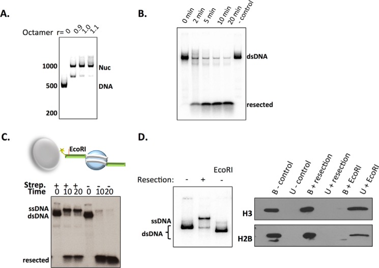
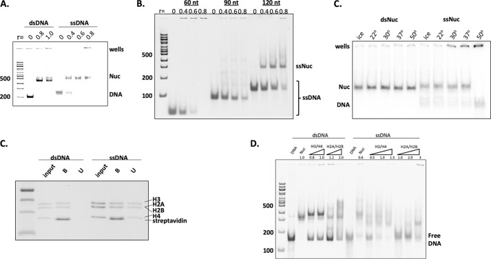
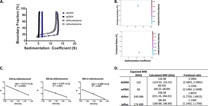
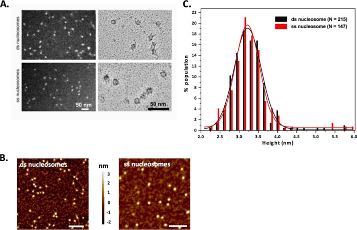
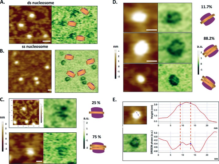
Repair of DNA double strand breaks (DSBs) is key for maintenance of genome integrity. When DSBs are repaired by homologous recombination, DNA ends can undergo extensive processing, producing long stretches of single-stranded DNA (ssDNA). In vivo, DSB processing occurs in the context of chromatin, and studies indicate that histones may remain associated with processed DSBs. Here we demonstrate that histones are not evicted from ssDNA after in vitro chromatin resection. In addition, we reconstitute histone-ssDNA complexes (termed ssNucs) with ssDNA and recombinant histones and analyze these particles by a combination of native gel electrophoresis, sedimentation velocity, electron microscopy, and a recently developed electrostatic force microscopy technique, DREEM (dual-resonance frequency-enhanced electrostatic force microscopy). The reconstituted ssNucs are homogenous and relatively stable, and DREEM reveals ssDNA wrapping around histones. We also find that histone octamers are easily transferred in trans from ssNucs to either double-stranded DNA or ssDNA. Furthermore, the Fun30 remodeling enzyme, which has been implicated in DNA repair, binds ssNucs preferentially over nucleosomes, and ssNucs are effective at activating Fun30 ATPase activity. Our results indicate that ssNucs may be a hallmark of processes that generate ssDNA, and that posttranslational modification of ssNucs may generate novel signaling platforms involved in genome stability.

Keywords: DNA repair; DSB processing; analytical ultracentrifugation; atomic force microscopy (AFM); chromatin remodeling; nucleosome; ssDNA.


Conflict of interest statement

The authors declare that they have no conflicts of interest with the contents of this article

Figures

FIGURE 1.
FIGURE 1.
Histone fate after in vitro chromatin resection by the Sgs1-Dna2 pathway. A, native PAGE of a 500-bp DNA fragment harboring a central, 601-positioning sequence reconstituted into mononucleosomes by salt step dialysis at different ratios (r) of histone octamers to DNA. Note that the minor nucleosome species is likely to represent a nucleosome assembled on the DNA end. B and C, chromatin resection time course with 3′-radiolabeled chromatin and reactions that contain Mre11-Rad50-Xrs2, Sgs1, Top3-Rmi complex, Dna2, and RPA. C, addition of streptavidin-coated magnetic beads inhibit chromatin resection on one strand. Note the appearance of slower migrating ssDNA. D, analysis of DNA and protein content following magnetic DNA pulldown after chromatin resection. Left panel, radiolabel analysis of DNA before (−) and after (+) resection. One sample was also treated with EcoRI prior to resection and the released DNA was analyzed. Right panel, histone immunoblotting of bead bound (B) and unbound (U) fractions before and after chromatin resection.
FIGURE 2.
FIGURE 2.
Biochemical characterization of reconstituted mononucleosomes. A, native PAGE of the indicated nucleic acid with increasing histone ratios (r) after reconstitution by salt step dialysis. Note that ssDNA stains less intensely with ethidium bromide. B, 4% native PAGE of ssDNA-histone octamer reconstitutions, using radiolabeled DNA fragments of varying length. r, histone octamer:DNA molar ratio. C, the stability of double-stranded and single-stranded nucleosomes after a 1-h incubation at the indicated range of temperatures, analyzed by separation on a 4% native PAGE. D, 200-nt ssDNA or 200-bp dsDNA chromatin reconstitutions were immobilized on streptavidin-coated magnetic beads, and histone content was analyzed after magnetic pulldown by SDS-PAGE. E, the indicated histone complexes were used in chromatin reconstitution reactions with 150-bp or 150-nt DNA fragments. Reconstitutions were analyzed by 4% native PAGE.
FIGURE 3.
FIGURE 3.
Analysis of ssDNA-histone complexes by sedimentation velocity analyses. A, van Holde Weischet analysis of analytical ultracentrifugation sedimentation profiles for 200-bp dsDNA, 200-nt ssDNA, ssNucs, and mononucleosomes. B, 2DSA/GA-MC modeling of samples analyzed in A. Note the presence of a single, major solute for both ssNucs (lower panel) and nucleosomes (upper panel). C, experimental determination of the ¯v parameter. Plots of sedimentation coefficient versus solvent density of the indicated particles sedimented in three different ratios of heavy oxygen to distilled water. Numbers in brackets represent the ¯v of the respective molecule as predicted by UltraScan3. D, native molecular weights (MW) and f/f0 ratios (frictional ratio) derived from 2DSA/GA-MC modeling. The 95% confidence intervals are shown in parentheses.
FIGURE 4.
FIGURE 4.
EM and AFM imaging of ssNucs and dsnucleosomes. A, electron microscopy of ds (top) and ss (bottom) nucleosomes. Samples were stained with uranyl acetate and processed for dark-field (left panels) or bright-field (right panels) imaging. B, AFM topographic images of ds (left) and ss (right) nucleosomes. x-y scale bar = 100 nm. C, the AFM height distributions for double and single strand nucleosomes. The solid lines show the Gaussian fit to the data. The average heights of double and single strand nucleosomes are 3.2 ± 0.7 nm (S.D.) (n = 215) and 3.2 ± 0.6 nm (S.D.) (n = 147), respectively.
FIGURE 5.
FIGURE 5.
DREEM phase images of nucleosomes and ssNucs. A and B, AFM topography (left panels) and DREEM phase images (right panels) of double (A) and single (B) strand nucleosomes. The models depict the orientation of DNA based on DREEM images. C and D, AFM topography (left panels) and DREEM phase images (right panels) of double strand (C) and single strand (D) nucleosomes showing one strand (top panels) or two strands of DNA (bottom panels). x-y scale bar = 10 nm. The panels on the right show the models based on DREEM phase images and percentages of nucleosomes with one strand or two strands of DNA revealed in DREEM images. The total number of nucleosomes analyzed are 44 and 222 for double and single strand nucleosomes, respectively. E, cross-section analysis of one of the single strand nucleosome shown in D.
FIGURE 6.
FIGURE 6.
Single strand nucleosomes transfer histone octamers between DNA strands. A, native PAGE of radiolabeled naked DNA (5 nm) incubated with increasing concentrations of unlabeled, ssNucs (5, 10, 20, 50, 100 nm) (left) or unlabeled, nucleosomes (right). Lanes 1 and 8 of each panel contains radiolabeled nucleosome or ssNuc as a marker. B, native PAGE of radiolabeled, single strand nucleosomes (1 nm) incubated with increasing concentrations of circular and linear naked DNA (0.2, 1, 2, 10, and 20 nm). Lanes 1 and 8 contain radiolabeled, free nucleosome template DNA as a marker.
FIGURE 7.
FIGURE 7.
The ATP-dependent remodeling enzyme Fun30 prefers single-stranded chromatin. A, SDS-PAGE analysis of purified Fun30 and RSC enzymes. MVM, molecular weight marker. B, electrophoretic mobility shift assay of RSC (left) and Fun30 (right) binding to nucleosomes (dsNuc) or ssNucs. Michaelis-Menten kinetics (C) and Km parameters (D) for ATPase activities of RSC and Fun30 with the indicated substrates. Units for Km values are nm.

References

    1. Khanna K. K., and Jackson S. P. (2001) DNA double-strand breaks: signaling, repair and the cancer connection. Nat. Genet. 27, 247–254 - PubMed
    1. Ciccia A., and Elledge S. J. (2010) The DNA damage response: making it safe to play with knives. Mol. Cell 40, 179–204 - PMC - PubMed
    1. Papamichos-Chronakis M., and Peterson C. L. (2013) Chromatin and the genome integrity network. Nat. Rev. Genet. 14, 62–75 - PMC - PubMed
    1. Pâques F., and Haber J. E. (1999) Multiple pathways of recombination induced by double-strand breaks in Saccharomyces cerevisiae. Microbiol. Mol. Biol. Rev. 63, 349–404 - PMC - PubMed
    1. Niu H., Chung W.-H., Zhu Z., Kwon Y., Zhao W., Chi P., Prakash R., Seong C., Liu D., Lu L., Ira G., and Sung P. (2010) Mechanism of the ATP-dependent DNA end-resection machinery from Saccharomyces cerevisiae. Nature 467, 108–111 - PMC - PubMed

MeSH terms

LinkOut - more resources