Review
. 2017 Feb 15;220(Pt 4):517-530.
doi: 10.1242/jeb.134056.

Mussel adhesion - essential footwork

Affiliations
Review

Mussel adhesion - essential footwork

J Herbert Waite. J Exp Biol. .

Abstract

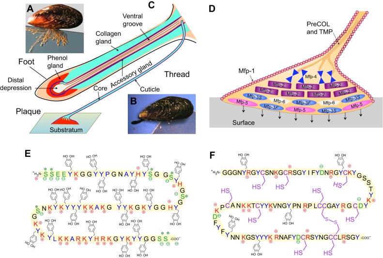
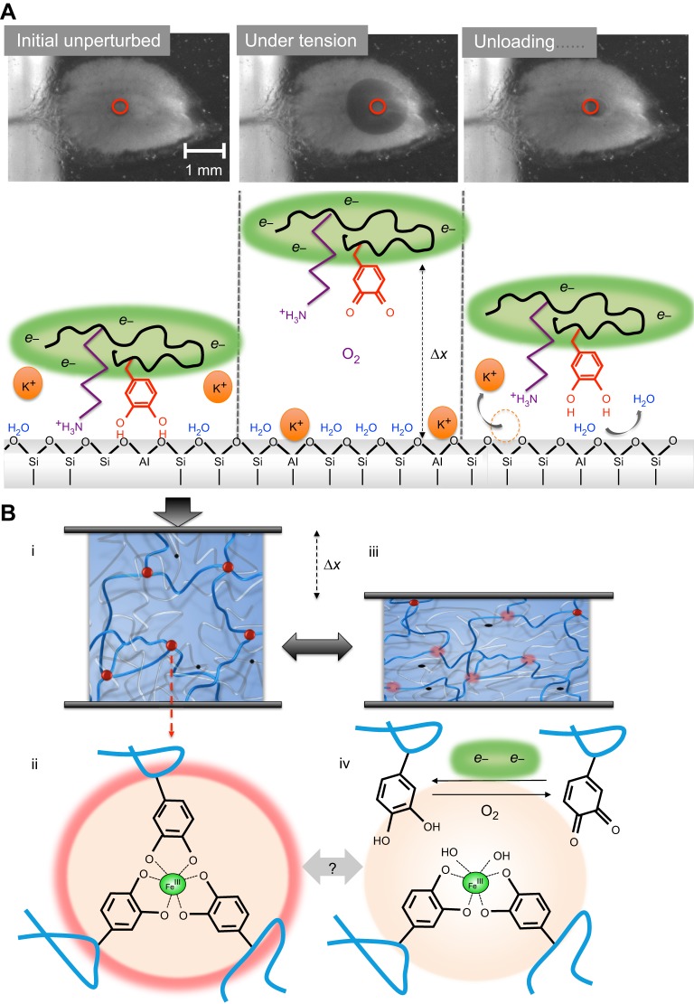
Robust adhesion to wet, salt-encrusted, corroded and slimy surfaces has been an essential adaptation in the life histories of sessile marine organisms for hundreds of millions of years, but it remains a major impasse for technology. Mussel adhesion has served as one of many model systems providing a fundamental understanding of what is required for attachment to wet surfaces. Most polymer engineers have focused on the use of 3,4-dihydroxyphenyl-l-alanine (Dopa), a peculiar but abundant catecholic amino acid in mussel adhesive proteins. The premise of this Review is that although Dopa does have the potential for diverse cohesive and adhesive interactions, these will be difficult to achieve in synthetic homologs without a deeper knowledge of mussel biology; that is, how, at different length and time scales, mussels regulate the reactivity of their adhesive proteins. To deposit adhesive proteins onto target surfaces, the mussel foot creates an insulated reaction chamber with extreme reaction conditions such as low pH, low ionic strength and high reducing poise. These conditions enable adhesive proteins to undergo controlled fluid-fluid phase separation, surface adsorption and spreading, microstructure formation and, finally, solidification.

Keywords: Dopa; Foot behavior; Interfacial chemistry; Mussel foot proteins.


Conflict of interest statement

Competing interests

The author declares no competing or financial interests.

Figures

Fig. 1.
Fig. 1.
Attachment process of Mytilus mussels to a surface. (A) The mussel byssus contains hundreds of threads proximally fused to muscle at the base of the foot and distally attached to the substratum. (B) To make a new thread, the foot emerges from the living space within the mussel shell and touches a surface. (C) Reminiscent of reaction injection molding, molecular precursor proteins of byssus are conducted to and assemble within the ventral groove and distal depression of the foot. Three gland clusters – phenol, collagen and accessory glands – synthesize and stockpile specific byssal proteins. (D) Schematic representation of the distribution of known proteins in the byssal plaque and distal thread. (E) Sequence of Mfp-5 from Mytilus edulis, showing the prominence of Dopa (Y-methyl catechol), Lys (K), Ser (S) and Gly (G). (F) Sequence of Mfp-6 from M. californianus with abundant Cys (C), Arg (R) and Lys (K), Gly (G) and Tyr (Y). Color key: Tyr/Dopa (blue), cationic side chains (red), anionic side chains including phosphoSer (green) and thiols (purple). Sequences from Lee et al., 2011.
Fig. 2.
Fig. 2.
Plaque protein deposition by the foot. (A) The inverted cup shape represents a 2D cross-sectional view of the distal depression in contact with a target surface for adhesion. Labeled panels show the stages of plaque protein deposition. (B) Cavitation or creation of negative pressure between foot and surface. (C) Secretion of acid to pH as low as ∼2. (D) Redox regulation. (E) Release of adhesive proteins (red and blue) and adsorption to target surface. Because Dopa-rich proteins help to adjust redox, the difference in timing of D and E may not be significant. (F) Redox activity driven by the difference between the high pH and O2 concentration of seawater versus the low pH and abundance of electron donors in the plaque: (i) Adhesive protein Mfp-3 (red) or Mfp-5 is deposited and (ii) binds to the target surface; as Fe3+ or O2 leaks in, some Dopa is oxidized to (ii′) Dopa-quinone, which is poor in adhesion but active in cross-linking (cohesion). (iii) At increasing pH, Dopa-quinone can self-reduce to Δ-Dopa (with conformational effects). (iv) Dopa can also be rescued by reduction using electrons from thiolates in Mfp-6. Not shown is the favorable scavenging reduction of O2 by Mfp-6 (blue) thiolates. (G) Coacervation: proteins undergo fluid–fluid phase separation. (H) Coacervate/water phase inversion. (I) Plaque assembly is completed and a protective cuticle is added over the plaque. (J) Solidification of fluid.
Fig. 3.
Fig. 3.
Measuring and comparing the molecular adhesion of plaque proteins on mica (muscovite) surfaces. (A) Two configurations for testing the adhesion and cohesion of proteins in the surface forces apparatus (SFA). Asymmetric means that a protein monolayer (blue) is applied to one surface only, whereas in the symmetric configuration protein is applied to both surfaces. D, distance; R, radius. (B) Adhesive (asymmetric) performance of Mfp-3F in the SFA at different pH values. ‘In’ denotes the approach of two surfaces, whereas ‘Out’ denotes separation. Adapted from Yu et al., 2011a,.
Fig. 4.
Fig. 4.
Plaque adhesive chemistry under the foot at acidic pH. (A) Interfacial interactions: H-bonds, and electrostatic, hydrophobic and coordinative interactions. The latter are uncommon at pH 2–3. (B) Cohesive interactions: H-bonds, cation-π interactions, electrostatic (e.g. salt bridges) and hydrophobic interactions.
Fig. 5.
Fig. 5.
Higher length scales of structure also improve adhesion performance. (A) Radial distribution of threads having a distal to proximal stiffness gradient. The stiff portion is in blue and the compliant portion is in orange. The compliant portion is typically concealed within the valves. (B) The spatulate geometry of a byssal thread and plaque [dashed line shows orientation of section for scanning electron microscopy (SEM) shown in C]. (C) The trabecular (spongy) structure of a plaque in SEM section. (D–F) The fracture mechanics of individual whole plaques in tension. (D) Schematic diagram of a plaque cross-section during tension. (E) Tensile force–deformation plot of a single plaque. (F) Photographic side (top) and underside (bottom) views of deformation in a plaque at point marked by asterisk in E are shown. Scale bars: 1 mm. Red circle indicates locus of plaque separation from the surface as sketched in D. The fracture energy Gc is derived from the force Fc by Fc/b=Gc/(1−cosθ), where b and θ denote width and pull angle, respectively. Adapted from Desmond et al., 2015.
Fig. 6.
Fig. 6.
Examples of plaque chemistry that change with pH. (A) Interfacial catechol bonding to metal oxide surfaces changes from H-bonds at acid pH to bidentate coordination at seawater pH. (B) Metal coordination by catechol (Dopa) increases in valency from none or one (no cross-linking) at acid pH to three at pH ∼8.0 (cross-linking). (C) Covalent cross-links (Yu et al., 2013a,b; Holten-Andersen et al., 2011). These are formed after the catechols are oxidized to quinones in a pH-dependent enzyme-catalyzed reaction (optimum pH 8). The blue, green, orange and purple squiggles denote different protein chains. The reactions shown are not the only pH-sensitive changes taking place.
Fig. 7.
Fig. 7.
Model of chemical adaptations during cyclic loading of the plaque. (A) Interfacial events as viewed from below using transparent mica. Initial unperturbed: the interface is intact, the substratum is dry, hydrated cations (K+) are desorbed, Dopa is engaged by bidentate H- or coordination bonding, and the local environment is strongly reducing (green, e). Under tension: upon loading, bidentate Dopa and other interactions are debonded from the surface, the interface is displaced from the substrate by some distance Δx and the surface is reinvaded by O2, salt and H2O. Oxidation of Dopa (shown) and/or thiols follows and is repaired by the reducing e reservoir. Unloading: upon unloading, material relaxes, cations are evicted and the surface is dehydrated by Dopa, which rebonds to bidentate sites as under initial conditions. (B) Cohesive events. (i) The scheme of a double polymer network such as that in the byssal cuticle or plaque. The red balls denote tris-catecholato–iron cross-linking sites (ii) that unite three stiff (blue) Mfp-1 chains, for example. The black dots are covalent cross-linking sites that unite two compliant (silver) chains. (iii) During tensile or compressive deformation (gray arrows) by Δx (shown in compression), the tris links are at least partially disrupted (pale pink balls) allowing load transfer from stiff blue chains to compliant silver chains (iv). During disruption, catechols (Dopa) become prone to oxidation. Self-healing depends on the repair of oxidative damage. Dopa-quinone could go on to become covalent cross-links leading to embrittlement.

References

    1. Ahn B. K., Lee D. W., Israelachvili J. N. and Waite J. H. (2014). Surface-initiated self-healing of polymers in aqueous media. Nat. Mater. 13, 867-872. 10.1038/nmat4037 - DOI - PubMed
    1. Allen J. A., Cook M., Jackson D. J., Preston S. and Worth E. M. (1976). Observations on the rate of production and mechanical properties of the byssus threads of Mytilus edulis. J. Mollusc. Stud. 42, 279-289.
    1. Arnold A. A., Byette F., Séguin-Heine M.-O., LeBlanc A., Sleno L., Tremblay R., Pellerin C. and Marcotte I. (2012). Solid-State structure determination of whole anchoring threads from the blue mussel Mytilus edulis . Biomacromolecules 14, 132-141. 10.1021/bm301493u - DOI - PubMed
    1. Ashby M. F. (1983). The mechanical properties of cellular solids. Metall. Trans. 14A, 1755-1769. 10.1007/BF02645546 - DOI
    1. Ashton N. N., Taggart D. S. and Stewart R. J. (2011). Silk tape nanostructure and silk gland anatomy of Trichoptera. Biopolymers 97, 432-445. 10.1002/bip.21720 - DOI - PubMed

Publication types